Tissue type
Benign
Malignant
Bone
Osteoid osteoma
Osteosarcoma
Osteoblastoma
Ewing sarcoma
Osteochondroma
Cartilage
Enchondroma
Chondrosarcoma
Chondromyxoid fibroma
Fibrous tissue
Fibrous cortical defect
Fibrosarcoma
Fibrous dysplasia
Fibrocartilaginous dysplasia of the tibia
Miscellaneous
Simple bone cysts
Metastatic disease
Anuerysmal bone cyst
Giant cell tumour
Langerhan cell histiocytosis
Massive osteolysis
Haemangioma of bone
Symptoms/Signs
Bone tumours commonly present with persistent pain, classically worse at night. Diagnosing ‘growing pains’ is a common mistake and this should be a diagnosis of exclusion. This is in contrast to soft tissue sarcomas, which commonly manifest as painless masses. Often patients only seek advice when the mass becomes painful, fails to resolve or enlarges.
A history of trauma or persistent pain following a traumatic incident is a common mechanism of presentation. Trauma may raise awareness of lesion, lead to a pathological fracture or a radiograph which itself may result in an incidental finding. There is no evidence that trauma precipitates bone or soft tissue tumours. Tumours may also cause neurological symptoms secondary to compression, erosion or tumours of nerve origin. They may also present with symptoms of metastasis to distant site such as the lung.
Investigations
Diagnosis of the musculoskeletal tumours starts with a good clinical history including family history and clinical examination. Investigations for suspected neoplasia may include any of the following:
Plain Radiographs
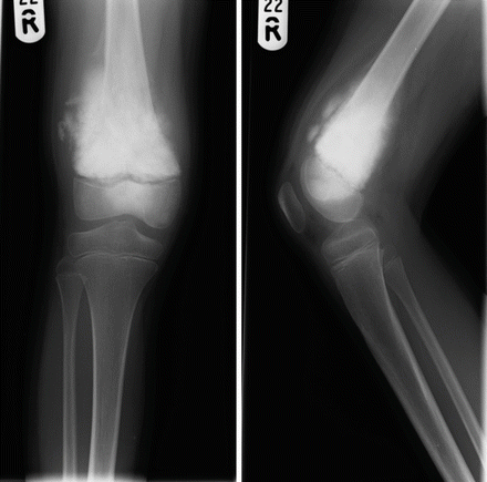
Radiographs provide valuable diagnostic information and some tumours have very characteristic appearances. Lesions are described by their location within the bone (diaphyseal, metaphyseal, epiphyseal) and their relationship with the cortex. Lesions may be lytic, sclerotic or demonstrate a mixed pattern. Slow growing tumours generally have a narrow zone of transition with a well-demarcated appearance in comparison to fast growing tumours, which more commonly have a permeative, wide zone of transition ± a periosteal reaction or elevation (endosteal scalloping or Codman’s triangle which can be seen in Fig. 16.1). Aggressive tumours may have cortical erosion (scalloping) where as slower growing tumours may result in bony expansion.


Figure 16.1
Plain radiographs demonstrating an extensive densely osteoblastic distal femoral metaphyseal osteosarcoma, which has spread into the central aspect of the epiphysis but does not appear to involve the joint. There is also a circumferential extraosseous extension. A Codman’s triangle is also visible
Cross Sectional Imaging & Scintography
Magnetic resonance imaging (MRI) and computerised tomography (CT) allow detailed information on local and distant staging. MRI can identify when tumours cross compartments, clarify the extent of oedematous change, the relationship to neurovascular structure and joints as well as identifying skip lesions. Positron emission tomography (PET CT) can help assess tumor activity and metabolism, differentiate between benign and malignant tumours, plan operative procedures, monitor response to chemotherapy and help to predict a patient’s prognosis. Bone scintography and radionuclide scanning can be useful in detecting small lesions, skip lesions or metastases.
Biopsy
Histological specimens can be gained through incisional, percutaneous or excisional biopsy. Primary malignant bone tumours are complex and rare and, as such, should be managed at regional specialist centres to minimise the chance of incomplete excisional biopsy and ensure the incision/tract is well planned and marked so that it can be excised when the tumour is removed. A biopsy should only be performed after appropriate cross sectional imaging has been undertaken as the biopsy can lead to haemorrhage, fracture or infection, which could significantly alter the diagnostic accuracy.
Percutaneous biopsy can be performed via a core biopsy or fine needle aspiration cytology (FNAC). They are commonly performed under image guidance to avoid a necrotic core and a non-diagnostic specimen. Histological analysis, immunohistochemistry and cytogenetics can achieve excellent diagnostic accuracy. Samples in the form of fresh specimens should be sent urgently to specialist centres as certain decalcification protocols can reduce the diagnostic accuracy.
Blood Tests
Blood tests may be important in the diagnosis, establishing a baseline prior to potent cytotoxic therapy as well as guiding prognosis such as alkaline phosphatase in osteosarcoma.
Classification Systems
Despite the rarity of bone tumors there is a very wide spectrum of entities. Classification is based on the recognition of the dominant tissue, the architecture, and type of matrix produced by the tumor.
Soft tissue tumours are staged using the Enneking Staging system; G = histological grade (1–3), T = Size, N = Lymph node involvement and M = Metastasis (Table 16.2).
Table 16.2
Enneking staging of bone tumours
Grade: |
G1 – Low grade, moderate cytological atypical and a low risk of metastasis,
Stay updated, free articles. Join our Telegram channel
Full access? Get Clinical Tree
 Get Clinical Tree app for offline access

|


