40 Neuromuscular disorders are a group of diseases that affect the normal operation of the pathways that allow the brain to communicate with and control the function of the muscles. Neuromuscular scoliosis is a common complication of these diseases, with an incidence of 25–90% depending on the underlying cause. The accepted classification scheme for neuromuscular disorders has been described by the Scoliosis Research Society (Table 40.1). In North America the most frequent diseases contributing to the development of neuromuscular scoliosis are cerebral palsy, myelomeningocele, muscular dystrophies, and post paraplegia. In the developing world, poliomyelitis is still occasionally seen. The exact cause of neuromuscular scoliosis is unknown. Contributing factors include paraplegia, mechanical forces, congenital abnormalities, and intraspinal abnormalities, such as tethering of the spinal cord, diastematomyelia, and syringomyelia. Asymmetric tone of the paraspinal muscles may result in increased tone or strength on one side with decreased tone on the other side, resulting in a coronal plane deformity. Mechanical forces causing spinal deformity may result from unilateral hip dislocation or pelvic obliquity with consequential compensatory deformity of the spine.1 As the spine collapses, pressure builds on the concave side of the vertebral body, increasing the load and decreasing growth on that side (Heuter–Volkmann law). The resultant “wedging” of the vertebral body compounds the curvature and increases the speed of scoliotic progression. In neuromuscular scoliosis, the curves tend to be early-onset, progress after skeletal maturity, and be long sweeping curves that may include the sacrum, with resultant pelvic obliquity. Curves are worse in patients who are nonambulatory, and the incidence and severity increase with the degree of neurologic involvement, with the severity of mental retardation, and with decreased functional status.2 Patients with curves greater than 40 degrees before the age of 15 tend to have larger curves in the future, and fusion should be considered in these patients. Spinal deformity, if left untreated, may result in compromise of walking and independent transfers, increased energy consumption in gait, and loss of trunk balance, which may require patients to use their hands for stability, effectively reducing their function because they cannot use their hands for tasks. The deformity may also result in pain and compromise of respiratory function and nutrition as well as pressure sores over the hips and/or ischium. These pressure sores may make sitting difficult and may require surgical correction of the curve even in patients who are nonambulatory. Table 40.1 Classification of Neuromuscular Scoliosis Established by the Scoliosis Research Society
Neuromuscular Scoliosis
![]() Classification
Classification
Etiology and Natural History
Neuropathic | Myopathic |
Upper motor neuron | Muscular dystrophy |
Lower motor neuron | |
Combined |
 Workup
History
Because of the early onset and rapid progression of neuromuscular scoliosis, rapid diagnosis and treatment are critical. Central nervous system abnormalities, premature birth, seizure disorders, and hydrocephalus are important to note. A family history of neurologic or muscular disease can provide insight into the likely clinical course. Information about developmental milestones and conditions during the perinatal period, such as maternal diabetes or perinatal anoxia, aids in identifying underlying conditions. For example, nonprogressive neuromuscular disorders like cerebral palsy are apparent early in life, whereas progressive neuromuscular conditions like muscular dystrophies tend to present later with loss of developmental milestones. Finally, a complete review of systems with information about past pulmonary, cardiac, renal, or nutritional disorders as well as symptoms of bladder or bowel dysfunction, weakness, or numbness aids in diagnosis and affects treatment options.
Physical Examination
A complete physical examination is important, as scoliosis may be the first clinical appearance of neuromuscular disease. Examination begins with the inspection of the skin lesions, such as sacral dimpling or hairy patches, which may indicate subtle underlying spinal abnormality. A thorough neurologic examination, including patterns of weakness, reflexes and joint motion, tone, contractures and atrophy, should be performed.
From this one should be able to differentiate spastic-type scoliosis from flaccid neuromuscular. It is not uncommon for patients with myelomeningocele to have a neuromuscular scoliosis that is the consequence of a mixture of spasticity and flaccid paralysis, with possible congenital malformation of the spine as well. The spinal examination should be conducted in a supine position and, if possible, sitting and standing positions to assess overall rigidity of the spine. Sagittal plane abnormalities should also be noted as hyperlordosis of the lumbar spine or kyphosis, as these patients may have a “collapsing spine” secondary to muscular weakness. Other deformities, such as rib prominence, shoulder asymmetry, trunk imbalance, or pelvic tilting, should be identified. Pelvic obliquity is often present in neuromuscular curve, and its cause (suprapelvic/spinal deformity, infrapelvic/hip contracture) can be evaluated with a simple test. The patient is placed prone on the examination table with the hips flexed over the edge of the table. This flexed position of the hips typically eliminates the hips as a cause of pelvic obliquity. It is then possible to visualize whether the pelvis is perpendicular to the trunk and assess whether there is any pelvic obliquity arising from the spine. In the case where the pelvis is oblique to the spine, one can assess the reducibility of the pelvic obliquity by abduction and adduction of the hips. This will determine whether the pelvic obliquity is fixed or not. In this position one will also assess whether there is any pelvic obliquity arising from the hips by looking at the pelvis and extending and abducting and adducting the hips. Functional assessment of the patient is important to help plan treatment choice. The patient’s ambulatory status, whether the patient is wheelchair bound, and upper extremity function need to be assessed. If the patient is ambulatory, it is essential to assess gait and see whether the patient is utilizing the lumbosacral junction, as fusion to the sacrum may impede mobility later.
Spinal Imaging
Plain radiographs standing or sitting, including posteroanterior (PA) or lateral views, are most useful for evaluation of curve progression. Supine films or traction films are also useful in assessing curve flexibility. Magnetic resonance imaging (MRI) of the entire spine is performed in cases of neuromuscular scoliosis where one suspects intraspinal lesions, such as syringomyelia, cord tethering, myelomeningocele, or spinal tumor.
 Treatment
The goal of treatment in neuromuscular scoliosis is to restore and maintain a spine balanced in the coronal and sagittal planes over a level pelvis, to preserve respiratory function, and to halt progressive loss of functional independence. Management may include observation, orthotic treatment, or surgery.
In general, curves less than 20 degrees can be observed. In nonambulatory patients, seating supports or customized seating systems may help improve trunk support and accommodate spinal or pelvis deformity, especially when curves are smaller or in patients with severe mental retardation and those patients unlikely to gain any functional improvement from surgery.
Bracing should be considered in curves > 20 degrees. Although brace management is rarely a definitive measure, it may be used in younger patients to slow curve progression and to delay surgical treatment. Bracing has been shown to be of some benefit in patients with short curves and those < 40 degrees with muscle hypotonia or mild spasticity. Types of bracing include the thoracolumbosacral orthosis (TLSO) and the suspension trunk orthosis (STO) for patients who lack voluntary muscle control. The STO attaches to the patient’s chair and controls trunk shift and pelvic obliquity without compromising respiratory function or causing pressure ulcers.3 The Milwaukee brace is rarely used nowadays for ambulatory patients who can maintain trunk control.
Indications for surgery include a progressive deformity, deformity that compromises respiratory or cardiac function, and curve-related pain that is unresponsive to nonoperative measures. Compared with idiopathic curves, neuromuscular curves tend to be more rigid and are often associated with pelvic obliquity (Fig. 40.1). Historically, these curves have been treated with posterior or circumferential fusion. The curve severity and rigidity and pelvic obliquity will affect the distal extent of the fusion. Inclusion of the pelvis is usually considered when there is truncal decompensation, when there is fixed pelvic obliquity > 15 degrees, and if the sacrum is part of the curve.2,4 Luque wire segmental instrumentation, often combined with Galveston pelvic fixation and hybrid hook–pedicle screw construct, has been employed (Fig. 40.2A–D). Hitesh et al.5,6 have looked at the posterior approach using all-pedicle screw fixation for neuromuscular scoliosis compared with adolescent idiopathic scoliosis. In their studies they obtained a comparable correction of Cobb angle, pelvic obliquity, and apical derotation without the need for anterior release.
Anterior surgery should be considered for patients with rigid deformities, in skeletal immaturity where there is potential for crankshaft phenomenon, or where risk of pseudarthrosis is high (e.g., myelomeningocele). It is usually performed in combination with posterior fusion and instrumentation and has been traditionally shown to have greater correction of scoliosis compared with either procedure alone. However, Basobas et al. and Tokala et al.7,8 have stated that in select patients, anterior fusion and instrumentation alone may have comparable results while preserving motion segments and, in some cases, avoiding instrumentation of the pelvis or sacrum.
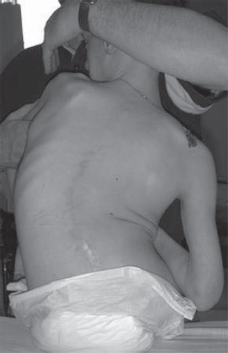
Fig. 40.1 Scoliosis in a patient with spastic quadriparetic cerebral palsy. The curve is long, sweeping and rigid, associated with pelvic obliquity which has affected his sitting balance and function.
Related posts:

Stay updated, free articles. Join our Telegram channel

Full access? Get Clinical Tree


