Musculoskeletal Neoplasms


Musculoskeletal Neoplasms


Mark J. Kransdorf

Thomas H. Berquist


INTRODUCTION

The imaging evaluation of musculoskeletal tumors has undergone dramatic evolution with the advent of computer-assisted imaging, specifically, computed tomography (CT) and magnetic resonance imaging (MRI). Despite these sophisticated imaging modalities, the objectives of initial radiologic evaluation remain unchanged: detecting the suspected lesion; establishing a diagnosis or, more frequently, formulating an appropriate differential diagnosis; and radiologic staging of a lesion.1 This chapter reviews techniques required for MRI in the evaluation of soft tissue and bone tumors, including coil selection, imaging planes, and pulse sequence selection. Particular emphasis is on those soft tissue and bone diagnoses that may be confidently made or suggested by MRI and lesions that are frequently encountered as incidental findings on examinations obtained for unrelated reasons. The use of MRI for differentiating benign from malignant soft tissue lesions, follow-up evaluation for differentiation of recurrent tumors from postoperative or radiation change, and response to therapy are also covered. Imaging evaluation for diagnosis, staging, and biopsy approaches is also discussed.


TECHNIQUES

MRI has emerged as the preferred modality for evaluating musculoskeletal lesions and should be obtained after radiographic evaluation. Critical assessment by multiple investigators2,3,4,5,6,7,8 has demonstrated the superiority of MRI over CT in delineating the extent of a musculoskeletal lesion and in defining its relationship to adjacent neurovascular structures. Recently, however, the superiority of MRI in the staging of musculoskeletal tumors has come into question. In a multi-institutional study of 316 patients with primary bone and soft tissue malignancies, the Radiology Diagnostic Oncology Group found no statistically significant difference between CT and MRI in determining tumor involvement of muscle, bone, joint, or neurovascular structures.9 Despite this caveat, most radiologists are comfortable with the use of MRI in the evaluation of musculoskeletal tumors, and we believe it is the modality of choice and, when used in conjunction with a systematic approach, can correctly diagnose most masses.

This is documented in recent American College of Radiology Appropriateness Criteria.10,11,12 Modalities are categorized as 1-3 for usually not appropriate, 4-6 for may be appropriate, and 7-9 for usually appropriate with 9 being the highest score.10,11,12 The criteria for osseous neoplasms suggest that initial radiographs (scored the highest or 9) are still important for morphologic detail and selection of additional studies.10 MRI is recommended when radiographs are indeterminate or normal in the presence of symptoms (scored 9, with technetium bone scans as an alternative,
but scored at 4). MRI is also scored at 9 if radiographs are suspicious for malignancy with CT scored at 5 and FDG-PET imaging also scored at 5.10 CT is still recommended if osteoid osteoma is suspected (scored 9 with bone scans as an alternative and scored 6 of 9).10

Patients with suspected soft tissue masses are still recommended to have initial radiographs to assess soft tissue features and select the next imaging studies (scored 9).11 In the setting where radiographs are normal or equivocal additional studies are often required. Under these circumstances MRI with and without contrast is scored at 9 and ultrasound of the region of interest is scored at 7. If a calcified mass is identified on radiographs both CT and MR are scored at 9 as the next imaging study to be performed. Superficial or juxta-articular masses should also be imaged with MR with and without contrast (scored at 9) with ultrasound as an alternative (scored at 7).11

The basic principles of MRI (see Chapter 1) and general techniques (see Chapter 3) have been previously discussed. Certain technical factors, however, require special attention and need to be emphasized: Patient positioning, coil selection, imaging planes, pulse sequence selection, and imaging limitations.


Positioning and Coil Selection for MRI

Positioning of patients and the choice of proper coils for studies of musculoskeletal neoplasms are essential to minimize motion artifacts and obtain optimal signal-to-noise ratios. Evaluation of the trunk and thighs is most efficacious using the torso coil with the patient supine. If a gluteal soft tissue lesion is suspected, the prone position may be helpful to avoid distortion of the posterior soft tissues. The torso coil also allows comparison of both lower extremities and can provide more complete evaluation of the entire osseous or soft tissue region of interest (i.e., femur, tibia, etc.). For certain skeletal neoplasms, imaging the entire bone or region of interest is important to be certain that skip lesions are not overlooked (Fig. 12.1).13,14,15,16,17

The peripheral lower extremities distal to the knee and the upper extremities are usually evaluated using flexible coils or circumferential volume coils. These coils are important in achieving superior image quality and are selected on the basis of the body part and patient position needed for the examination. For example, a lesion in the shoulder might be easily evaluated using the conventional shoulder coil. A lesion in the humerus should be studied using a larger multichannel phased array coil to include a larger area of the upper extremity (Fig. 12.2). The circumferential volume extremity coil provides a more uniform signal, but it must be positioned in the center of the gantry with most imagers. Positioning is not difficult for evaluating the knee, calf, or foot and ankle, but evaluation of the forearm and wrist may require that the patient place his or her arm above the head (see Chapter 11). The latter position is uncomfortable, leading to motion artifacts that reduce image quality.14,15


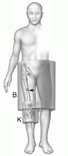
Figure 12.1 Illustration of a primary sarcoma in the knee with a mid femoral skip lesion. The knee coil would be optimal for imaging the knee, but the skip lesion cannot be identified unless the entire femur is examined.

New coils are now becoming available that allow superior image quality and more flexibility in the amount of area covered. These array coils are commonly used for the spine and are very useful for evaluating large segments of the spine in patients with suspected lymphoma, myeloma, or metastatic disease.13,14,15,16,17,18


Imaging Planes and Pulse Sequence

In a given clinical setting, numerous pulse sequences could be used to evaluate the musculoskeletal system. These include the commonly used spin-echo(SE) sequences (short echo time [TE], repetition time [TR], and long TE, TR), short TI inversion recovery (STIR) sequences, gradient-echo (GRE) sequences, and fast spin-echo techniques.13,14,15,16,17,18 For most patients with suspected bone or soft tissue neoplasms, spin-echo sequences are well suited for lesion detection and characterization.13,14,15,16,17,19,20,21,22 With these sequences, normal tissues have predictable signal intensity. Fat and bone marrow have high signal intensity on T1-weighted and intermediate signal on T2-weighted spin-echo MR images. Muscle has intermediate signal intensity, whereas cortical bone, ligaments, tendon, calcium, air, and fibrocartilage appear dark or black. Flowing blood usually gives no signal, but this finding is inconsistent and varies with the flow rate and pulse sequences used. Nerves are usually of slightly lower intensity than muscles.14


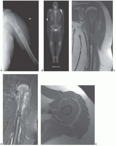
Figure 12.2 High-grade osteosarcoma. A: Radiograph demonstrates a destructive lesion in the upper humerus with periosteal reaction. B: Technetium-99m MDP whole body bone scan demonstrates intense uptake in the proximal humerus with no distant metastasis. Coronal T1- (C) and STIR (D) images and axial T1-weighted (E), T2-weighted (F), and postcontrast (G) images clearly demonstrate the extent of the tumor. H: Specimen radiograph of the resected proximal humerus. Note the significant (>5 cm) margin of normal bone adjacent to the distal tumor extent. I: The patient was treated with a long stem hemi-arthroplasty component and proximal allograft with cortical bone graft at the allograft-humeral junction. (From Berquist TH. Imaging of Orthopedic Fixation Devices and Prostheses. Philadelphia: Lippincott-Williams and Wilkins; 2009.)


Figure 12.2 (continued)

Lesions should be imaged in at least two orthogonal planes, using conventional T1- and T2-weighted spin-echo MR pulse sequences in at least one of these. Standard spin-echo imaging is most useful in establishing a specific diagnosis, when possible, and is the most reproducible technique, and the one most often referenced in the tumor imaging literature. It is the imaging technique with which we are most familiar for tumor evaluation, and it has established itself as the standard by which other imaging techniques must be judged.21 The main disadvantage of spin-echo imaging remains the relatively long acquisition times, especially for double-echo T2-weighted sequences.21 We most commonly use conventional T1-weighted spin-echo, fast spin-echo T2-weighted with orwithout fat suppression or STIR sequences, and post-contrast fat-suppressed T1-weighted images in the two optimal image planes based upon lesion location (Figs. 12.2 and 12.3).

Radiologists are most familiar with conventional axial anatomy, and axial T1- and T2-weighted spin-echo images should be obtained in almost all cases. The choice of
additional imaging plane or planes will vary with the involved body part, the lesion location, and its relationship to crucial structures. In general, the additional plane is sagittal with anterior or posterior masses and coronal with medial or lateral lesions. Oblique planes may also be a useful adjunct to reduce the problems from partial volume effects (Figs. 12.4 and 12.5). In these additional planes, a combination of conventional T1- and T2-weighted spin-echo (SE) images, turbo (fast) spin-echo images, gradient images, and STIR imaging is useful, as the cases require.


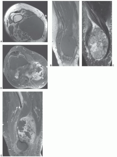
Figure 12.3 High-grade leiomyosarcoma. Axial (A) and sagittal (B) T1-weighted and sagittal turbo spin-echo T2-weighted sequences demonstrate a large soft tissue mass (arrows) with inhomogeneous signal intensity on the T2-weighted sequence (C). Axial (D) and sagittal (E) post-contrast fat suppressed T1-weighted images demonstrate areas of non-enhancement (arrows) due to tissue necrosis.


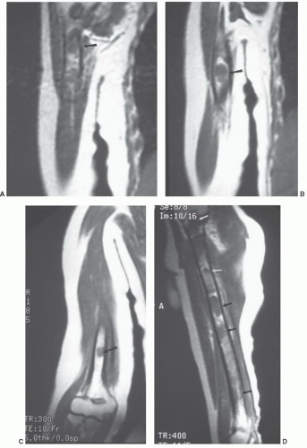
Figure 12.4 Middle-aged man with lymphoma and arm pain. A phased array coil was used to evaluate the right humerus. Coronal T1-weighted images (A-C) do not include the entire structure on a single image plane. Diffuse involvement (arrows) is apparent when a sagittal image (D), which shows the entire length of the humerus, is selected.


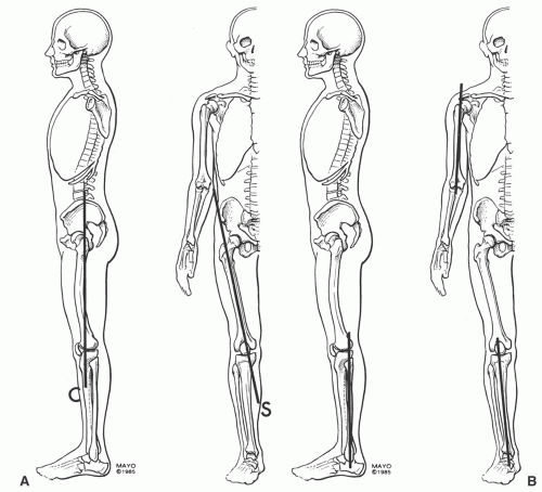
Figure 12.5 Skeletal illustrations demonstrating oblique planes required to accurately access the long bones. A: The femur is most easily evaluated in the oblique sagittal plane (S) due to the normal anterior bowing, which creates partial volume problems in the coronal plane (C). B: The oblique sagittal plane is also most useful for the humerus. Either the sagittal or the coronal plane can be used for the tibia and fibula.

Field of view (FOV) is dictated by the size and location of the lesion. In general, a small FOV is preferred; however, the field of view must be large enough to evaluate the lesion and allow appropriate staging (Fig. 12.1). When an extremity is being evaluated, it is not usually necessary to obtain the contralateral extremity for comparison, unless no lesion is detected on initial sequences. It is useful to place a marker over the area of clinical concern to ensure it is appropriately imaged. This marker becomes important in evaluation of lesions such as a subcutaneous lipoma or lipomatosis, in which the lesion may not be appreciated as distinct from the adjacent adipose tissue (Fig. 12.6). When small superficial lesions are being evaluated, care should be taken to ensure that the marker or patient position does not compress the mass.

Fast scanning techniques allow for shorter imaging times, decreased motion artifact, and increased patient tolerance and patient throughput.14,22 They may add additional information and may be helpful in specific instances, although fast scanning techniques have not replaced standard spin-echo imaging. Gradient-echo imaging may be a useful supplement in demonstrating hemosiderin because of its greater magnetic susceptibility, and in general, susceptibility artifacts related to metallic material, hemorrhage, and air are accentuated on gradient-echo images (Fig. 12.7).23 Gradient-echo images may also be better in some instances to demonstrate the lesion-fat interface and to depict small surrounding vessels.23 Short TI inversion recovery sequences are very useful in evaluating subtle marrow abnormalities and are frequently added in addition to the conventional spin-echo sequences. This technique suppresses fat signal while providing an additive effect on T1 and T2 signal enhancement. The superior contrast achieved with this sequence can allow subtle lesions to be more easily identified.17,19 Some authors prefer short TE/TR spin-echo (500/20) and STIR sequences to conventional or fast spin-echo T1- and T2-weighted spin-echo sequences.26 Short TI inversion recovery imaging can be an adjunct in selective cases but should not replace conventional spin-echo sequences. Lesions are generally well seen on standard imaging, and in our opinion, STIR imaging tends to reduce the variations in signal intensities identified on conventional spin-echo MR images that are most helpful in tissue characterization.


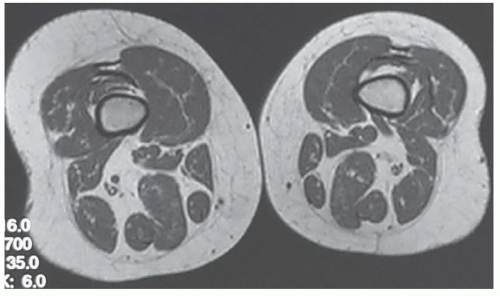
Figure 12.6 Lipomatosis of the right lower extremity in a 54-year-old woman presenting with “fullness” around the knee. Axial T1-weighted MR image of both distal thighs shows increased adipose tissue on right as compared with contralateral side. Images of both distal thighs were obtained after no cause for clinical findings was found on axial images of right knee.

Use of gadolinium as a contrast medium has been suggested to improve characterization of musculoskeletal neoplasms and for evaluating recurrence after surgery, radiation, or chemotherapy.25,26,27,28,29,30,31,32,33 Gadolinium diethylenetriamine pentaacetic acid (Gd-DTPA) produces increased signal intensity on T1-weighted spin-echo images because of the reduction in T1 relaxation time. Therefore, zones in neoplasms that show a marked increase in signal intensity correlate with highly vascular regions, whereas zones that show low intensity or no intensity are thought to be due to necrosis (Fig. 12.3).14,30,31 These data suggest that gadolinium may be valuable in identifying the areas of viable tumor within a mass, which is useful in determining whether residual tumor is still present. In addition, this information can be useful for selecting biopsy sites.31 One should try to avoid areas of necrosis and biopsy to concentrate only on those areas more likely to contain viable tumors.14,26,28,31


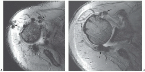
Figure 12.7 Gradient-echo axial images of the shoulder (A and B) following rotator cuff repair. There are multiple blooming artifacts (arrows) due to metal debris.

Early studies with gadolinium have suggested several techniques (static or dynamic using bolus injection and fast scan techniques) to evaluate enhancement rates, enhancement patterns, tumor volume,andother parameters.Results have been inconsistent and confusing, especially to radiologists who have learned morphologic tumor patterns that occur with specific lesions using conventional sequences (Fig. 12.8).14,30,31 Benedikt et al.32 studied 30 patients with soft tissue masses (22 benign and 8 malignant). Eighty-seven percent of lesions enhanced after gadolinium. This occurred in 82% of benign and 100% of malignant lesions. The pattern of enhancement (homogeneous vs. inhomogeneous) was not useful for differentiating benign and
malignant lesions.30,32 Dynamic studies using fast scan techniques were initially reported by Erlemann et al.27,28 Others have also studied these techniques, but with inconsistent results. In our practice, we reserve the use of dynamic gadolinium studies for selected cases (see below, limitations of MRI) (Figs. 12.8 and 12.9).


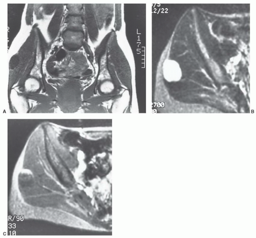
Figure 12.8 Myxoma. Coronal T1-weighted (A) and axial T2-weighted (B) images demonstrate a well-defined lesion with low intensity on T1-weighted (A) and high uniform signal intensity on T2-weighted (B) images. These features are characteristic of myxoma. After gadolinium injection (C), the enhancement pattern is inhomogeneous and nonspecific.

Spectroscopy or combined imaging and spectroscopic techniques have also been explored to improve lesion characterization and evaluate response to therapy.34,35 Spectroscopic techniques are not commonly used in most clinical settings. These techniques are summarized in Chapter 16.


Limitations of MRI

An important limitation of MRI is its relative inability to detect soft tissue calcification3,4,13,36,37; consequently, diagnoses that may be readily apparent on radiographs frequently remain nonspecific on MRI. As an additional caveat,13 noted that MRI failed to demonstrate soft tissue gas in 1 of 32 patients being evaluated for a soft tissue mass. CT may be useful in specific instances to identify the presence and pattern of subtle soft tissue mineralization and in those cases in which lesions are not adequately evaluated by radiographs.38 Although initial investigations maintained that CT is superior to MRI in detecting destruction of cortical bone,3,5,11 more recently it has been suggested that these two modalities are comparable in this regard.2,11,39 In fact, the ACR appropriateness criteria rank CT and MR equally (both scored 9) in the presence of radiographic evidence of soft tissue calcification.11

It has also been our experience that nonmetallic foreign bodies may be difficult to identify on MRI. In such cases,
MR images will show the changes associated with the foreign body, although the foreign body itself may have no signal and may be difficult to identify. We have found ultrasound a useful adjunct in such cases (Fig. 12.10).40


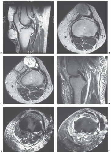
Figure 12.9 Sagittal T2-weighted (A), axial proton density-weighted (B) and T2-weighted (C) images demonstrate an inhomogeneous lesion in the anterior knee. The lesion was surgically removed and was found to be a high-grade pleomorphic sarcoma. Sagittal (D) and axial (E) T2-weighted images 1 year later show an area of increased signal intensity (asterisk) anterolaterally. There is a large amount of edema due to radiation therapy. Post-gadolinium image (F) shows peripheral enhancement with low intensity in the region of interest (asterisk) indicating a fluid collection and not a solid mass.


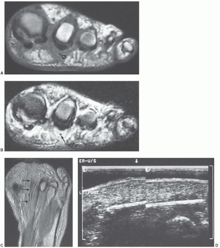
Figure 12.10 Toothpick foreign body in the foot of a 49-year-old woman. Coronal T1- (A) and T2-weighted (B) MR images show poorly defined abnormal signal intensity with a small mass below the second toe. The small signal void within the mass is the toothpick foreign body (arrow). C: Axial proton density-weighted MR image shows the foreign body as a linear signal void (arrows). D: Follow-up ultrasound shows the toothpick to better advantage.


It must be emphasized that it is essential to interpret MR images in conjunction with appropriate corresponding radiographs (and other imaging studies, if available). Unbending adherence to this principle will minimize interpretive errors.


STAGING OF MUSCULOSKELETAL NEOPLASMS

Decisions regarding the approach to musculoskeletal neoplasms may be relatively simple in the case of a localized benign lesion or more complex. The therapeutic decisions may be more complex depending upon the histology, location, bone and soft tissue involvement, and patient factors such as age, general health status, activity, and prognosis. Limb-saving procedures, often with extensive reconstruction, are usually considered when the lesion is located in a region that allows complete resection with wide surgical margin. This would require a 6 cm margin for bone and soft tissue sarcomas. Preoperative chemotherapy is often used preoperatively in conjunction with limb-saving procedures.41,42,43,44,45,46

Limb-saving procedures are usually contraindicated in patients with neurovascular encasement, which would result in functional impairment in the extremity following resection, and in patients with significant pathologic fractures that may result in dissemination of tumor cells. Lower extremity procedures may compromise growth in an immature skeleton. However, new prosthetic designs and other surgical options do not totally exclude limb-saving procedures in children in whom the growth plate may be at risk. Additional considerations for limb-saving procedures include the risk of local recurrence, long-term survival, functional outcome compared to amputation, psychological benefit, and immediate and delayed morbidities of the surgical procedure.45,46

Preoperative assessment includes multiple clinical and imaging factors. Enneking et al.,44 developed a functional system for pre- and postoperative evaluation that scored seven categories (0-5 points) for a total of 35 points. The categories included motion, pain, stability, deformity, strength, functional activity, and emotional acceptance.


Table 12.1 Enneking System: Staging of Musculoskeletal Neoplasms


Stage


Grade


Site


Metastasis


MR Features


IA


G1


T1


No


Abnormal signal intensity confined to bone or soft tissue compartment


IB


G1


T2


No


Abnormal signal intensity extends beyond the compartment


IIA


G2


T1


No


Abnormal signal intensity with cortex or capsular involvement


IIB


G2


T2


No


Abnormal signal intensity extending beyond bone or compartment


IIIA


G1-G2


T1


Yes


As above with distant metastasis


IIIB


G1-G2


T2


Yes


As above with distant metastasis


G1, low grade; G2, high grade; T1, intracompartmental; T2, extra-compartmental. (Adapted from Berquist TH. Magnetic resonance imaging of musculoskeletal neoplasms. Clin Orthop Relat Res. 1989;244:101-118; Berquist TH. MRI of the Musculoskeletal System. 3rd ed. Philadelphia, PA: Lippincott-Raven; 1996:735-840; Enneking WF. Staging of musculoskeletal neoplasms. Skeletal Radiol. 2115;13:196-207; and Enneking WF, Spanier SS, Goodman MA. A system for surgical staging of musculoskeletal sarcoma. Clin Orthop Relat Res. 1980;153:106-120.)


Lesion identification may be accomplished with radiographs or other imaging modalities.9,14,17 Once identified, multiple approaches are used to identify the type of lesion and extent of involvement. This may be initiated with lesion biopsy, followed by additional imaging for staging if the lesion is malignant. Planning the biopsy requires close communication with the orthopedic surgeon so that the needle path can be resected along with the tumor during the procedure.47,48 In addition, knowledge of the anatomic compartments is essential when planning bone or soft tissue biopsies (Fig. 12.11). Inappropriate needle approaches can unnecessarily contaminate compartments that could interfere with the planned limb-saving procedure. Biopsies are most often performed using CT guidance, but ultrasound or fluoroscopy can also be used to guide needle placement (Fig. 12.12).

Tumor staging typically involves the lesion (T), grade (G), nodal involvement (N), and metastasis (M). Thorough and accurate staging requires the use of multiple imaging modalities. Multiple staging systems have been used. Enneking et al.,44 proposed a staging system in 1980 that incorporated prognostic factors of local recurrence and metastasis, included surgical implications, and provided guidelines for adjuvant therapy (Table 12.1). Malignant bone lesions were staged on the basis of grade, interosseous and extraosseous extent, and whether there was associated metastasis. This system was later endorsed by the Musculoskeletal Tumor Society(MSTS) and the American Joint Committee on Cancer (AJCC). The MSTS staging system remains the most commonly used system (Table 12.2).46,49,50

Using the MSTS system, low-grade bone lesions are considered Stage I and high-grade lesions, Stage II. Metastases are considered Stage III regardless of the histology. The stages are subdivided based upon local extent. Lesions confined to bone are sub-classified A (intra-compartmental). Lesions that extend through the cortex into the soft tissues are sub-stage B (extra-compartmental).


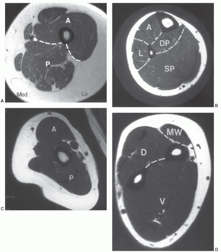
Figure 12.11 Compartmental anatomy. A: Thigh divided into anterior (A), posterior (P), and medial compartments. The anterior compartment contains the quadriceps and sartorius muscles; the posterior compartment contains the semimembranosus, semitendinosus, and biceps group; and the medial compartment contains the adductors and gracilis muscles. B: The calf is divided into, anterior (A), lateral (L), superficial posterior (SP), and deep posterior (DP) compartments. The anterior compartment contains the extensors hallucis longus and digitorum longus and the tibialis anterior. The lateral compartment contains the peroneus brevis and longus. The superficial posterior compartment contains the gastrocnemius and soleus and the deep posterior compartment comprises the tibialis posterior, flexor hallucis longus, and flexor digitorum longus. C: The arm is divided into anterior (A) and posterior (P) compartments. The anterior compartment contains the biceps brachii and brachialis and the posterior compartment the triceps. D: The forearm is divided into the volar (V), dorsal (D), and mobile wad (MW). The volar compartment contains the flexor muscle groups, pronator teres, and palmaris longus, and the dorsal compartment contains the extensor muscle groups and abductor pollicis longus. The mobile wad contains the brachioradialis and extensor carpi radialis longus and brevis. (From Berquist TH. Imaging of Orthopedic Fixation Devices and Prostheses. Philadelphia: Lippincott-Williams and Wilkins; 2009.)


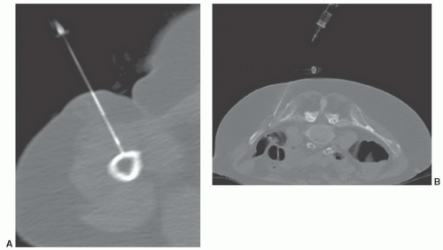
Figure 12.12 Needle biopsy. CT-guided needle biopsies of the humerus (A) in a patient with lymphoma. The needle enters through the anterior compartment. CT-guided biopsy (B) of an iliac lesion entering posteromedially. (From Berquist TH. Imaging of Orthopedic Fixation Devices and Prostheses. Philadelphia: Lippincott-Williams and Wilkins; 2009.)

Imaging of primary skeletal or soft tissue lesions and potential metastasis requires multiple modalities to optimize staging. Over the years accuracy has greatly improved with the additions of CT, MRI, and PET imaging.9,14,17,51 To assist the clinician, the radiologist must identify all of the following features so they can be correlated with the staging system:


Imaging Features for Tumor Staging

Tumor location and size (greatest diameter)

Intra- or extra-compartmental

Soft tissue involvement

Joint involvement

Neurovascular displacement or encasement

Skip lesions

Local and distant metastasis


Table 12.2 Staging of Malignant Musculoskeletal Neoplasms46,49,50


Stage


Grade


Site


Metastasis


I


IA


G1


T1


No metastasis


IB


G1


T2


No metastasis


II


IIA


G2


T1


No metastasis


IIB


G2


T2


No metastasis


III


G1or G2


T1 or T2


Metastasis


G1, low grade histologically; G2, high grade histologically; T1, intra-compartmental; T2, extra-compartmental.


Primary Skeletal Lesions

Radiographs or CR (computed radiography) images are still useful for detection of benign and malignant skeletal lesions. Radiographic features are extremely useful for characterizing the aggressiveness of the lesion.14,17 In addition, CT is useful in patients with subtle lesions, soft tissue calcification, tumor matrix, and thin cortex. MRI with contrast is most useful for evaluation of local bone, soft tissue compartment, and neurovascular involvement. The bone and soft tissue anatomy for at least 6 cm about the lesion should be evaluated to assess the ability to perform and the extent of resection for limb-saving procedures (Fig. 12.2).

For skeletal lesions the entire structure (i.e., entire femur and proximal and distal articulations) should be included on the image for operative planning and to exclude skip lesions (Figs. 12.1 and 12.2). Images should be obtained in at least two planes using T1- and T2-weighted sequences. Additional imaging with STIR may be indicated in some cases. Post-contrast-enhanced (Gadolinium) images are routinely obtained unless there is a history of allergy or renal compromise. The same approach should be used postoperatively to establish a baseline for patient follow-up.14,17

Angiography may be necessary in some cases to define neovascularity and vascular displacement or encasement. Angiography can also be used preoperatively to deliver chemotherapy and embolize tumor vessels.


Soft Tissue Neoplasms

Radiographs demonstrate bone changes and soft tissue calcification. Fatty lesions may also be evident on radiographs.
MRIis the technique of choice for detection and local staging of soft tissue lesions. Certain benign lesions, such as lipomas, myxomas, hemangiomas, and cysts have characteristic appearances that may obviate the need for biopsy or surgical intervention (Fig. 12.13). Malignant or indeterminate lesions are evaluated using T1- and T2-weighted conventional or fast spin-echo sequences with or without fat suppression. Gadolinium-enhanced T1-weighted images are also obtained. Two image planes are necessary to fully evaluate the extent, compartment, and neurovascular involvement (Fig. 12.3). Dynamic gadolinium techniques may be of value for differentiating benign form malignant lesions. Angiography is not often performed, but can be used for indications noted above.


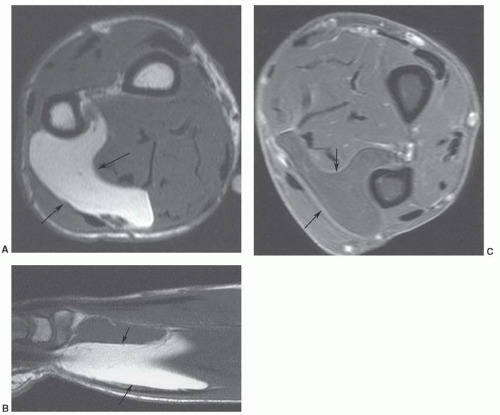
Figure 12.13 Benign lipoma. Axial (A) and sagittal (B) T1-weighted and axial fat-suppressed turbo spin-echo T2-weighted (C) images demonstrate a large fatty tumor in the distal forearm with no signal changes to suggest low-grade malignancy.


Metastasis

Radionuclide bone scans with technetium-99m MDP have long been the standard for detection of skeletal metastasis. With a few exceptions (myeloma, aggressive lytic lesions) this technique is still useful. MRI can be used for axial skeleton screening with axial and sagittal spine images and coronal images of the pelvis and hips using T1-weighted and/or STIR images. PET, especially PET/CT, plays an important role in tumor staging.51 Table 12.3 summarizes the staging for metastatic disease.


SOFT TISSUE NEOPLASMS

Patients with a soft tissue tumor or tumorlike mass usually present with nonspecific clinical findings, such as soft tissue swelling or a discrete palpable mass, sometimes with accompanying tenderness or pain.1,14,17 When clinical findings are equivocal, however, imaging evaluation can confirm the presence of a soft tissue lesion or reassuringly identify a suspected “bump” or “mass” as normal tissue or clearly distinguish it as nonneoplastic (Fig. 12.14).


Initial Evaluation

Despite dramatic technologic advances in computer-assisted imaging, the radiologic evaluation of a suspected soft tissue mass must begin with the radiograph. Radiographs may be diagnostic of a palpable lesion caused by
an underlying skeletal deformity (such as exuberant callus related to prior trauma) or bony exostosis that may masquerade as a soft tissue mass. Radiographs may also reveal the presence and nature of soft tissue calcifications, which can be suggestive and at times very characteristic of a specific diagnosis. For example, they may reveal the phleboliths within a hemangioma, the juxta-articular osteocartilaginous masses of synovial osteochondromatosis, the peripherally more mature ossification of myositis ossificans, or the characteristic bone changes of other processes with associated soft tissue involvement (Fig. 12.15).


Table 12.3 Staging System for Metastases44,45


Tumor sites


Intra-compartmental (T1)


Extra-compartmental (T2)


Intraosseous


Extraosseous extension


Intra-articular


Extra-articular extension


Intrafascial compartments


Extrafascial extension


Posterior calf


Anterolateral leg


Anterior thigh


Posterior thigh, etc.


Tumor grade


Low grade (G1)


High grade (G2)


Parosteal osteosarcoma


Classic osteosarcoma


Secondary chondrosarcoma


Paget sarcoma


Giant cell tumor


High-grade pleomorphic sarcoma


Myxoid liposarcoma


Chondroma


Angiosarcoma


Adamantinoma


Neurofibrosarcoma


In addition, plain radiographs are the best initial method of assessing coexistent bony involvement, such as osseous remodeling, periosteal reaction, or overt osseous destruction.45 Unlike its intraosseous counterpart, however, the biologic activity of a soft tissue mass cannot be reliably assessed by its growth rate. A slow-growing soft tissue mass that may remodel adjacent bone (causing a scalloped area with well-defined sclerotic margins) may still be highly malignant on histologic examination.46


Figure 12.14 Atrophied adductor muscle in a 24-year-old woman presenting with soft tissue asymmetry, suggesting a soft tissue mass. Axial T1-weighted MR image shows the marked atrophy to the left adductor longus muscle (asterisk). Atrophy was secondary to a previous muscle injury.

A soft tissue mass may also be the initial presentation of a primary bone tumor or inflammatory process. In such cases, the radiograph may also be useful. The diagnosis of a malignant bone tumor, such as Ewing sarcoma or primary lymphoma of bone, should be considered when a large circumferential soft tissue mass is present in association with an underlying destructive bone lesion. A subtle radiologic feature that may be used to separate inflammatory and neoplastic processes is that inflammatory processes typically obliterate fascial planes, rather than displace them.

Initial radiographs should be obtained with a low kilovoltage technique (i.e., less than 50 kV peak), thereby enhancing radiographic density differences between soft tissues such as fat and muscle.14


Specific Diagnoses

Despite the superiority of MRI in identifying, delineating, and staging soft tissue tumors, it remains limited in its ability to precisely characterize soft tissue masses, with most lesions demonstrating prolonged T1 and T2 relaxation times.52,53,54 There are instances, however, in which a specific diagnosis may be made or strongly suspected: Lipoma (Fig. 12.13); liposarcoma; benign vascular lesions such as hemangioma, arteriovenous malformation, and pseudoaneurysm; hemosiderin-laden lesions such as pigmented villonodular synovitis (PVNS); fibromatosis; subacute hematomas; and certain tumorlike lesions.21,55 Clearly, the percentage of cases in which MRI may correctly suggest the diagnosis will vary with the referral population.21 In general, a correct histologic diagnosis can be reached on the basis of imaging studies in approximately one-fourth to one-third of cases.21,37,56,57 Significantly higher levels of accuracy may be achieved with common benign lesions and by using a systematic approach to evaluation.


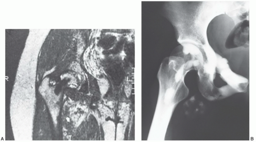
Figure 12.15 Melorheostosis in a 26-year-old woman presenting with hip pain and a soft tissue mass in the right groin. A: Coronal T2-weighted MR image of the hip shows a heterogeneous nonspecific soft tissue mass (arrow). The lesion showed signal intensity similar to that of skeletal muscle on corresponding T1-weighted image (not shown). An associated interosseous abnormality is noted in the proximal femur. B: Corresponding radiograph readily confirms the diagnosis of melorheostosis with soft tissue involvement.


Lipomatous Tumors


Lipoma

Soft tissue tumors are derived predominantly from primitive mesenchyme. Probably the most common mesenchymal tumor is lipoma, a benign tumor composed of mature adipose tissue.59,60,61,62 Patients with a soft tissue lipoma typically present in middle age (fifth and sixth decades). After an initial period of discernible growth, a lipoma usually stabilizes in size.58 A soft tissue lipoma is categorized by anatomic location as either superficial (cutaneous) or deep. The superficial lipoma occurs more commonly and is more sharply circumscribed and smaller in size than its deep-seated counterpart. A superficial (subcutaneous) lipoma may be inapparent, blending in with the adjacent subcutaneous fat on MRI. Unless a marker is placed over the mass before imaging, it may appear only as a thickening of the subcutaneous fat (Fig. 12.16). The deep-seated lipoma occurs most commonly in the retroperitoneum, chest wall, and deep soft tissue of the hands and feet. Retroperitoneal lipomas are relatively rare. Most large lipomatous retroperitoneal tumors are liposarcomas. The pathogenesis of a lipoma is unknown, although it is thought to represent a true mesenchymal neoplasm. It is usually a solitary lesion, although a small percentage of patients (5% to 7%) demonstrate multiple tumors, which can vary in number from a few to several hundred.63 A rare entity of familial multiple lipomas has also been reported.63,64,65 Interestingly, the fat within the lipoma is unavailable for systemic metabolism and, paradoxically, may actually increase in size during starvation.66

Depending on the size and location of the lesion, the plain radiograph may be either unremarkable or demonstrate a mass of fat density. The lipoma is well characterized on MRI, with the lesion having an appearance identical to that of subcutaneous fat on all pulse sequences, without discernible enhancement after the administration of intravenous gadolinium (Figs. 12.13, and 12.16, 12.17, 12.18).4,27,59,60,61,62,67,68,69,70

Lipomas occasionally contain other mesenchymal elements. In fact, the World Health Organization Committee for Classification for Soft Tissue Tumors defines nine distinct benign lipomatous lesions.60,62 These include simple lipoma, lipomatosis, lipomatosis of nerve, lipoblastoma/lipoblastomatosis, angiolipoma, myolipoma, chondroid lipoma, spindle cell lipoma, and hibernoma.62 In a review of 126 consecutive fatty masses by Gaskin and Helms,59 they found 13% simple lipomas, 50% benign variants, 13% chondroid lipomas, 6% osteolipomas, 6/5 hibernomas, 6% myolipomas, 6% angiolipomas, and 13% infarcted lipomas.59 The most common tissue variant is fibrous connective tissue, which may be in the configuration of septa and therefore appear as linear densities on CT70 or linear areas of decreased signal on MRI, regardless
of pulse sequence (Fig. 12.17).59,60,61,62,70 When significant fibrous tissue is present, these lesions may be termed fibrolipomas. It is important to remember that when a fatty lesion does not meet the imaging requirements for a lipoma, liposarcoma is typically the diagnosis of exclusion; however, lipoma variants are encountered more commonly than liposarcoma.59,71


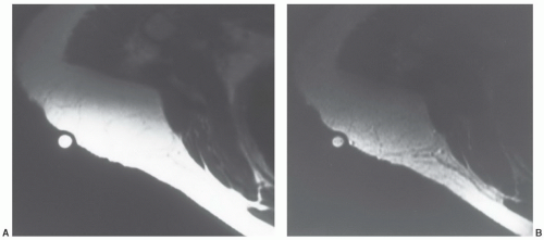
Figure 12.16 Superficial lipoma in the subcutaneous tissue of the shoulder of a 65-year-old woman. A: Axial T1-weighted MR image of the right shoulder shows a poorly defined mass, imaging identical to that of subcutaneous fat. Such lesions may be inapparent on MRI unless a marker is placed over the palpable abnormality. B: Corresponding non-fat suppressed T2-weighted MR also shows the lesion to image identical to that of subcutaneous fat.


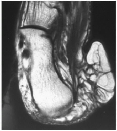
Figure 12.17 Recurrent superficial lipoma in the subcutaneous tissue of the heel in a 69-year-old woman. T1-weighted MR image shows the mass to have a lobulated contour with signal intensity identical to that of subcutaneous fat. The lesion has multiple linear septations of decreased signal intensity coursing through its substance. These showed similar decreased signal intensity on T2-weighted images (not shown). They corresponded to fibrous tissue on histologic examination. Lesions such as these are often referred to as fibrolipoma.

Soft tissue lipoma may be associated with changes in the skeleton.Cortical thickening may be seen in associationwith adjacent parosteal lipoma, and congenital osseous anomalies have been described adjacent to deep lipomas.72,73 Chondroid or osseous metaplasia is occasionally encountered within a lipoma, particularly if the lipoma is long standing. The term “benign mesenchymoma” is occasionally used to describe this type of lesion (Fig. 12.19).

A few cases of malignant transformation of lipomas have been reported, but these may represent cases where the subtle histologic features of malignancy were initially overlooked.52,74


Intramuscular and Intermuscular Lipomas

Intramuscular and intermuscular lipomas are relatively common benign lipomatous tumors that arise, respectively, either within or between skeletal muscles. They are members of a subgroup of lesions referred to as “lipomatous tumors” in which the fatty mass is intimately associated with specific nonadipose tissue. The remaining members of this subgroup of lesions are uncommon and include lipomas of the tendon sheath and joint, and lipomatosis of nerve.62,74 Although it arises within the muscle, intramuscular lipoma may actually involve both muscular and intermuscular
tissues; however, involvement isolated to the intermuscular region (intermuscular lipoma) is less common. Intramuscular lipoma occurs in patients of all ages but predominantly in adults, with most cases presenting in patients between 30 and 60 years of age.52 There is a slight male predominance. Patients typically present with a mass in the large muscles of the extremities, especially the thigh, shoulder, and upper arm. The fat within the intramuscular lipoma may infiltrate between skeletal muscle fibers, giving the intramuscular lipoma a striated appearance on gross inspection.


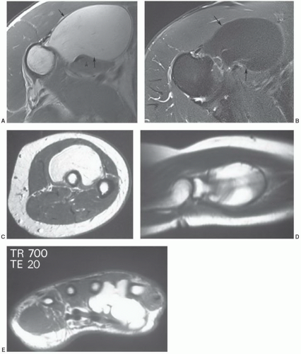
Figure 12.18 Benign lipomas, multiple patients. Axial T1-weighted (A) and T2-weighted (B) images of a benign lipoma along the proximal humerus with (arrows). C and D: Axial and sagittal T1-weighted images of a benign lipoma in the supinator muscle. E: Axial T1-weighted MR image of a lobulated lipoma in the hand.

Radiographs may reveal an intramuscular mass of fat density. An intramuscular lipoma may be identified on
MRI as a predominantly fatty mass (with signal intensity equal to that of the subcutaneous fat), infiltrating the adjacent skeletal muscle. The mass is usually well defined and sharply circumscribed, with imaging characteristics similar to that of an “ordinary lipoma.” This lesion has also been referred to as “infiltrating lipoma.” Despite being well-defined radiologically, margins are frequently infiltrating at microscopy, with adipose tissue intermingled with skeletal muscle fibers that are variably atrophic (Figs. 12.20 and 12.21). Matsumoto et al.75 reported the MR appearance of intramuscular lipoma in 17 cases and found the lesion to be homogeneously pure fatty tissue in 12 (71%), with the remainder being fat with intermingled muscle fibers, the latter showing a signal intensity identical to that of skeletal muscle on T1- and T2-weighted pulse sequences. An infiltrative margin was seen in 7 cases (41%).


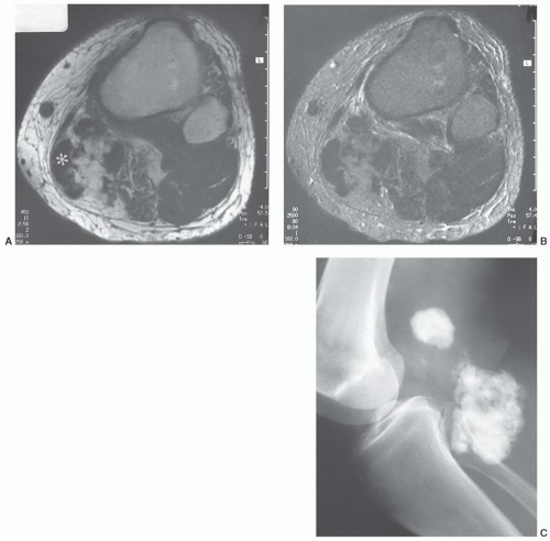
Figure 12.19 Benign mesenchymoma in the popliteal fossa of a 79-year-old man. A: Axial T1-weighted MR image shows a fatty mass with a central region of markedly decreased signal intensity (asterisk). B: Corresponding T2-weighted image shows similar findings. C: Radiograph shows densely mineralized mass.


Lipoma of Tendon Sheath and Joint

There are two variants of these rare tumors: A discrete solid fatty mass that extends along the affected tendon or within the affected joint and a “lipoma-like” lesion composed of hypertrophic synovial villi distended with fat. Synonyms for the latter include diffuse synovial lipoma or lipoma arborescens. Lipoma of tendon sheath most commonly arises in the hand and wrist and less commonly in the ankle
and foot. Lipoma arborescens usually involves the knee.60,76 About 20% of the time, knee involvement is bilateral.77


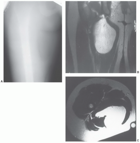
Figure 12.20 Intramuscular lipoma in the thigh of a 29-year-old woman. A: Anteroposterior radiograph shows a fat density mass in the medial aspect of the proximal right thigh. B: Corresponding coronal T1-weighted MR image shows the signal intensity of the mass to be identical to that of the subcutaneous fat. C: Axial T1-weighted localizes the mass to the adductor compartment.

Although it may arise de novo, it is frequently associated with degenerative joint disease, chronic rheumatoid arthritis, or prior trauma involving the affected joint.60 It may be a reactive process related to chronic synovitis. Lipoma arborescens of the subacromial-subdeltoid bursa has been reported in association with rotator cuff tears.78 Although all these lesions are rare, the lipoma arborescens form of synovial lipoma is encountered more frequently than the discrete form of synovial lipoma.

Radiologically, a lipoma of tendon sheath or discrete synovial lipoma is a focal lipomatous mass, similar to a superficial or deep lipoma. Patients afflicted with the lipoma arborescens form of synovial lipoma present with soft tissue swelling around the knee, which may or may not be radiolucent on plain radiographs.60 MRI examples of lipoma arborescens are rare, but limited experience has shown a fatty proliferation of the synovium with an associated joint effusion (Fig. 12.22). Osseous erosions at the articular margins have been reported in up to 38% of cases, associated synovial cysts are seen in 25%, and degenerative change in 13%.77,79


Neural Fibrolipoma

Fibrofatty enlargement of the median nerve was initially described in English literature in 1953, with the presentation of two cases to the American Society for Surgery of the Hand.80 This lesion has been reported under a variety of names, which include neural fibrolipoma, fibrolipomatous
hamartoma of nerve, perineural lipoma, fatty infiltration of the nerve, and intraneural lipoma.80,81 The term “neural fibrolipoma” was originally preferred because it better describes the underlying pathology.80 More recently, the WHO has adopted the term lipomatosis of nerve.62 The cause of this disorder remains unclear; it may be related to hypertrophy of mature fat and fibroblasts in the epineurium.81


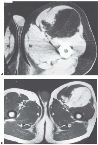
Figure 12.21 Intramuscular lipoma in the thigh of a 50-year-old man. A: Axial CT shows a fatty mass in the anterior aspect of the left thigh. There are some small areas of increased attenuation within the lesion. B: Corresponding axial T1-weighted MR image nicely shows the fatty nature of the mass. The area of increased attenuation on CT on the medial aspect of the mass images is identical to skeletal muscle and is compatible with muscle infiltrating the margin of the lesion. Similar findings were seen on T2-weighted images (not shown).

Patients with neural fibrolipoma typically present during early adulthood with a soft slowly enlarging mass occurring in the volar aspect of the hand, wrist, or forearm.60,81 Males and females are affected equally. Majority of lesions (78% to 96%) involve the upper extremity with the median nerve being affected in majority (80%) of the cases.60,81 The lower extremity is involved much less frequently (4% to 22% of cases).82,83 The lesion is typically present at birth or presents within the first 2 years of life.82,83 There is no familial predisposition.81 As noted above, approximately 80% of upper extremity lesions originate in the distribution of the median nerve.82,83 Accompanying symptoms include pain, tenderness, decreased sensation, and paresthesia. Carpal tunnel syndrome may be a late symptom.83 Patients may demonstrate macrodactyly,83,84 referred to as macrodystrophia lipomatosa, usually involving the second and third digits of the hand or foot. Macrodactyly was noted in 7 of 26 (27%) cases reported by Silverman and Enzinger81 and 12 of 18 (67%) lesions reported by Amadio et al.82 Multiple digits may be involved. Surgical excision is not without risk, and motor and sensory deficits have been reported after resection.83,84

Grossly, the lesion is described as a fusiform, sausage-like enlargement of the nerve by fibrofatty tissue,81
appearing as a tan yellow mass within the nerve sheath.84 Microscopy demonstrates infiltration of the epineurium and perineurium by fibrofatty tissue.81 Cases in which there is macrodactyly are histologically indistinguishable from those in which there is no macrodactyly.81


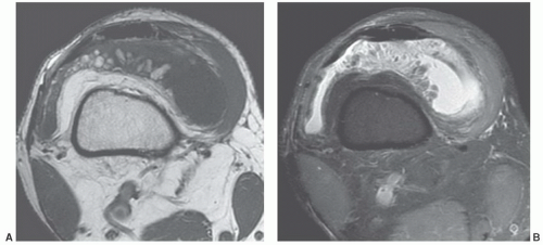
Figure 12.22 Lipoma arborescens in a 58-year-old man. Axial T1-weighted (A) and T2-weighted (B) MR images show a joint effusion with fat in a frond-like pattern, representing the synovial villi, distended with adipocytes.

Radiographs of patients with macrodystrophia lipomatosa demonstrate both bone and soft tissue abnormalities. The phalanges are long, broad, and oftensplayed at their distal ends. The osseous overgrowth may be marked and disproportionately large with extensive secondary degenerative change (Fig. 12.23).60,85 The MR appearance of the nerve is characteristic, reflecting the morphology of the lesion. MRI demonstrates small longitudinally oriented cylindrical areas, approximately 3 mm in diameter. These cylindrical areas demonstrate a decreased signal intensity on a background of increased signal intensity, thought to represent the nerve fascicles with epineural and perineural fibrosis on a background of fatty tissue (Fig. 12.24).84 The amount of fat present varies; however, it tends to be more prominent between the nerve fibers rather than surrounding them peripherally.86

Amadio et al.82 reviewed the Mayo Clinic institutional experience with macrodactyly from 1950 to 1985. Neural fibrolipoma was the most common condition associated with macrodactyly of the upper extremity, seen in 10 of 22 cases. Other lesions included five vascular cases, five idiopathic cases, and two cases of neurofibromatosis. In contradistinction, it was noted that neural fibrolipoma was the least common cause associated with macrodactyly of the lower extremity, being identified in 1 of 43 cases. In the remaining cases, the cause was vascular in ten, neurofibromatosis in one, and idiopathic in the remaining cases.87 In this study, idiopathic cases were defined as those patients with no stigmata of neurofibromatosis or congenital malformation. The authors also noted nine cases of hemihypertrophy. The differential diagnosis of localized gigantism would also include Proteus syndrome.88


Parosteal Lipoma

Parosteal lipoma is an unusual lesion that represents about 0.3% of all lipomas.73 The lesion was originally described as a “periosteal lipoma” by Seering89 in 1966, with the term “parosteal lipoma” subsequently suggested by Power90 to indicate that the lesion does not arise within the periosteum. The term “parosteal lipoma” is generally accepted over periosteal lipoma because the former indicates the juxtaposition of the lesion to the surface of the bone without identifying the tissue of origin.62 Patients are usually adults with an average age of approximately 50 years (range, 4 to 64 years). There is a male predilection.73,91 Lesions are most common in the thigh, forearm, calf, and arm adjacent to the diaphysis or metadiaphysis of bone.73,91 Virtually all lesions are singular, with the exception of a case reported by Goldman et al.68 in which there was a coincident intramuscular lipoma. Patients typically present with a painless soft tissue mass. Muscle atrophy is not uncommon.91

Lesions are encapsulated and adherent to the underlying periosteum.91 Histologically, a parosteal lipoma is identical to a superficial or deep lipoma.68 Cartilage and bone metaplasia may bepresent. Cartilage is typically hyaline, but small foci of fibrocartilage may be seen at the periphery of large osseous excrescences.91 The septa seen within the lesion contain fibrovascular tissue.91 At the point of attachment there may be a bony excrescence or cortical thickening. The cortical thickening is likely to be secondary to tugging on the periosteum.

Radiographs reveal a well-defined radiolucent mass.68,91 Variable fibrovascular septa may be present within the
mass.86 The adjacent bone may demonstrate solid periosteal reaction, cortical thickening, saucerization, or osseous excrescences.68,91 Osseous changes are seen in 67% to 100% of cases,68,91,92 although these may be subtle. The periosteal reaction in two cases reported by Murphey et al.91 was minimal, being identified only on magnification radiography. The osseous excrescences do not demonstrate the cortical and medullary continuity or hyaline cartilaginous cap seen with a true osteochondroma.68


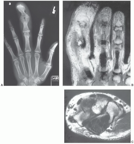
Figure 12.23 Macrodystrophia lipomatosa. A: Radiograph of the hand demonstrates abnormalities of both bone and soft tissue. The phalanges are long, broad, and splayed at their distal ends. The osseous overgrowth is marked and disproportionately large with extensive secondary degenerative change. Coronal (B) and Axial (C) MR images in a different patient demonstrating fatty change in the finger and a neural fibrolipoma of the median nerve in (C).

MRI demonstrates the fatty nature of the mass with signal intensity identical to that of subcutaneous fat on all pulse sequences. Fibrovascular tissue septa may demonstrate increased signal intensity on long TR images, as may hyaline cartilage, which may also be found within lesions.91 In addition, MRI can identify muscle atrophy as increased striations of fat within muscle (Fig. 12.25).


Lipomatosis

Diffuse lipomatosis is an entity characterized by diffuse overgrowth of mature adipose tissue infiltrating through the soft tissues of an affected extremity or body trunk.60,62 Microscopically, this lesion is indistinguishable from lipoma or intramuscular lipoma (Fig. 12.26).

Patients affected with lipomatosis usually present during the early years of life, often by age 2, although there have been scattered reports of presentation in adulthood.52,93 Coode et al.93 noted that the designation of diffuse congenital lipomatosis has been suggested on the assumption
that adult cases represent delayed presentation. Diffuse lipomatosis typically affects the limbs, although involvement of the trunk and chest wall may be seen.93 Lipomatosis may be associated with coexistent osseous hypertrophy, but unlike macrodystrophia lipomatosa, the nerve is unaffectedand the disease is not confined to an extremity. Reported as rare,52 we believe that mild cases of lipomatosis are not uncommon and may be easily overlooked (Fig. 12.6).


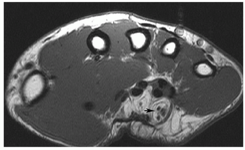
Figure 12.24 Neural fibrolipoma. Axial T1-weighted image of the wrist demonstrating a fibrolipoma of the ulnar nerve (arrow).


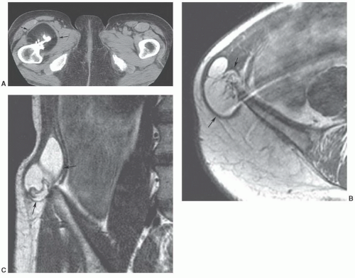
Figure 12.25 Parosteal lipomas. A: Axial CT of a parosteal lipoma of the upper femur (arrow). Axial (B) and coronal (C) T1-weighted images in a different patient demonstrating a parosteal lipoma of the ilium (arrows).

Lipomatosis may be distinguished from the rare symmetric lipomatosis, which is also known as Madelung disease or benign symmetric lipomatosis. Symmetric lipomatosis is seen almost exclusively in middle-aged men, often, though not always, with a history of alcoholism or liver disease.94 In such cases, the masses may appear suddenly and grow rapidly, infiltrating the neck and extending into the axilla or extending into the back.94 The growth rate is irregular, and the lesion may cease to grow spontaneously or become intermittently quiescent.94 Involvement may occur in the groin as well (Fig. 12.27).60,94


Liposarcoma

Liposarcoma is the second most common soft tissue sarcoma encountered in adults, accounting for 16% to 18% of all malignant soft tissue tumors.52 Liposarcomas are classified into four histologic subtypes: well differentiated, myxoid, pleomorphic, and dedifferentiated.53,62 The well-differentiated variant is considered to be a low-grade malignancy, whereas the pleomorphic and dedifferentiated types are considered to be high grade, with a high rate of local
recurrence and metastasis. While the round cell liposarcoma was previously identified as distinct subtypes by the WHO Classification of Soft Tissue Tumors, the myxoid and round-cell liposarcoma are now combined under the designation of myxoid liposarcoma.62 These lesions were known to form a histological continuum, and represented the ends of a common spectrum.58 Now under a single diagnosis, the pure myxoid lesion is considered an intermediate grade tumor at the low-grade end of this spectrum, while the hypercellular (round cell) morphology represents the histologically similar, high-grade counterpart. The presence of the hypercellular component is associated with a more aggressive clinical course and a significantly worse prognosis.95 Well-differentiated liposarcomas are most common, accounting for about 54% of all classified liposarcomas. Myxoid liposarcoma is next most common,
accounting for 28%, followed by dedifferentiated (10%), and pleomorphic liposarcomas.96 Liposarcomas tend to occur in both the retroperitoneum and extremities, with extremity lesions presenting about 10 years earlier than those in the retroperitoneum. Dedifferentiated liposarcomas are most common in the retroperitoneum, whereas the other subtypes are more common in the extremities.96


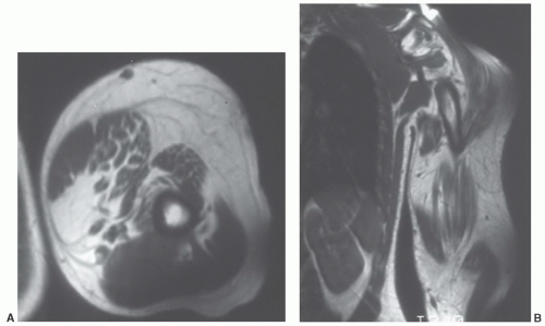
Figure 12.26 Upper extremity lipomatosis in a 23-year-old man. Axial (A) and coronal (B) T1-weighted SE MR images of the left upper extremity show diffuse overgrowth of adipose tissue.


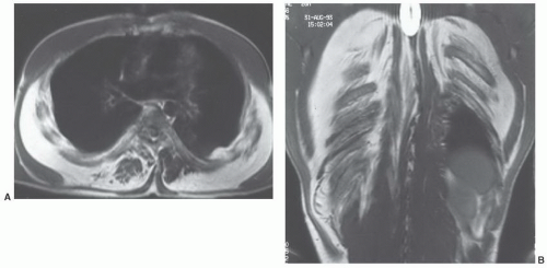
Figure 12.27 Symmetric lipomatosis in a 28-year-old man. Axial (A) and coronal (B) T1-weighted MR images show extensive, but symmetric, lipomatosis of the chest wall.


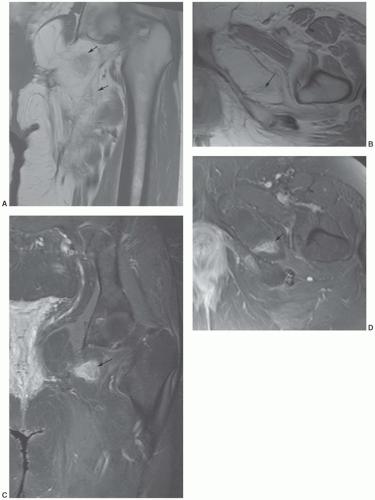
Figure 12.28 Well-differentiated liposarcoma of the left groin and upper thigh. Coronal (A) and axial (B) T1-weighted images demonstrate a fatty tumor with globular areas of low signal intensity (arrows). Coronal (C) and axial (D) T2-weighted images demonstrate corresponding areas of high signal intensity (arrows).


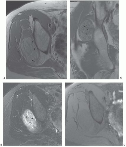
Figure 12.29 Well-differentiated gluteal liposarcoma. Axial T1- (A) and T2-weighted (B) images demonstrate a well defined (<10 cm) lesion with thickened septa (arrows). Coronal T1-weighted image (C) demonstrates similar findings (arrows). Post-contrast fat-suppressed T1-weighed image (C) shows no enhancement.

OnMRI,awell-differentiated (lipoma-like) liposarcoma will image as a predominantly fatty mass with irregularly thickened linear or nodular septa, which demonstrates a nonspecific decreased signal on T1-weighted and increased signal on T2-weighted spin-echo images (Figs. 12.28,12.29,12.30).97,98 Although a lipoma that is completely homogeneous and images identical to fat may be easily distinguished
from a well-differentiated liposarcoma, lipoma variants occur with an imaging appearance that will overlap that of a well-differentiated liposarcoma. The distinction of lipoma and well-differentiated liposarcoma is simple when the former is homogeneous with an imaging appearance identical to that of the subcutaneous adipose tissue. When nonadipose elements are present, however, this distinction may be quite problematic. Recent literature has documented awider spectrum for the imaging features of lipoma than had been previously appreciated, with a small but significant number of lipomas demonstrating prominent nonadipose areas and an imaging and appearance that may mimic that traditionally ascribed to well-differentiated liposarcoma.96 In these cases, the nonadipose areas represent fat necrosis and associated calcification, fibrosis, inflammation, and myxoid change. As a generalization, lesion size may also be useful, in that well-differentiated liposarcoma tends to be significantly larger than lipoma. In a recent review of 60 well-differentiated fatty tumors, the average largest dimension of malignant lesions was nearly twice that of benign lipomas (24 cm vs. 13 cm).96 Enhancement pattern may also be useful, with well-differentiated tumors showing contrast enhancement.98 Kransdorf et al.,61 noted that statistically significant features of liposarcoma included lesions more than 10 cm in size (p <.001), thick septa (p = .001), lesion less than 75% fatty tissue (p <.001), and globular or nodular areas of non-fatty tissue.61


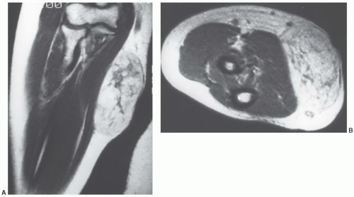
Figure 12.30 Well-differentiated liposarcoma of the upper extremity in a 34-year-old woman. A: Coronal T1-weighted MR image of the forearm shows a fatty subcutaneous mass. The mass is predominantly fatty but shows linear and globular areas of non-fatty tissue within it. B: Axial T2-weighted image shows linear areas of non-fatty tissue, although these are not as conspicuous as those seen in A. This is the type of lesion sometimes referred to as an atypical lipoma.

The dedifferentiated liposarcoma is a rare, interesting variant of the well-differentiated liposarcoma. A “dedifferentiated” sarcoma is best defined as a bimorphic neoplasm in which a borderline or low-grade malignant neoplasm is juxtaposed with a high-grade histologically different sarcoma.99,100 Although the concept of dedifferentiation was initially described in chondrosarcoma, the term “dedifferentiated liposarcoma” was introduced by Evans99 in 1979 to describe a histologically distinctive lesion in which a well-differentiated liposarcoma is juxtaposed with a high-grade sarcoma, such as a malignant fibrous histiocytoma or fibrosarcoma. Dedifferentiated liposarcoma is probably the most common of all the dedifferentiated sarcomas, with up to half of the deeply situated liposarcomas demonstrating this phenomenon.100 Dedifferentiated liposarcoma occurring outside of the mediastinum, retroperitoneum, or inguinal regions (the inguinal regions can be considered to be an extension of the retroperitoneum) is, however, quite rare. Although the different types of liposarcoma cannot be reliably distinguished with imaging studies, a well-defined nonlipomatous mass juxtaposed with a predominantly fatty tumor is suggestive of a dedifferentiated liposarcoma (Figs. 12.31 and 12.32).101

Currently, an atypical well-differentiated lipomatous tumor of the extremity is often termed either an atypical lipoma or a well-differentiated liposarcoma. Atypical lipomas and well-differentiated liposarcomas are essentially
histologically indistinguishable, and the term “atypical lipoma” has been advocated by some to spare the patient a malignant diagnosis and prevent unnecessary radical surgery for well-differentiated lipomatous tumors of the extremity. However, other investigators prefer the term “well-differentiated liposarcoma” for deep fatty tumors of the extremities because of the propensity of these tumors to recur and because of the remote possibility of dedifferentiation; either de novo or in recurrences. One could theoretically categorize both atypical lipoma and well-differentiated liposarcoma as atypical lipomatous tumors, because both have a propensity to recur locally but no tendency to metastasize.99 Lesions with similar histology in the retroperitoneum have retained the designation of well-differentiated liposarcoma because of their association with multiple local recurrences (presumably because they are frequently incompletely resected) and because such lesions may eventually be fatal.102,103,104,105


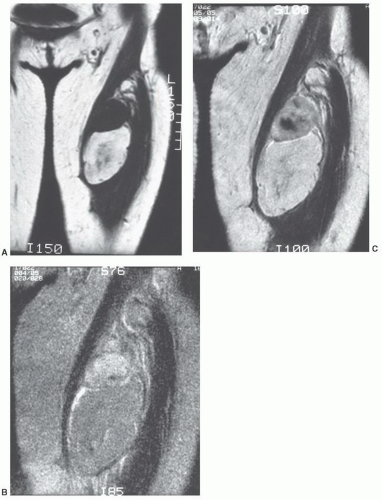
Figure 12.31 Recurrent dedifferentiated liposarcoma in the thigh of a 33-year-old woman. A: Coronal T1-weighted image shows a mass in the anterior left thigh. The well-differentiated portion of the mass images similarly to subcutaneous fat. B: Corresponding T2-weighted image shows the non-fatty component to have signal intensity higher than that of fat. Note areas of increased signal intensity in well-differentiated portion. C: Gd-DTPA-enhanced T1-weighted (650/20) image shows significant enhancement in the high-grade component and mild enhancement within the well-differentiated portion of the tumor. (From Kransdorf MJ, Meis JM, Jelinek JS. Dedifferentiated liposarcoma of the extremities: imaging findings in 4 patients. AJR Am J Roentgenol. 1993;161:127-130, with permission.)


Accordingly, the new WHO Classification of Tumors notes that atypical lipomatous tumor and well-differentiated liposarcoma are identical morphologically and karyotypically, and recommends that the term “well-differentiated liposarcoma” be retained for lesions located at sites at which a wide surgical margin cannot be obtained.62 Such sites include the retroperitoneum and mediastinum.62 Although there is no agreement on the terminology for
lesions in the deep somatic soft tissues, we would agree with Weiss and Goldblum and use the term “atypical lipoma” only for subcutaneous extremity lesions, reserving the term “well-differentiated liposarcoma” for lesions with similar histologies in all remaining sites.52,58


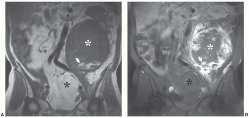
Figure 12.32 Dedifferentiated liposarcoma in the retroperitoneum of a 74-year-old man. Coronal T1-weighted (A) and postcontrast fat-suppressed coronal T1-weighted (B) images show a large mass (white asterisk) with a juxtaposed poorly defined predominantly non-fatty component (black asterisk) (fat much less than 25% of the lesion). Note central nonenhancing area in B (asterisk). Area of high signal intensity inferiorly (arrow) represents subacute blood, in keeping with previous hemorrhage and necrosis. Also note superior displacement of kidney.


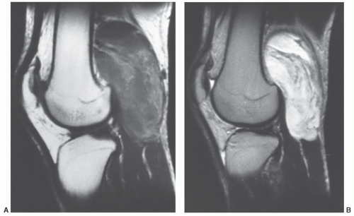
Figure 12.33 Myxoid liposarcoma in the popliteal fossa of a 22-year-old man. Sagittal T1-weighted (A) and T2-weighted (B) images show thickened linear and amorphous fatty areas within an otherwise nonspecific mass.

The myxoid, pleomorphic, and round-cell liposarcomas often do not contain substantial amounts of fat, and only approximately 50% to 80% will demonstrate fat radiologically (Figs. 12.32,12.33,12.34).106,107 When fat is present, it is usually in a lacy, amorphous, clumplike, or linear pattern (Fig. 12.35).54,107,108,109 The pleomorphic and round-cell types are more heterogeneous. Myxoid liposarcoma is typically more homogeneous and may appear deceptively benign on MRI, and an appearance similar to that of a cyst has been reported in as many as 20% of cases (Fig. 12.36).37,67,106,110


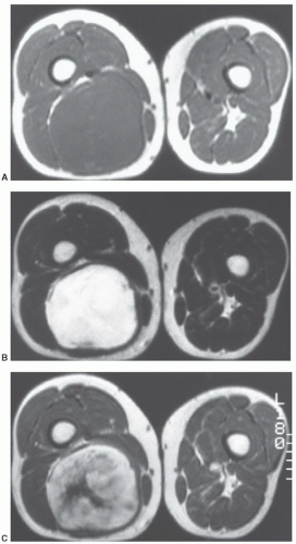
Figure 12.34 Myxoid liposarcoma in the posterior thigh of a 49-year-old man. Axial T1-weighted (A) and T2-weighted (B) images show a nonspecific mass in the posterior right thigh. C: Corresponding nonfat suppressed T1-weighted image after Gd-DTPA administration shows marked enhancement.


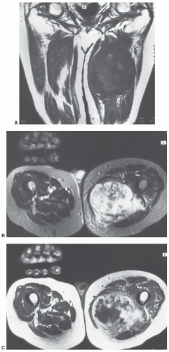
Figure 12.35 Pleomorphic liposarcoma in the posterior thigh of an 85-year-old man. A: Coronal T1-weighted image shows a large predominantly non-fatty inhomogeneous mass with central areas of increased signal intensity. B: Axial T2-weighted image is very inhomogeneous, but otherwise nonspecific. C: Corresponding axial T1-weighted non-fat suppressed image after Gd-DTPA administration shows marked irregular peripheral enhancement.


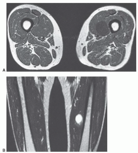
Figure 12.36 Myxoid liposarcoma in the thigh of a 56-year-old woman. Axial T1-weighted (A) and coronal T2-weighted (B) images show a round well-defined lesion in the medial aspect of the left thigh. The lesion has imaging characteristics identical to a cyst or myxoma.


Lipoblastoma

The “ordinary” lipoma is overwhelmingly the most frequently encountered fatty soft tissue tumor; however, there are numerous lipoma variants. Although these variants are well described in the pathology literature, they have received scant attention in the radiology literature. These lipoma variants differ from the classic soft tissue lipoma with regard to both clinical presentation and microscopic appearance. Lipoblastoma is a relatively immature cellular lipoma that occurs almost exclusively in infancy and early childhood, usually in children under 3 years of age; however, rare cases have been reported in adults.111,112,113,114 Most lipoblastomas are situated in the superficial soft tissue or subcutis of the extremities, although they have also been reported in the neck, trunk, perineum, and retroperitoneum.113 Males are affected two or three times more frequently than are females.112,113 Two-thirds of these masses are circumferentially well circumscribed, comprising the classic lipoblastoma. The remaining cases are diffuse, infiltrating both the musculature and the subcutis, and are referred to as diffuse lipoblastomatosis.115

Radiologically, lipoblastoma and liposarcoma may be indistinguishable (Figs. 12.28, 12.29, and 12.37). Although the radiologic differential diagnosis is that of liposarcoma, this entity is exceedingly rare in children. In a review of more than 2,500 cases of liposarcoma at the Armed Forces Institute of Pathology, only 2 (0.08%) occurred in children younger than 10 years of age. Fifteen additional cases were identified in children between the ages of 11 and 15 years.116


Vascular Lesions


Hemangioma

Soft tissue hemangiomas represent a broad spectrum of benign neoplasms that histologically closely resemble normal blood vessels.Hemangiomas often contain considerable amounts of nonvascular tissue, the most common of which is adipose tissue. Other nonvascular elements encountered in hemangiomas include smooth muscle, fibrous tissue, thrombi, and bone. Lesions may intermittently change size and may be painful.117 The overlying skin may have a bluish discoloration.117 Hemangiomas may be superficial (Fig. 12.38) or deep (Fig. 12.39), with deep-seated lesion more frequently a diagnostic dilemma.

Classification and nomenclature for benign vascular lesions has been confusing andvaries on the basis of specialty (surgeon, pathologist, or radiologist).118 Enzingerand Weiss supported a classification suited to pathologists that emphasized the size of the major vessels and tissue involved.119 Though there have been many classifications, more recently, the WHO120 and the most recent edition of Enzinger and
Weiss authored by Weiss and Goldblum121 have devised a more anatomic approach to benign vascular lesions. The subtypes include synovial hemangiomas arising in the synovium, intramuscular which is a proliferation of benign vessels in muscle, venous hemangiomas, and arteriovenous hemangiomas with shunts and a mix of venous and arterial structures.118,119,120,121 Other categories include the epithelioid hemangioma, a feeding artery, and well-defined mature vessel lined by epithelioid endothelial cells and stroma mimicking lymph nodes.118 Angiomatosis is another category with a diffuse hemangioma affecting large anatomic regions that crosses tissue planes and compartments.118,122


Figure 12.37 Lipoblastoma in a 1-year-old boy. Axial T1- (A) and T2-weighted (B) images show a large predominantly fatty mass with a significant non-fatty component. In an adult, this appearance would suggest a liposarcoma.

Kransdorf et al.118 summarizes the issue of nomenclature and controversy in a recent article in 2011 suggesting that working together (pathology, radiology, and clinicians) could result in a uniform approach for optimal patient care.

This discussion is limited to intramuscular hemangioma in that it is the lesion most likely to be confused clinically with a soft tissue mass. Most occur in youngadults,with 80% to 90% presenting by 30 years of age.123 The long duration of symptoms and relatively young age at presentation suggests that many of these lesions are congenital. Both males and females are affected equally.52,123,124 Hemangiomas are usually classified as either cavernous (large-caliber vessels) or capillary (small-caliber vessels), depending on the size of the blood vessels comprising most of the lesion. However, admixtures or mixed capillary and cavernous hemangiomas are not uncommon.123 Nonvascular elements are most commonly encountered in cavernous hemangiomas.125

Radiographs of patients with soft tissue hemangioma are frequently nonspecific but may reveal phleboliths within a soft tissue mass. Osseous changes may be seen in as many as one-third of cases, consisting of periosteal or cortical thickening.126 Phleboliths are most common in cavernous hemangiomas and are seen in 30% to 50% of cases. The MRI appearance of an intramuscular hemangioma is frequently characteristic. On T1-weighted images, the intramuscular hemangioma is typically poorly marginated and isointense to skeletal muscle (Fig. 12.40).124 Within the lesion are areas of increased signal4,127 approximating that of subcutaneous fat.125 These areas vary in appearance from fine, delicate, or lacelike strands to thick, coarse bands. On T2-weighted images, the intramuscular hemangioma is typically well marginated and markedly hyperintense as
compared with subcutaneous fat (Fig. 12.41).125,128 Segments of the lesion are isointense to either fat and/or muscle. MR angiography is useful for evaluation of the vascular supply of the lesion (Fig. 12.41). Phleboliths (within the hemangioma) may be detected as small rounded areas of signal void on MRI, but these are more readily apparent on radiographs or CT.116 Marrow signal abnormalities may be seen adjacent to large hemangiomas. Although their nature is not known, they are hypothesized to represent either marrow edema or hematopoietic conversion with localized hyperemia.126


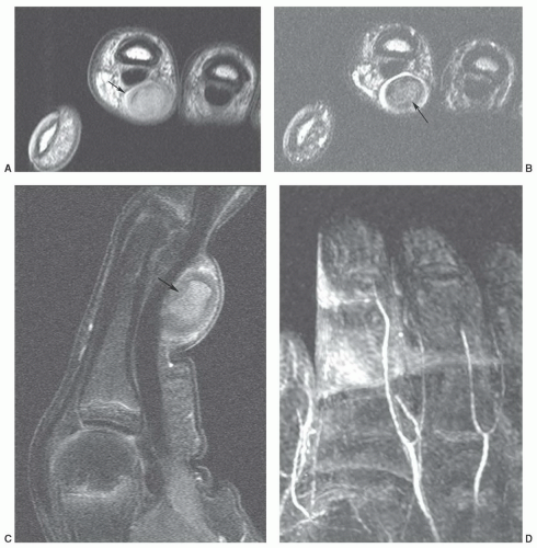
Figure 12.38 Thrombosed superficial hemangioma in the hand. Axial T1-weighted (A) and T2-weighted (B), sagittal fat-suppressed post-contrast T1-weighted (C) and MR angiogram (D) demonstrate the thrombosed hemangioma (arrow) with occlusion of several digital arteries (D).

Naturally, the MR appearance reflects the underlying morphology. Areas of hyperintensity on T1-weighted images reflect fatty tissue interspersed between the vessels. The hyperintense signal on T2-weighted spin-echo images reflects the slowly flowing relatively stagnant blood within the hemangioma.125,128 A cavernous hemangioma may be larger than a capillary hemangioma and contains greater amounts of nonvascular tissue, especially adipose tissue. In fact, a cavernous hemangioma can contain such large amounts of adipose tissue that portions may be indistinguishable from a lipoma.123


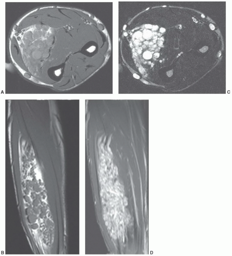
Figure 12.39 Deep intramuscular hemangioma of the forearm. Axial (A) and sagittal (B) T1-weighted and axial (C) and sagittal (D) T2-weighted images demonstrate a large hemangioma in the forearm.


Angiolipoma

Angiolipoma is a lesion characterized histologically by adipose tissue, small vessels, and capillaries. Angiolipoma is a cutaneous lesion that typically occurs on the trunk and extremities of young adults, with the forearm being the most common location (Fig. 12.42). As a cutaneous lesion, it is typically not subject to radiologic examination, and the diagnosis is usually established clinically. A rare variant, the infiltrating angiolipoma, has been described as a nonencapsulated infiltrating lesion composed of mature adipose tissue and benign vascular elements.129 This lesion has been separated from the cutaneous (encapsulated) form because of its tendency to recur locally.129,130,131,132,133 Calcium salts, heterotopic bone, and phleboliths may be found within the infiltrating nonencapsulated angiolipoma.64,134 These lesions fall within the spectrum of benign vascular lesions and are probably best classified as an intramuscular hemangioma.135


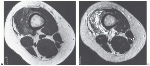
Figure 12.40 Intramuscular hemangioma in the thigh of a 23-year-old woman. A: Axial T1-weighted image shows areas of increased signal, in a lacelike pattern, coursing through the lesion. B: Corresponding axial T2-weighted image shows the lesion to have a lobular configuration with areas markedly hyperintense to subcutaneous fat, and others isointense to fat and skeletal muscle.


Arteriovenous Hemangioma (Vascular Malformation)

Although many authors separate benign vascular lesions into hemangiomas and vascular malformations, we do not. The two are not necessarily mutually exclusive, and attempting to separate them assumes that these lesions are always histologically and clinically distinguishable, which they frequently are not. Consequently, we prefer to classify all these benign vascular soft tissue lesions as hemangiomas, reserving the term “arteriovenous hemangioma” for those lesions demonstrating unequivocal arterial and venous components. Clearly, those lesions in which there is a significant arterial and/or venous component will behave differently.129 MRI will reflect the rapidly flowing blood within the arterial component as multiple serpiginous flow voids, interdigitating within the interstices of the mass (Fig. 12.43).


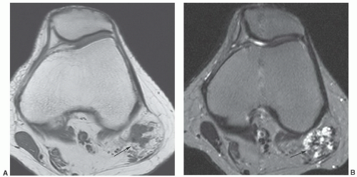
Figure 12.41 Biceps femoris hemangioma. Axial T1-weighted (A) and T2-weighted (B) and sagittal fat-suppressed turbo spin-echo (C) images demonstrate the hemangioma at the myotendinous junction (arrow). MR angiogram (D) clearly demonstrates the vascular supply and lesion vascularity.


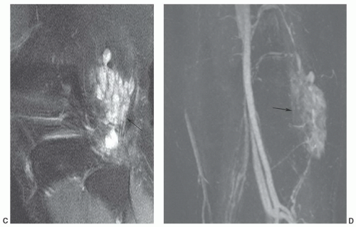
Figure 12.41 (continued)


Lymphangioma

A lymphangioma is a lesion made up of tissue resembling normal lymphatic channels, composed of endothelial cells and supporting connective tissue.136 Other mesenchymal elements, typically fat, fibrous tissue, and smooth muscle, are also frequently present.136,137 The etiology of lymphangioma is unknown. It may represent a developmental anomaly of the lymphatic vessels137 or may be the sequela
of congenital obstruction of lymphatic drainage.138,139 Its progressive nature, however, has suggested that it may be a benign mesenchymal neoplasm.136


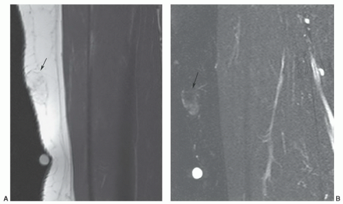
Figure 12.42 Superficial angiolipoma in the thigh. Sagittal T1-weighted (A) and post-contrast fat-suppressed T1-weighted images (B) demonstrate the subtle angiolipoma (arrow).


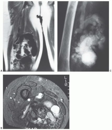
Figure 12.43 Arteriovenous hemangioma (arteriovenous malformation) in the thigh of a 35-year-old man. A: Coronal T1-weighted MR image of the thigh shows prominent flow voids representing rapidly flowing blood in the lesion. B: Axial gradient image shows the lesion to have an infiltrative growth pattern. C: Arteriogram shows the marked vascularity of the lesion, with markedly enlarged, tortuous, draining veins.

Lymphangiomas are subclassified by the size of the lymphatic vessels that comprise the lesion into simple (capillary) lymphangiomas, cavernous lymphangiomas, and cystic lymphangiomas (cystic hygromas).136,139,140 The sizes of the vessels within these lesions range from thin-walled capillary-sized vessels, to dilated lymphatic channels, to cysts from a few millimeters to several centimeters in diameter.139 Additionally, one may see vascular lymphatic malformations.141 Lymphangiomas are often an admixture of all histologic subtypes and should be considered as a pathologic spectrum.117,139,141

The cystic lymphangioma (cystic hygroma) is the most common type of lymphangioma and is characterized by
large uniloculated or multiloculated cystic spaces, lined by lymphatic endothelium.136,141 It contains serous or chylous fluid.137 Cystic lymphangiomas are most common in the neck (typically in the posterior cervical space) and axilla, with these locations accounting for 75% and 20% of lesions, respectively.142,143 The prevalence of these locations has been suggested to be the result of sequestered lymphatic anlage, which lack adequate drainage. Other rare locations include the mediastinum, retroperitoneum, bone, omentum, and mesentery.143 Up to 10% of cervical cystic lymphangiomas will extend into the mediastinum.144 Cystic lesions (cystic hygromas) are typically found in regions in which the loose fatty connective tissue allows relatively unlimited growth.139,141 The overwhelming majority of lesions present in children, with more than half presenting at birth and 90% discovered by the age of 2 years.136,137,144,145,146 Fewer than 10% are found in adults.141 Retroperitoneal cystic lymphangiomas are usually found in older children and adults. Acute symptoms result from infection, rupture, hemorrhage, or pressure on adjacent structures.137 Cystic lymphangiomas are usually isolated lesions, although posterior neck cystic lymphangiomas may be associated with Turner syndrome.147


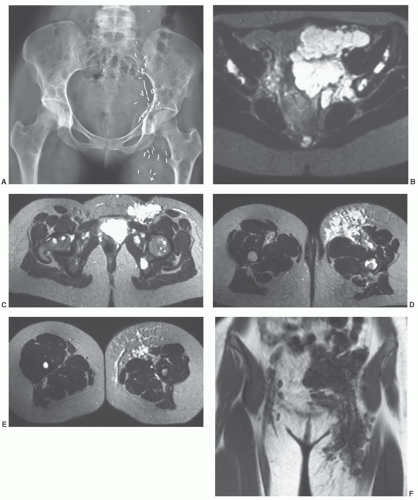
Figure 12.44 A 22-year-old woman with diffuse lymphangiomatosis involving bone and soft tissue. A: Anteroposterior radiograph of the pelvis demonstrating diffuse lucent areas in the femurs, pelvis, and sacrum. B-E: Axial T2-weighted images demonstrating diffuse pelvic and left thigh involvement. There are also bone changes in the iliac bones, both femoral heads, and the upper femur. Coronal (F) and sagittal (G) T1-weighted images demonstrating the anterior and posterior lymphangiomas.


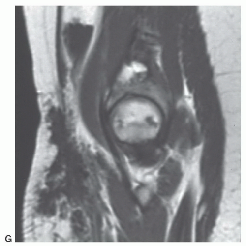
Figure 12.44 (continued)

A cavernous lymphangioma is typically a subcutaneous lesion composed of dilated lymphatic spaces, intermediate in size between those of the cystic hygroma and simple lymphangioma.136,141 Cavernous lymphangiomas are more common in areas in which there is a more limited potential for expansion, such as in the floor of the mouth, lips, tongue, cheek, salivary glands, and intramuscular septa.136,139,141

A simple or capillary lymphangioma is a rare tumor composed of small capillary-sized vessels, lined by a flat or cuboidal epithelium.136,138 It is usually small, well circumscribed, and localized to the dermis and epidermis.136,141 These lesions may be seen in patients of any age, and approximately one-fourth are found in patients older than 45 years of age.141 Because of its superficial location and small size, capillary lymphangioma is rarely imaged.

Patients with lymphangioma most commonly present with a discrete soft tissue mass,136,139 although they are otherwise asymptomatic.138 In children, large cervicomediastinal lymphangiomas are commonly associated with respiratory distress.139 Deviation of the trachea and compression of the esophagus may be seen in adults.144 Rarely, infiltrating lesions may give rise to elephantiasis.134 Lymphangiomas have no malignant potential. Surgery remains the treatment of choice, although recurrence is not rare and has been reported to be as high as 15%.139,141,146 Frequently, complete excision may not be possible because of infiltration of adjacent essential structures (Fig. 12.44).139 The most common postoperative complication is edema and may be seen in up to 50% of cases.139

Radiographs may reveal a soft tissue mass. Calcification is rarely seen.148 The lesion may be associated with secondary bone changes and consequently may cause increased tracer accumulation on bone scintigraphy.149 MRI shows a cystic lymphangioma as a unilocular or multilocular mass of water density. Siegel et al.138 reported the MRI appearance of 17 lymphangiomas in 15 patients, describing a typical appearance: Heterogeneous with a low signal intensity, similar to that of muscle, on T1-weighted images and high signal intensity, larger than that of fat, on T2-weighted images, reflecting the preponderance of fluid-filled cystic spaces. Focal inhomogeneities within the lesions are present in nearly all cases and appear as low-intensity linear structures of variable thickness, representing fibrous septa within the lesion (Fig. 12.45). Four of these lymphangiomas demonstrated signal intensity similar to that of fat on T1-weighted images. In two of these cases, the lesions were composed of small lymphatic vessels, separated by thick fibrous-fatty septa. One other lesion was composed of both small and large cysts, one filled with fat and the other filled with clotted blood and necrotic debris. Rim and septal enhancement may be seen on MRI after gadolinium administration.150 It is our experience that the fibrous septa within lymphangiomas are seen to better advantage on sonography (Fig. 12.45).


Synovial Lesions

Benign proliferative lesions of the joint, bursa, and tendon sheath are common in clinical practice. The most frequent of these lesions is the localized giant cell tumor of tendon sheath (nodular tenosynovitis), representing the localized form of a spectrum of benign synovial proliferations, which when diffuse and intra-articular is termed pigmented villonodular synovitis (PVNS).151,152


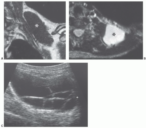
Figure 12.45 Lymphangioma in the neck of a 41-year-old woman. Coronal T1-weighted (A) and axial T2-weighted (B) images show the lesion (asterisk) to image similar to fluid. C: Ultrasound showed the lesion to be cystic with multiple septations. The MR appearance is nonspecific, and a myxoid tumor could have a similar appearance. The diagnosis was suggested preoperatively on the basis of both the MR and ultrasound appearances.


Giant Cell Tumor of Tendon Sheath

Giant cell tumor of tendon sheath occurs in either a localized or a diffuse form.52 The localized form is often termed nodular tenosynovitis and, as its name would imply, this lesion clinically presents as a nodular or polypoid mass, most commonly in the hand and wrist. In its diffuse form, the lesion is less well defined and grossly characterized by shaggy beardlike projections (representing hypertrophic synovial villi). Clearly, the distinction between the localized and diffuse form is on occasion blurred and a function of its gross and microscopic appearance. The term “PVNS” is usually reserved for those cases in which there is diffuse involvement of a large joint. The diffuse form of giant cell tumor of tendon sheath usually occurs adjacent to large weight-bearing joints and in most cases, although not all, represents extraarticular extension of PVNS. Ushijima et al.,153 in reporting a 20-year experience with 220 cases, found nodular tenosynovitis to be more than seven times more common than PVNS.

Localized nodular tenosynovitis is one of the most common masses of the hand,154 second in frequency only to a ganglion. Patients are typically adults with a peak incidence in the third and fourth decades,153,155,156 and there is a slight female predominance (1.5-2.1:1). The lesion affects the volar aspect of the digits, more commonly than the dorsal surface, although lesions may be lateral or circumferential.153,156 Involvement is most usually seen in the first three fingers and first two toes.153,156

Most patients present with soft tissue swelling153,155 or a slowly enlarging painless soft tissue mass, which is freely mobile under the skin but attached to deeper structures.157 Pain is not uncommon and may be aggravated by activity.155
Multiple lesions are unusual but have been reported.153 Local recurrence is not uncommon and may be seen in approximately 9% to 20% of cases.153,156 A malignant giant cell tumor of tendon sheath with metastases was reported by Carstens and Howell158; however, such lesions are quite rare.

Radiographs most usually demonstrate a soft tissue mass.155,156 Pressure erosions are seen on the underlying bone in about 15% of cases.153 Because the diagnosis is usually suggested clinically, CT and MRI are rarely used, and experience with this lesion is quite limited. MRI typically demonstrates a nonspecific well-defined mass adjacent to a tendon, isointense with muscle on T1-weighted, and more inhomogeneous and hyperintense on muscle (but less than fat) on T2-weighted MR spin-echo images (Fig. 12.46).108,159,160


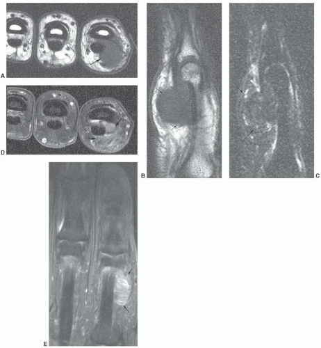
Figure 12.46 Giant cell tumor of tendon sheath. Axial (A) and sagittal (B) T1-weighted and sagittal fat-suppressed turbo spin-echo (C) images demonstrate the low signal intensity surrounding the flexor tendon (arrows). Post-contrast fat-suppressed T1-weighted images in the axial (D) and coronal (E) planes demonstrate inhomogeneous enhancement.

Some investigators have suggested that fibroma and giant cell tumor of the tendon sheath make up a spectrum
of histiocytic-fibroblastic-myofibroblastic lesions.161,162 Maluf et al.161 noted that the lesions have an overlapping clinical presentation, with similar patient age and gender as well as lesion location and distribution. In addition, these lesions share similar growth patterns, showing a lobulated architecture. Although the microscopic appearance varies, both lesions contain spindle cells and multinucleated giant cells and share similar immunohistochemical attributes. Hence, the fibroma and giant cell tumor of tendon sheath may represent end points of a spectrum of cellular proliferation.162,163 It has been our experience that these lesions will show a similar appearance atMRI (Fig. 12.47).163


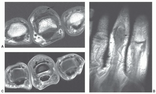
Figure 12.47 Fibroma of tendon sheath. Axial (A) and coronal (B) T1-weighted images demonstrate a well-defined low intensity lesion involving the extensor tendon (arrow). Post-contrast fat-suppressed T1-weighted axial image (C) demonstrates inhomogeneous enhancement (arrow).


Pigmented Villonodular Synovitis

Pigmented villonodular synovitis (PVNS) is a relatively uncommon disorder characterized by synovial proliferation with hemosiderin deposition in the involved synovial tissues.152,164,165 This condition may involve joints, bursae, and tendon sheaths. There are diffuse forms (80% involve the knee) and localized forms such as giant cell tumor of the tendon sheath. As noted, joint involvement most commonly involves the knee with other large joints affected in order of decreasing frequency including the hip, ankle, shoulder, and elbow.164,165

Pigmented villonodular synovitis occurs most commonly in the second through fifth decades with an age range of 10-90 years, though most patients present in their early 20s. The disease is usually monoarticular, which assists in diagnosis.166 Involvement of more than one joint is distinctly unusual,166 although Cotton et al.167 reported 58 patients, 2 (3%) of whom probably had bilateral hip involvement. Malignant transformation is exceedingly rare. A case of malignant PVNS was reported by Kalil and Unni,168 in which the metastases were identified 64 years after initial presentation. Childhood PVNS has been reported in conjunction with synovial hemangioma, and recurrent hemorrhage within the hemangioma has been suggested as a cause.154

Patients usually complain of intermittent pain, swelling, and recurrent joint effusions with decreased motion.169,170 The time interval from onset of symptoms to clinical presentation varies from months to years.164,171 There is typically an associated joint effusion with serosanguineous or xanthochromic fluid.164,170,171 Surgery remains the preferred treatment. Recurrence rates are typically quite high, approaching 50%.172 Joint fusion may be required in cases with advanced disease.7 Total joint replacement will relieve pain and restore function in selected patients.164

Lesions are usually much larger and more irregular in shape than those seen in nodular tenosynovitis. Grossly, the lesion has been likened to a “shaggy red beard” to emphasize the villous or frond-like synovial projections. The reddish or rust color is the result of iron pigment (hemosiderin) within the lesion.171,173

Radiographs may be normal or show a noncalcified soft tissue mass. Well-defined bone erosions on both sides of a joint and joint effusion may also be seen.152,164 Erosive bone lesions are seen in about 50% of all cases,169 being most common in joints with tight capsules such as the hip (93%) and shoulder (75%) and least common in
the knee (26%).164 Bone erosive changes are usually geographic lytic lesions with well-defined thinly sclerotic margins. They are most characteristic when they are multiple and are seen on both sides of the joint. The joint space is usually preserved, as is bone density.164 Uncommonly, radiographs will demonstrate an osteoarthritis appearance, with typical osteophytes, sclerosis, cysts, and joint narrowing, or an arthritis-like appearance, with concentric joint space loss, osteoporosis, and erosions.167 Radiologic calcification within the mass has been reported174 but is extremely
unusual and should suggest an alternative diagnosis. Calcification has also been reported in diffuse giant cell tumor of tendon sheath.175


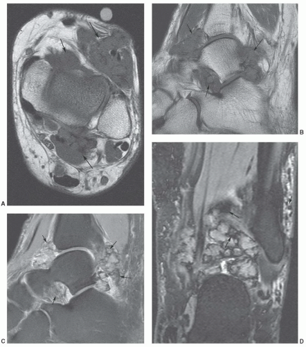
Figure 12.48 Pigmented villonodular synovitis in the ankle. Axial (A) and sagittal (B) T1-weighted images demonstrate a lobulated low signal intensity mass (arrows) about the ankle. Sagittal T2-weighted turbo spin-echo image shows areas of low signal intensity (arrows). Coronal DESS image (D) demonstrates multiple areas of blooming due to hemosiderin deposition (arrows).

The MRI appearance of PVNS is often characteristic. It presents as a heterogeneous synovial process that usually extends away from the joint space.152 The lesions contain areas of intermediate signal intensity and/or hypointensity when compared with skeletal muscle on T1-weighted spin-echo MR images (Fig. 12.48). pattern may be seen on T2-weighted images.152,176 The decreased signal intensity is usually more pronounced on long TR/TE images, due to the preferential shortening of T2 relaxation times of hemosiderin (Fig. 12.48).171 This is more pronounced at high field strengths.171,176,177 An important finding on gradient-echo sequences is susceptibility artifact (blooming) created by hemosiderin in the synovial tissues.169 Contrast enhancement on fat suppressed T1-weighted images is irregular and nonspecific due to hypervascularity with
synovial inflammation and proliferation.152,169 The lytic bone lesions seen on radiographs and joint effusions are typically seen well on MRI.152 The diffuse giant cell tumor of tendon sheath has a skeletal distribution similar to that of PVNS and is often considered an extra-articular extension of PVNS. Its MRI signal intensity characteristics are similar to PVNS (Fig. 12.49).


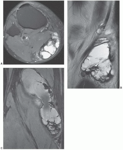
Figure 12.49 Diffuse giant cell tumor of tendon sheath. Axial (A) and sagittal (B) T2-weighted turbo spin-echo images demonstrate a large soft tissue mass below the knee with areas of low signal intensity (arrows). Coronal gradient-echo image (C) demonstrates blooming due to hemosiderin deposition (arrows).


Popliteal (Synovial) Cysts

Popliteal cysts, also known as synovial cysts, probably result from a slit-shaped communication of the knee joint with the normally occurring gastrocnemius semimembranosus bursa.178 This communication is more common in older individuals because of degeneration and reduced elasticity of the joint capsule.178,179 In a study of adult cadaver knees, a communication between the semimembranosus and gastrocnemius bursa was found in more than half the cases.180 The incidence of popliteal cyst increases with age,178,181 being demonstrated arthrographically in 16% of patients in the second decade, 36% in the third decade, and 54% beyond the fifth decade.179 The term “Baker cyst” is usually reserved for those cases in which this gastrocnemius semimembranosus bursa is distended by fluid.178 Baker described eight cases of swelling in the popliteal region in 1877 and 1885, hypothesizing that it was the result of synovial membrane herniation and cyst formation due to osteoarthritis (Fig. 12.50).178 While involvement of the gastrocnemius semimembranosus bursa is necessary for diagnosis, popliteal cysts may dissect between muscle planes, and may rarely dissect into the vastus medialis or gastrocnemius muscle (Fig. 12.51).182

In a study of the MRI examinations of 1,113 patients referred for evaluation of internal derangement, the incidence of popliteal cysts was 5%.181 The prevalence of cysts demonstrated by arthrography ranges between 7% and 42%.179,181,183,184 This higher incidence is likely due to the distension of the normally collapsed bursa during arthrography.181


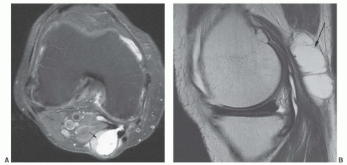
Figure 12.50 Popliteal cyst. Axial (A) and sagittal (B) fat-suppressed proton density-weighted images demonstrating fluid extending into the gastrocnemius semimembranosus bursa (arrow).

Only gold members can continue reading. Log In or Register to continue

Stay updated, free articles. Join our Telegram channel

May 25, 2016 | Posted by in RHEUMATOLOGY | Comments Off on Musculoskeletal Neoplasms

Full access? Get Clinical Tree

Get Clinical Tree app for offline access