Miscellaneous Conditions


Miscellaneous Conditions


Thomas H. Berquist


INTRODUCTION

As the techniques for magnetic resonance imaging (MRI) have improved, the applications have continued to expand.1,2,3,4,5,6 Many of these applications have been discussed in the previous pathologic and anatomic chapters. However, certain evolving applications or applications with overlap into multiple anatomic regions deserve mention here.


MYOPATHIES

The MRI applications and characteristics of soft tissue infection, trauma, and neoplasms have been discussed in previous chapters. MRI also plays a significant role in evaluation of other primary muscle and neuromuscular disorders. Both imaging and spectroscopy may be of value in this regard.2,6,7,8,9,10,11,12,13,14,15,16

There are numerous overuse syndromes, inflammatory and metabolic myopathies, neural myopathies, and forms of muscular dystrophy that potentially could be studied with imaging techniques and/or MR spectroscopy.6,7,8,9,10,11,12,13,14,15,16,17 To date, none of these techniques have demonstrated specific histologic diagnosis though morphologic appearance and specific muscle involvement have been helpful for confirming the clinical diagnosis. MRI is also useful for selection of biopsy sites for histologic diagnosis.7,8,9 Conventional imaging techniques provide limited information regarding nonneoplastic muscle diseases. Several authors have studied computed tomographic (CT) features of myopathy.18,19 Patterns of muscle replacement have been described using CT. Both localized and diffuse low-density areas were noted in patients with pseudohy pertrophic muscular dystrophy.18,19,20,21,22 Changes in selected muscle groups have been reported in Duchenne muscular dystrophy. Hawley et al.19 demonstrated that CT scans of patients with neuromuscular diseases initially showed atrophy of muscles followed by decreased muscle density. Primary myopathies revealed similar changes but in reverse order, with decreased density preceding muscle atrophy. The low-density areas in muscle seen on CT are likely due to fat and/or connective tissue replacement.19

MRI has superior soft tissue contrast compared with CT. Therefore, detection of early muscle changes can be more easily accomplished. From an imaging standpoint, the findings seen on MRI may be no more specific than CT. However, certain important changes can be noted with MRI. The muscle groups can be easily distinguished and the extent of involvement determined by using multiple image planes. Using T1-weighted, T2-weighted, fat-suppressed, and short TI inversion recovery (STIR) sequences makes it possible to distinguish early edema or inflammation (high signal intensity on T2-weighted and STIR and low intensity on T1-weighted images) from fatty infiltration or replacement. Other changes, such as increase or decrease in muscle volume and fibrous replacement, can also be identified (Figs. 15.1 and 15.2).3,22,23

Borghi et al.10 studied T1 relaxation times. This group reported that T1 relaxation times were considerably reduced in patients with myopathy (normal, 450 to 800 milliseconds; myopathy, ≤500 milliseconds). This is undoubtedly due to replacement of muscle with fat and fibrous tissue. Although further studies are needed, these data may be useful in differentiating myopathy from other pathology, such as neoplasms where T1 values are elevated.10


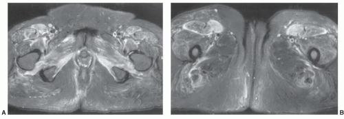
Figure 15.1 Axial fat-suppressed fast spin-echo T2-weighted images at the level of the ischial tuberosities (A) and the proximal thighs (B) demonstrating an infiltrative process in the adductor muscles and anterior musculature, predominantly the tensor fasciae latae due to polymyositis.

Shellock et al.24 studied T2 data on muscles during exercise that allowed differentiation between eccentric (lengthening or stretching) and concentric (shortening) of muscles with exercise. T2 relaxation times increase with exercise due to changes in intra- and extracellular water. Muscles performing eccentric activity have lower T2 values than muscles performing concentric exercises.24,25

The role of MRI for evaluating myopathies include clearly defining the muscle groups involved, differentiating atrophy and fatty replacement for more acute inflammatory changes, following treatment phases and progression of disease, and localizing optimal sites for biopsy. The current use of imaging parameters alone does not obviate the need for clinical and histochemical studies to define the pathologic process completely.7,8,9


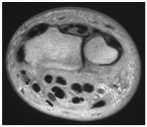
Figure 15.2 Axial proton density image of the wrist demonstrating complete fatty replacement of the muscles due to a chronic neuropathy.

Significant progress has been made in evaluating the utilization of spectroscopy for defining muscular and neuromuscular inflammatory diseases. With higher magnetic field strengths (3 to 7 T), imaging and spectroscopic studies with 31P, 21Na, 13C, and other nuclei may be possible.26,27,28,29 Conditions such as inflammatory myopathies, metabolic myopathies, muscular dystrophies, and neuropathic muscular changes have been studied that demonstrate changes in organic phosphate and phosphocreatine ratios as well as other phosphate metabolites. These studies may not only be useful in more specifically identifying the pathologic process, but also show potential in monitoring the response of patients to drug therapy.26,27,28,29,30 Further data are necessary before the role of spectroscopy in evaluating myopathies and neuromuscular disorders is completely understood (see Chapter 16).

In recent years, several articles have reviewed the role of MRI and MR spectroscopy in specific myopathies and muscular dystrophies. A review of these reports is warranted to demonstrate the current role of MR techniques in evaluating specific myopathies.


Duchenne Muscular Dystrophy

Duchenne muscular dystrophy is one of the most common inherited skeletal muscle diseases.31,32,33,34,35 The condition affects about 1 in 3.500 males.34 The condition is the result of an X-linked dystrophin gene. Diagnosis of Duchenne muscular dystrophy is generally accomplished using clinical and laboratory data. The disease most commonly affects young males (<5 years) who present with muscle weakness and markedly elevated serum creatine kinase levels.31 Diagnosis is confirmed with muscle biopsy.18,31 Treatment has been difficult due to disease complexity and lack of consistent
standardized documentation of disease activity and tissue damage.34


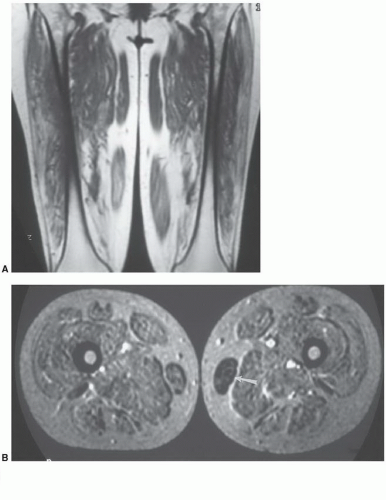
Figure 15.3 Duchenne muscular dystrophy. Coronal T1-weighted (A) and axial T2-weighted (B) images in a patient with Duchenne muscular dystrophy. All muscles are involved except for partial sparing of the gracilis (arrow) on the left.

Imaging has played a role in determining the extent of muscle involvement and following the progression of the disease process. Prior to MRI, CT was used to evaluate muscle volume and fatty degeneration.19,31 MRI, using axial and coronal or sagittal image planes with T2-weighted, fat-suppressed, T2-weighted, or STIR sequences, is a more sensitive technique due to its superior soft tissue contrast.3,30,31,32,33,34,35 Therefore, MRI has demonstrated utility in demonstrating the distribution and extent of muscle involvement.17,19 Liu et al.31 demonstrated that certain muscles in the lower extremity were spared. The gracilis (100%), sartorius (83%), semitendinosus (69%), and semimembranosus (48%) were uninvolved most commonly (Fig. 15.3). An MR grading system was developed that correlated with the clinical (Brooke) system. The system was based on muscle involvement and the degree of fatty infiltration. The system, summarized in Table 15.1, was based on the number of muscles involved in the pelvis and thighs as well as severity of fatty infiltration and increase in subcutaneous fat.31 The score correlated with disease progression and response to therapy.31


Table 15.1 MR Grading of Duchenne Muscular Dystrophy


Degree of Involvement


Score


Pelvic muscles spared


≤3


3


<7


2


≥7


1


Normal muscles


0


Thigh muscles spared


≤2


3


<6


2


≥6


1


Normal muscles


0


Fatty muscle infiltration


Severe


2


Mild


1


None


0


Subcutaneous fat


Severe involvement


2


Mild


1


None


0


From Liu GC, Jong YJ, Chiang CH, et al. Duchenne muscular dystrophy: MR grading system for functional correlation. Radiology. 1993;186:475-480.


More recently, additional MR approaches have begun to evolve in an attempt to better quantify muscle involvement and disease activity or progression.33,34,35,36 Multiple techniques including pre- and postcontrast following exercise, the three-point Dixon approach to fat quantification, T2-mapping, and spectroscopy have been explored. Kim et al.33 used T2-mapping correlated well with fatty infiltration and clinical assessments in 34 boys (mean age, 8.4 years) with Duchenne muscular dystrophy.33 Spectroscopy is still not commonly employed in daily practice. However, Hsieh et al.34 measured trimethyl ammonium (TMA) and total creatine (tCr) and compared these levels to water and calculated the TMA/tCr ratios. Decrease in TMA/tCr ratios correlated with the degree of decreased muscle function in patients with Duchenne muscular dystrophy compared with normal volunteers.34


Neurotrophic Myopathy

There are multiple causes of denervation myopathy, including spinal cord injury, nerve compression syndromes, Grave disease, and neuritis.9,17 Traditionally, diagnosis of denervation myopathy has been accomplished with clinical findings and electromyography.9,37,38,39,40,41 MR evaluation of myopathies related to loss of motor neuron function has also been studied.9,37,38,39,40,41 MRI has been evaluated in acute and chronic conditions. Fleckenstein et al.38 evaluated patients with acute neural injury and found MRI has little utility in the acute setting. Signal intensity may remain normal for up to 15 days, but increased signal intensity on T2-weighted or STIR sequences is typically noted in 15 to 30 days. Signal intensity may remain increased for up to 1 year.37,38 Obviously, if the nerve injury or the cause for neuropathy is not corrected, one can expect fatty infiltration and atrophy of the involved muscles, sometimes before 1 year, which may overlap with changes described earlier. Fatty infiltration typically occurs as soon as 3 months after nerve injury (Fig. 15.2).37

More recently, Kamath et al.9 demonstrated patterns of denervation myopathy in the acute (<1 month), subacute (1 to 6 months), and chronic (>6 month) stages. During the acute phase histology demonstrates edema-like changes. Muscle signal intensity is normal on T1-weighted and increased on T2-weighted and STIR sequences. Contrast enhancement may or may not be evident. In the subacute phase, histology demonstrates an increase in extracellular water. Signal intensity of muscles is normal to decreased on T1-weighted sequences and increased on T2-weighted sequences. STIR sequences may demonstrate increased or decreased signal intensity though the former is more common. Contrast enhancement is inconsistent and probably does not add significantly to muscle characterization. In the chronic phase (>6months), histologic changes demonstrate fatty infiltration and atrophy that increases over time (Fig. 15.4) and may lead to fibrotic changes. MR signal intensity is increased on T1-weighted sequences due to fatty infiltration. Signal intensity is increased on T2-weighted sequences and decreased on STIR sequences. There is no contrast enhancement during the chronic phases.9

More recently, diffusion-weighted MRI has been used to demonstrate early neuopathic changes due to increased diffusion coefficient and enlargement of the extracellular fluid space.41 We still prefer axial T1-weighted, fat-suppressed fast spin-echo (FSE) T2-weighted, and STIR images with comparison of both extremities. This allows subtle changes in muscle size or signal intensity to be more easily appreciated. We do not typically use gadolinium unless a mass is suspected.8


Dermatomyositis

Polymyositis is a rare paraneoplastic or autoimmune syndrome without skin involvement.22 Dermatomyositis involves the skin and skeletal muscle. Both conditions are caused by cell mediated (type IV) autoimmune involvement of striated muscle.17

Dermatomyositis is a multisystem disease with diffuse inflammatory changes in the skin and muscles. Dermatomyositis has a bimodal pattern with peak occurrence in childhood and in adults in the fifth decade. Symptoms tend to be more severe in childhood. The adult onset form includes increased incidence of multiple malignancies (breast, prostate, lung, and gastrointestinal).17 Diagnosis is based on clinical features of skin rash and progressive, potentially painful proximal muscle weakness. Electromyleogram (EMG) and biopsy are required to confirm the diagnosis.17,42,43,44,45,46


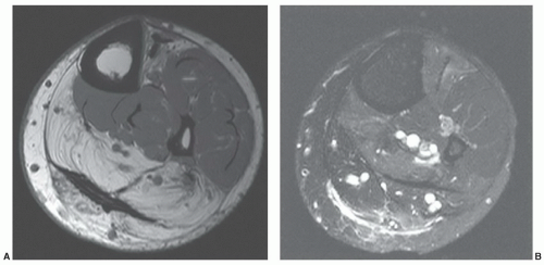
Figure 15.4 Neuropathic myopathy. Axial T1-weighted (A) and fast spin-echo T2-weighted images demonstrate increased signal intensity in the posterior compartments on the T2-weighted image (B) and fatty replacement on the T1-weighted image (A) due to tibial nerve compression.

Although MR features are not specific, the signal intensity changes are useful for monitoring, especially in children, where repeat biopsy and EMG studies are not practical.44 Classically, there is bilateral symmetrical muscle edema.17 Increased signal intensity on T2-weighted or STIR sequences has been described in the involved muscles and subcutaneous fat (Fig. 15.5). The degree of edema correlates with disease severity. Signal intensity is usually normal during inactive phases of the disease.42,43,46 Over time, there is progression to fatty infiltration and muscular atrophy.17,43


Polymyositis

Polymyositis (Figs. 15.1 and 15.6) is usually symmetrical, involving the proximal lower extremity muscles with progression to involve the proximal upper extremity, neck, and pharyngeal muscles.22,37,45,46,47 Polymyositis typically presents in the fourth decade. Females outnumber males by a 2:1 ratio.22,45 Onset is insidious, most often with associated muscle pain, and tenderness in 30% of patients. Articular symptoms, including pain and periarticular calcifications, occur in 20% to 50% of cases.48 Etiology may be related to viral infection or genetic. Up to one third of patients have associated connective tissue disease, and 10% have underlying malignancy. Malignancy is more common in patients older than 60 years.22,45,49

Diagnosis is based on clinical presentation, laboratory and EMG data, and muscle biopsy. MR has been demonstrated to be cost effective for diagnosis, selection of biopsy sites, and monitoring therapy.47 Techniques as described earlier using T1- and T2-weighted and STIR sequences are most commonly performed.47 O’Connell et al.22 described MR approaches to polymyositis. Coronal STIR images of the body were obtained using the body coil and a 50-cm field of view (FOV). Slice thickness was 8 mm with a 0.8-mm skip. The number of sections varied with patient size. Inflammatory changes in the muscles were easily identified with the STIR sequence. STIR sequences are 97% specific for demonstrating inflammatory myopathies. There is no advantage to contrast-enhanced imaging.17,22,42

STIR sequences can also be used to follow response to therapy. Differential diagnostic considerations include infectious myopathy, inclusion body myositis (IBM), HIV myositis, sarcoidosis, and eosinophilic myositis.22


Sarcoid Myopathy

Sarcoidosis is a systemic granulomatous disease that involves lung, lymph nodes, heart, central nervous system, liver, spleen, and, in 1% to 13%, bone, but rarely involves the muscles (Table 15.2).45,49,50,51,52,53,54,55 The exact incidence of muscle involvement is probably underestimated because patients are often asymptomatic.54 Proximal limb muscle involvement is most often asymptomatic. Symptomatic muscle involvement only occurs in about 1.4% of patients.21,52 However, muscle biopsies demonstrate granulomas in 50% to 80% of patients.52

Otake21 reported two types of muscular involvement— nodular and myopathic. The former involves the extremities. Others consider three categories: palpable nodules (type 1), acute myositis (type 2), and chronic myopathy (type 3). Acute myositis causes inflammation with myalgia and
tenderness. Muscle weakness and atrophy is common with chronic myositis (type 3).52 Patients typically present with single or multiple palpable nodules. Nodules may be tender to palpation. The myopathic type is symmetrical and presents with progressive weakness and muscle atrophy.21,55


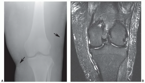
Figure 15.5 Dermatomyositis. A: AP radiograph of the knee demonstrating subtle subcutaneous soft tissue calcifications (arrows). Axial T1-weighted image (B) demonstrates low signal intensity in the subcutaneous fat (arrows) corresponding to the calcification on the radiographs. There is also significant muscle atrophy. Coronal STIR sequence (C) shows subcutaneous edema and edema along the ligaments and tendons.

An association of sarcoid myopathy with IBM has also been reported.56,57 Vattemi et al.57 reported that 7.4% of their patients with IBM also had sarcoidosis.

Imaging of sarcoid myopathy can be accomplished with CT, radionuclide scans, and MRI. Contrast-enhanced CT demonstrates peripherally enhancing lesions.54 Gallium-67 and technetium-99m scans have also been employed and provide the advantage of total body imaging to allow detection of multiple areas of involvement. Nodular lesions are demonstrated as focal areas of abnormal uptake compared with diffuse uptake of tracer with myopathic sarcoidosis.21 MRI features are useful with nodular lesions. Otake21 described stellate areas of low signal intensity with
surrounding high signal intensity on T2-weighted or STIR sequences (Fig. 15.7). These findings are not specific and can also be seen with fibromatosis or other local inflammatory and neoplastic lesions.51,54 Myopathic changes (types 2 and 3) have nonspecific MR features with diffuse increased signal intensity on T2-weighted or STIR sequences. Biopsy is required to confirm the diagnosis.21,45,51,54


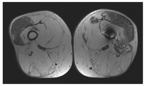
Figure 15.6 Polymyositis. Axial T1-weighted images demonstrating diffuse fatty infiltration and atrophy with relative sparing of the vastus lateralis.


Table 15.2 Sarcoidosis Organ System Involvement


Location


Incidence (%)


Pulmonary


90


Mediastinum


85


Ocular


80


Liver/spleen


50-80


Muscles


50-80a


Cardiac


25


CNS


25


Lung


20


Skeleton


5-13


Parotid glands


6


GU tract


5


GI tract


1


CNS, central nervous system; GU, genitourinary; GI, gastrointestinal.


a 1.4% symptomatic.


From references 45, 49, 50, 52, 53.


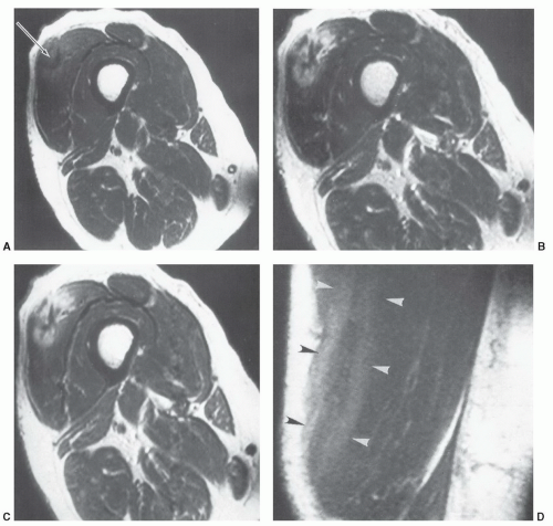
Figure 15.7 A 55-year-old woman with nodular sarcoid involving the vastus lateralis. A: SE 550/30 axial shows a stellate low intensity region (arrow) with slight increased intensity at the margin. SE 1,800/50 (B) and SE 1,800/100 (C) images show more obvious increased intensity surrounding the low intensity region. Coronal SE 550/30 image (D) shows three stripes (arrowheads) with outer increased and central decreased signal intensity. (From Otake S. Sarcoidosis involving skeletal muscle: imaging findings and relative value of imaging procedures. AJR Am J Roentgenol. 1994;162:369-375.)


Diabetic Myopathy

Diabetic myopathy occurs in patients with poorly controlled diabetes mellitus.17,58,59 Although characteristically reported in patient with type 1 diabetes, recent studies demonstrated that 88% of patients with myopathy had type 2 diabetes.60 Diabetic myopathy or infarction most commonly involves the lower extremities. The muscles of the thigh are involved in 80% and the calf in 20% of patients.60 Thigh muscle involvement is most common in the anterior compartment (100%) but, the posterior compartment is involved in 64% and the medial compartment in 55% of cases.58,59,60 Patients present with acute pain and swelling of the involved extremity or extremities. Pain may be relieved by rest but returns with activity.20 A painful mass is evident in 33% to 44% and fever may be evident in up to 10% of patients. Most patients have associated diabetic complications including nephropathy, neuropathy, and retinopathy that occur in 60% to 80% of patients with myopathy.60


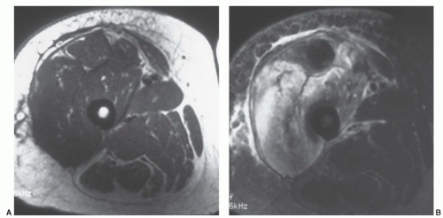
Figure 15.8 Diabetic muscle infarction. Axial T1-weighted (A) and T2-weighted (B) images of the thigh show fascial, subcutaneous, and vastus muscle inflammation. Changes are more easily appreciated on the T2-weighted image in B.

MRI should be accomplished using T1-and T2-weighted or STIR sequences (conventional or FSE) in two image planes to define the extent of involvement. Contrast enhancement may be useful, but care must be taken in the diabetic population due to the risk of nephrogenic system sclerosis.60 MR findings in the thigh and calf differ slightly. In the thigh, 100% demonstrate subcutaneous edema and 91% subfascial edema. In the calf, 100% demonstrate subcutaneous edema with subfascial edema in 60%.60 Infarction may be hemorrhagic (increased signal intensity on T1-weighted images) or ischemic with low signal intensity on T1-weighted images centrally with a rim of high signal intensity on T2-weighted sequences or peripheral enhancement on fat-suppressed T1-weighted images (Figs. 15.8 and 15.9).60


Infectious Myopathy

Myopathy may be related to bacterial, viral, mycobacterial, fungal, or parasitic organisms. Infections may be localized (pyomyositis, abscess, and gas gangrene) or generalized.52,61,62,63,64,65,66,67

Pyomyositis is an acute infection usually due to Staphylococcus aureus. Other bacteria and mycobacteria are less common. Patients typically have underlying disease such as malignancy, diabetes, or AIDS. In a series of 40 patients reported by Yu et al.67 77.5% of patients had some underlying disease with diabetes mellitus accounting for 40% of the cases followed by malignancy and other disorders such as cirrhosis. Patients present with pain, fever, and local swelling.52,63,66,67

As with the above myopathies, T1-weighted, T2-weighted, and STIR images are most useful. Contrast enhancement is also useful for abscess detection and the normal diffuse enhancement pattern.66 Care must be taken when considering gadolinium depending upon the underlying conditions, specifically diabetes and renal failure.66,67 MR images demonstrate increased signal intensity in the involved muscle and subcutaneous tissues on T2-weighted
or STIR sequences. Abnormal signal intensity in adjacent structure also occurs. The adjacent marrow may be involved in 32.5% cases, fascial planes in 35%, and adjacent joints in 26%.66 Fluid collections or abscesses are evident in 49% to 90% of cases. Yu et al.67 reported at least one abscess in 90% of patients (Fig. 15.10). Contrast-enhanced fat-suppressed T1-weighted images demonstrate peripheral enhancement of abscesses.61,64,66 Abscesses tend to be larger in patients with underlying diabetes.67 The majority of patients with abscesses are managed with surgical or image guided drainage procedures.66


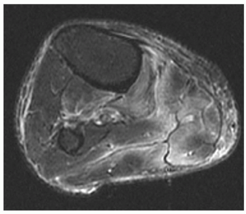
Figure 15.9 Ischemic myopathy in a diabetic. T2-weighted MR image demonstrating subcutaneous and subfascial increased signal intensity.


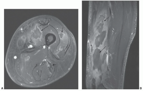
Figure 15.10 Bacterial pyomyositis. Post-contrast fat-suppressed T1-weighted axial (A) and sagittal (B) images demonstrating multiple abscesses with peripheral enhancement (arrows).

Gas gangrene is diagnosed clinically. This is a rapidly progressive, potentially life-threatening clostridial infection. Onset is acute with severely ill patients. Crepitus is present on physical examination and gas is usually obvious on radiographs.45

Generalized infectious myopathies are associated with HIV or related to rhabdomyolysis due to infections elsewhere.62,63


Miscellaneous Myopathies

Granulomatous myositis has been described with sarcoidosis (see above), but may also occur with Crohn disease or primary biliary cirrhosis. Patients with thymoma and myasthenia gravis may also develop granulomatous myositis.65 Granulomatous myositis has also been described with graft-versus-host disease (GVHD). This condition is seen in transplant patients. GVHD is reported in 25% to 40% of patients undergoing bone marrow transplant. Myositis of GVHD typically involves the proximal muscles of the extremity, but can involve distal muscles and the respiratory musculature.65

IBM affects both the proximal and distal muscles.56,57 This condition leads to severe disability. Elderly patients are most often affected. The etiology is uncertain. However, one theory is accumulation of abnormal proteins in muscles. The condition is progressive, resulting in fatty replacement in the involved muscles (Fig. 15.11). Muscle abnormalities are easily demonstrated on MR images.45,68


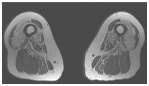
Figure 15.11 Inclusion body myositis. Axial T1-weighted images demonstrating marked atrophy and fatty replacement in the anterior compartment with partial sparing of the posterior compartment.


Focal myositis is rare. This is considered a benign inflammatory pseudotumor. Etiology is unknown. MR features are not clearly defined. However, muscle swelling with inflammation and fatty infiltration has been reported. These changes are clearly demonstrated on STIR and T1-weighted images.45,69


TRAUMA


Muscle Injuries

Traumatic muscle and myotendinous injuries were discussed in anatomic chapters. However, the complexity of these injuries and the fact that they may have features similar to other myopathies indicate that we should include them in this section, as well. Injuries may be due to overuse resulting in microtrauma, rhabdomyolysis due to intense exercise, focal acute tears, or delayed onset muscle soreness (DOMS).23,25,70,71,72,73,74,75,76

Muscle injuries are graded depending upon the extent of injury. First-degree strains result in injury of a few fibers, second-degree injuries involve 50% of muscle fibers, and third-degree strains are complete disruptions of a muscle (Fig. 15.12).4,73,74,77 Minor injuries have excellent prognosis and usually are not imaged. DOMS is muscle pain related to exertion that occurs hours to days after the initiating activity.70 This phenomenon is associated with eccentric (lengthening) contraction that results in ultrastructural damage, elevated plasma proteins, and edema on T2-weighted MR images. The extent of edema correlates with pain levels and elevations in creatine kinase.70,74,77 Symptoms related to DOMS usually begin 1 to 2 days following exercise and usually improve over the next 3 to 4 days.77 Symptoms are separated from grade 1 muscle strains clinically as in the latter the symptoms are acute and tend to resolve in 2 to 3 weeks.73,77

Muscle strains (grades 1 to 3) are classified due to the extent of injury. Trauma may be indirect or direct including laceration from penetrating injuries.71,77 Grade 1 strains result in microscopic injuries with disruption of about 5%, but certainly less than 50% of muscle fibers. There is little loss of function and may be minimal swelling of the involved muscle on MRI. Edema and hemorrhage cause focal poorly defined high signal intensity on T2-weighted and STIR sequences. MR images in the axial and either coronal or sagittal plains define the extent of muscle involvement.76,77


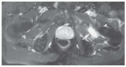
Figure 15.12 Adductor muscle tear. Axial fat-suppressed fast spin-echo T2-weighted image shows a grade 2 strain with increased signal intensity involving nearly 50% of the cross-sectional area and a focal hematoma (arrow).

Grade 2 strains result in loss of muscle function due to partial disruption (about 50% of the fibers) and frequently a hematoma at the myotendinous junction (Figs. 15.12 and 15.13). Once again signal intensity is increased on water-sensitive sequences with variable signal intensity in an associated hematoma depending on the age of the lesion.77 Contrast enhancement is useful when a hematoma is present to exclude underlying mass lesions. Hematomas enhance peripherally, whereas tumors tend to enhance centrally unless very necrotic.3,4,73,76,77

Grade 3 strains result in complete disruption of the muscle or myotendinous junction. Depending upon the involved muscle, there may be significant retraction of the muscle fragments.3,4,77 The site of injury almost always has a hematoma due to hemorrhage into the disruption site (Fig. 15.14). Hematomas tend to heal more slowly and may require evacuation if adjacent to neural structures (Figs. 15.14 and 15.15).3

Prognosis and repair vary with the extent of injury. Grade 1 strains similar to contusions typically heal within 2 weeks. Higher-grade strains may require 1 to 4 months to heal. Depending upon the activity (high-level athletes, etc.) operative intervention may be required for higher-grade strains and certain hematomas (Fig. 15.14).3,4,77

Complications of muscle sprains include hernias, compartment syndrome, fibrosis, atrophy, and heterotopic ossification. Muscle hernias most frequently occur in the lower extremity. They are most common in athletes and soldiers. Hernias may be multiple and bilateral, with muscle protruding through the fascial defect. Muscle signal intensity is often normal on MRI. Although often palpable, it may be necessary to image muscle herniations with active muscle contraction. Therefore, fast scan techniques with motion studies or active muscle contraction may be required to confirm the diagnosis and exclude other soft tissue lesions. The axial image plane is often most useful for detection of hernias.23,77


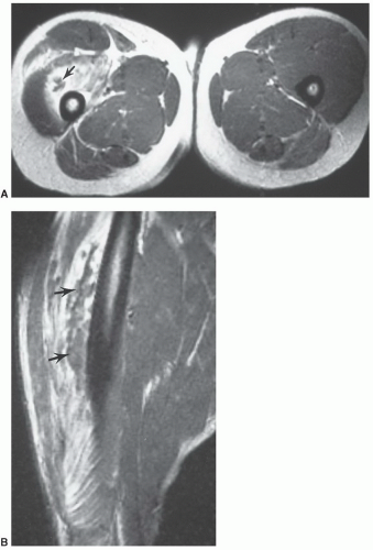
Figure 15.13 Grade 2 muscle strain with extensive edema and hemorrhage and low intensity hematoma (arrows) seen on axial (A) and sagittal (B) images.

Fibrosis is more common with grades 2 and 3 strains and more common in the calf than the thigh or upper extremities. Fibrotic tissue has low signal intensity on both T1- and T2-weighted sequences.3,4,77 Larger hematomas may ossify or develop thick fibrous capsules with central fluid collections.

Compartment syndrome may occur acutely or be a chronic phenomenon due to increased venous flow and increased interstitial pressure in a give fascial unit.77,78 Vascular and neural compression may occur with significant ischemic risk. Fasciotomy may be required if clinically measured pressures exceed 30 mm Hg.78

Rhabdomyolysis may be due to ethanol overdose, infection, crush injuries, collagen disease, or intense exercise. MR images typically show a more generalized process than local muscle tears (Fig. 15.16).3,75,79

Calcific myonecrosis is a rare sequelae of muscle trauma.80 Knowledge of this condition is important, as clinical and radiographic features may mimic an aggressive neoplasm.81,82 Calcific myonecrosis occurs with cystic muscle degeneration. The process can occur 10 to 64 years after trauma and results in a painful, expansive, calcified soft tissue mass. A remote history of compartment syndrome is common.80,82

Radiographs demonstrate plaque-like calcifications in the periphery of the mass. There may be adjacent bone erosion.81 Increased tracer accumulation is evident on technetium-99m bone scans. The fluid-filled mass with
calcification is easily appreciated on CT studies. MRI demonstrates cystic changes on T1- and T2-weighted images. Calcifications may be more difficult to define if radiographs or CT are not available for comparison.80,81 Contrast enhancement does not facilitate the diagnosis (Fig. 15.17).65


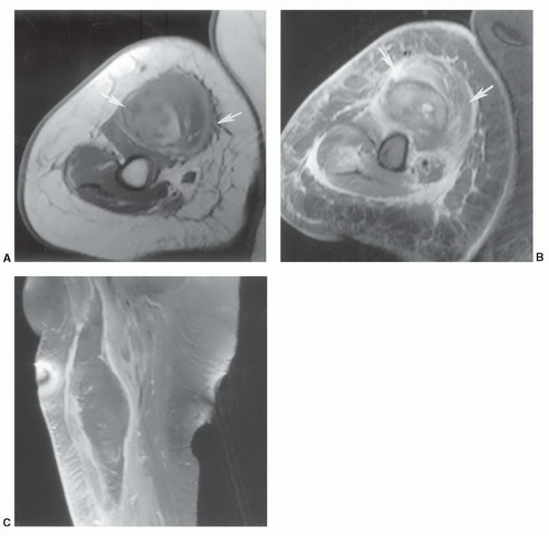
Figure 15.14 Grade 3 biceps tear with a large hematoma. Axial T1-weighted (A) and T2-weighted (B) images demonstrate a large hematoma (arrows) adjacent to the neurovascular structures. Sagittal contrast-enhanced T1-weighted image (C) shows the extent of the lesion with marginal enhancement.

Treatment typically requires debridement and excision of the entire region, with wound coverage using skin or muscle flaps. Secondary infection following treatment is common.80,82


Morel-Lavallée Lesions

Morel-Levallée lesions result from abrupt separation of the skin and subcutaneous tissues from the underlying fascia.83,84,85 The lesions are most common near the greater trochanter and thigh (Fig. 15.18), but have also been reported in the low back and knee.85 Lesions may occur with a variety of traumatic etiologies though most are reported in football players and wrestlers.85

Injuries usually occur where the vascular plexus passes through the fascia. With disruption of the fascia, blood, lymphatic fluid, and tissue debris form the fluid collection. MR features are that of a fluid collection with low signal intensity on T1-weighted images and scattered areas of high signal intensity due to blood products. On T2-weighted images, the lesion is largely high intensity with mixed areas of intermediate to low signal intensity, based on the type of fluid and chronicity of the lesion (Fig. 15.18).83,84,85 The lesions are typically well defined and located between the fascia and subcutaneous fat.85

Some lesions respond to conservative therapy whereas others require aspirations to achieve resolution. Lesions that do not respond to these measures can be treated with doxycycline sclerodesis.84


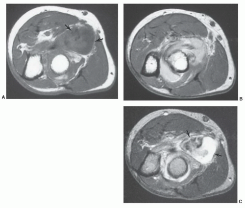
Figure 15.15 Hematoma in the proximal forearm related to biceps insertion tear. Axial T1-weighted (A), proton density (B), and T2-weighted (C) images show the inhomogeneous fluid collection (arrows). The hematoma compressed the radial nerve requiring surgical decompression.


EOSINOPHILIC FASCIITIS

Eosinophilic fasciitis is a relatively rare condition.86 There is no age predilection. Patients present with symmetric skin swelling and pain in the involved extremities. There is an absence of fever or systemic symptoms. Skin changes include edema and erythema. Symptoms of swelling, stiffness, and pain may be present acutely following strenuous exercise. Over time, contractures develop in 56% of patients. Joints most commonly affected in order of decreasing frequency include the elbow, wrist, ankle, and knees.87 Etiology is uncertain, but characterized by eosinophilia (63%), hypergammaglobulinemia (35%), elevated sedimentation rate (29%), and scleroderma-like skin findings (Table 15.3). Antinuclear antibodies are negative.84

Early diagnosis is important, as patients respond favorably to steroid therapy.86,87,88

Diagnosis may be delayed due to misdiagnosis as scleroderma (Table 15.3) or even congestive heart failure.88 Until recently, imaging has not played a significant role in
diagnosis of eosinophilic fasciitis. However, patients with active disease have characteristic MR features. These include fascial thickening, high signal intensity in the fascia on T2-weighted and STIR sequences, and enhancement following gadolinium administration (Fig. 15.19).88,89,90,91 These findings correlate with disease activity. Response to therapy and localization for biopsy are additional advantages of MRI imaging features. Currently, definitive diagnosis requires biopsy of cutaneous and muscle tissue.88,89,91


Table 15.3 Eosinophilic Fasciitis versus Scleroderma: Common Useful Distinguishing Features


Eosinophilic Fasciitis


Scleroderma


Sex


Males = females


Females


Symptoms with exercise


Common


Sometimes


Hand involvement


Uncommon


Common


Telangiectasia


Uncommon


Common


Visceral involvement


Rare


Common


ANA positive


Uncommon


Common


Fascial Bx


Inflammation


Normal


ANA, antinuclear antibodies; Bx, biopsy


From Lakhanpal S, Ginsburg WW, Michet CJ, et al. Eosinophilic fasciitis: clinical spectrum and therapeutic response in 52 cases. Semin Arthritis Rheum. 1988;17:221-231.


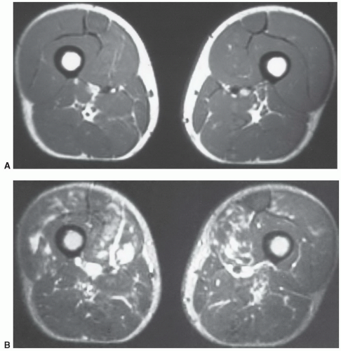
Figure 15.16 Bilateral rhabdomyolysis in an athlete. A: Axial T1-weighted image is normal. B: Axial T2-weighted image shows multiple areas of increased signal intensity in both thighs.


SARCOIDOSIS

Sarcoidosis is a multisystem disease (Table 15.2). Muscle involvement is symptomatic in 1.4% of patients, though muscle biopsies demonstrate granulomata in 50% to 80% of patients.52 Myopathy was discussed in a previous section. This section will focus on osseous and articular changes associated with sarcoidosis.

Inflammatory arthropathy is evident in up to 40% of patients with sarcoidosis.51 The knees, ankles, elbows, and wrists are most commonly involved. Lofgren syndrome is a well-known feature of sarcoidosis. This consists of arthralgias, erythema nodosum, and bilateral hilar adenopathy. Arthralgias seen with Lofgren syndrome are likely related to circulating cytokines rather than granulomas. Arthralgias occur more commonly in women.51,52,55,75,92

Patients present with pain and stiffness. Soft tissue swelling may be evident on radiographs, but osseous changes, except for osteopenia, are uncommon.92 Arthropathy at greater than 6 months typically involves two to three joints, including the knees, ankles, and phalanges of the hand. Sausage-like swelling of the fingers may be evident clinically and radiographically. Lacelike cystic changes may be evident in the phalanges on radiographs.92

MR features of arthropathy are nonspecific. Contrast-enhanced images may demonstrate synovial inflammation early. Tenosynovitis (Fig. 15.20), tendonitis, and bursitis may also be present. Biopsy is required to establish the diagnosis.51,52,92

Osseous involvement occurs in 5% to 13% of patients.45,51,52,55,93,94,95,96 The small bones of the hands and feet are classically involved. Cystic lacelike changes are characteristic. Deformities may result from pathologic fractures.92

MRI is not required for diagnostic purposes. However, occult marrow lesions and cortical involvement can be identified that are not apparent radiographically. Intramedullary lesions may be small (<1 cm) or as large as 3 to 4 cm.55

Only gold members can continue reading. Log In or Register to continue

Stay updated, free articles. Join our Telegram channel

May 25, 2016 | Posted by in RHEUMATOLOGY | Comments Off on Miscellaneous Conditions

Full access? Get Clinical Tree

Get Clinical Tree app for offline access