Musculoskeletal Infection


Musculoskeletal Infection


Thomas H. Berquist


Musculoskeletal infections may present with an acute, rapidly progressing course or in a more insidious fashion. The insidious onset of infection often follows trauma or surgery, such as after placement of orthopedic appliances. Presentation is also highly dependent upon age, virulence of the organism, patient condition, site of involvement, and circulation.1,2,3,4,5 Early treatment, particularly in children with articular infections, is essential to prevent growth defects, joint ankylosis, or other complications.3,7,8

Infections may involve osseous, articular, and soft tissues, either isolated or as multisite involvement. Terminology used for different categories of infection is important when considering imaging approaches and treatment (Table 13.1). Osseous infection may involve marrow, cortex, or periosteum. Multiple osseous structures are commonly involved.7,8,9,10,11,12 Similarly, it is not uncommon for soft tissue, osseous, and articular structures to be simultaneously involved (Table 13.1).


MECHANISMS OF INFECTION

Musculoskeletal tissue may become infected by hematogenous spread, spread from a continuous source, by direct implantation (i.e., skin puncture wound), or following surgery or trauma.7,8,11,12,13

Hematogenous spread is common in osteomyelitis and joint space infections. In children, osteomyelitis is almost always hematogenous. The source is commonly the throat, middle ear, or indwelling catheters in infants. Infections typically occur in the metaphysis of long bones or near the physis in flat bones such as the ilium.8,11,13 Involvement of the joint space depends upon capsular attachment in relation to the physis and patient age. In neonates or infants (up to age 2 years) and adults, the growth plate does not protect the epiphysis. Vascular channels cross the growth plate, allowing metaphyseal and epiphyseal involvement that increases the incidence of associated joint space
infection.6,7,11 In children aged 1 or 2 years and up to 16 years of age, the growth plate prevents spread to the epiphysis. Therefore, joint space involvement is less common unless the metaphysis is intracapsular (Fig. 13.1).7,8,13,16


Table 13.1 Infections: Terminology and Categories


Term/Conditions


Clinical/Image Features


Osteomyelitis


Infections of bone and bone marrow. Bacterial most common


Infective osteitis


Cortical infection. Often associated with marrow or soft tissue infection


Infective periostitis


Periosteal infection. Frequently, cortex and marrow also involved


Soft tissue infection


Involves shin, subcutaneous tissues, muscles, tendons, ligaments, fascia, or bursae


Sequestrum


Segment of necrotic bone separated from viable bone by granulation tissue


Involucrum


Living bone surrounding necrotic bone


Cloaca


Tract through viable bone


Sinus


Tract from infected region to skin


Fistula


Abnormal communication between two internal organs or internal organ and skin


Brodie abscess


Sharply delineated focus of osteomyelitis


Garre sclerosing osteomyelitis


Sclerotic nonpurulent infection with intense periosteal reaction


Chronic recurrent multifocal osteomyelitis (CRMO)


Subacute or chronic osseous infection common in children. May be associated with SAPHO


Synovitis, acne pustulosis, hyperostosis, and osteitis (SAPHO)


Palmoplantar pustulosis, articular and periosteal inflammation. Chronic course involving chest wall, spine, long and flat bones, large and small joints


From references 1, 13,14,15.


Spread from a contiguous source may result in osseous infection extending into the joint or soft tissues, or viceversa.16 In soft tissue infection, spread may be along tendon sheaths or in soft tissue compartments. Spread along the tendon sheaths in the upper extremity may result in involvement of the forearm to hand and wrist.6,17 Osseous involvement in the foot (see Chapter 8) is most commonly due to spread from a contiguous source.18,19 Most patients develop soft tissue infection due to diabetes, skin ulceration, or puncture wounds.20,21,22,23 Lederman et al.21 reported contiguous spread to osseous structures in the forefoot in 16% of 161 feet imaged by MRI. Joint space infection occurred in 33%. The first and fifth digits and metatarsals were most commonly involved.21

Direct implantation is usually related to a puncture wound, bite, or scratch (Fig. 13.2).7,8,13 Puncture wounds in the foot occur with stepping on sharp objects, usually when
barefoot. Puncture wounds in the hand (i.e., thorns) occur when working with plants or gardening. Direct implantation may also occur when working in contaminated water or soil in the presence of skin abrasions.17,24,25,26


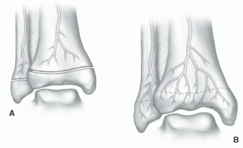
Figure 13.1 Illustrations of vascular patterns at the metaphyseal-epiphyseal junction in a child (A) and adult (B). The epiphysis is protected by the growth plate in patients 1 to 16 years of age. (From Berquist TH. Imaging of the Foot and Ankle. 3rd Ed. Philadelphia, Lippincott Williams & Wilkins, 2011.)


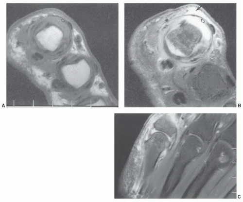
Figure 13.2 Fish hook injury with soft tissue infection extending into the fifth metacarpophalangeal joint. Axial T1- (A) and T2- (B) weighted images demonstrate soft tissue edema (arrow, B) and joint distention (open arrow). Fat-suppressed contrast-enhanced coronal T1-weighted image (C) shows enhancement of the soft tissues and synovial fluid.

Infection may also result from surgical or other minimally invasive procedures such as diagnostic injections.7,27,28,29 Symptoms are often insidious, resulting in delayed diagnosis. Imaging evaluation may also be more complicated when anatomy is distorted (trauma) or orthopedic implants are in place.


Organisms

Most musculoskeletal infections are bacterial (Table 13.2). Staphylococcal infections account for 80% to 90% of cases of osteomyelitis.7 In newborns and infants, Group B streptococcal infections are common.7,9,30,31 Other organisms, such as pseudomonas, have been associated with puncture wounds to the foot.7,8,16 Salmonella infections have been associated with sickle cell disease, hemoglobinopathies, systemic lupus erythematosus, leukemia, and lymphoma.31 Haemophilus influenza is most common in children 7 months to 4 years of age and in adults with diabetes or immunodeficiency.

Brucellosis, cryptococcosis, coccidiomycosis, histoplasmosis, and echinococcosis tend to be endemic in certain regions of the United States and the world. However, infections have become less geographic due to immigration and world travel.7,32,33,34,35

Typical (Mycobacterium tuberculosis) and atypical (M. marinum, M. avium, M. fortuitum, M. chelonaes, etc.) mycobacterial (tuberculosis) infections have become more common over the past decade due to immigration, human immunodeficiency virus (HIV), alcohol and drug abuse, and the aging population.17,23,25,26,36,37,38 Musculoskeletal tuberculosis is evident in 19% to 20% of infections with extrapulmonary pulmonary involvement.38 Atypical mycobacterial infections involve the musculoskeletal system in 5% to 10% of patients.25,26 Infections may be hematogenous or from dealing with animals or contaminated water or soil (Table 13.3).26,39 In the latter setting, infection is usually related to skin lesions or abrasions.26 Symptoms are nonspecific and insidious resulting in delayed diagnosis.17,25,26


Table 13.2 Common Organisms in Musculoskeletal Infections


Bacterial Infection


Gram-positive


Staphylococcal


Streptococcal


Meningococcal


Gonococcal


Gram-negative


Coliform bacterial infection


Proteus


Pseudomonas


Klebsiella


Salmonella


Haemophilus


Brucella


Mycobacteria


Tuberculosis


Atypical mycobacterial infection


Fungal and higher bacterial infection


Actinomycosis


Nocardiosis


Cryptococcosis


Coccidioidomycosis


Histoplasmosis


Sporotrichosis


Parasitic infections


Hookworms


Cysticercosis


Echinococcosis


From Resnick D. Osteomyelitis, septic arthritis and soft tissue infections: organisms. In: Resnick D, ed. Diagnosis of Bone and Joint Disorders. Philadelphia, PA: WB Saunders, 2002:2510-2624.


Table 13.3 Atypical Mycobacterial Infections


Mycobacterium


Reservoir/Transmission


Common Musculoskeletal Involvement


Avium


Soil, water, hogs, cattle, and birds


Osseous


Marinum


Water, fish


Soft tissue, hand and wrist


Bausaaii


Water, cattle, and hogs


Osseous


Fortuitum


Soil, water, animals, and marine life


Osseous


Scrofulaceum


Soil, water, and liquid foods


Osseous


Balnei


Water, fish


Soft tissue


From Theodorou DJ, Theodorou SJ, Kakitsubata Y, et al. Imaging characteristics and epidemiologic features of atypical mycobacterial infections involving the musculoskeletal system. AJR Am J Roentgenol. 2001;176:341-349. Wongworawt MD, Holton P, Learch TJ, et al. A prolonged case of Mycobacterium marinum flexor tenosynovitis: Radiographic, and histological correlation, and review of the literature. Skeletal Radiol. 2003;32:542-545. Amrami KK, Sundaram M, Shin AY, et al. Mycobacterium marinum infections of the distal upper extremities: Clinical course and imaging findings in two cases with delayed diagnosis. Skeletal Radiol. 2003;32:546-549.


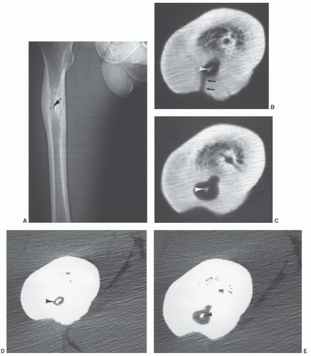
Figure 13.3 Chronic osteomyelitis with sequestrum and cloaca. A: CT scout images show medullary sclerosis and cortical thickening in the femur with a central lucency (arrow). Axial CT images with bone (B, C) and soft tissue (D, E) settings show a local abscess with a sequestrum (arrowheads in C and D) and cloaca (arrows). Beam hardening artifact degrades the image quality of the adjacent soft tissues.


Multiple imaging approaches may be required for detection and staging of musculoskeletal infections. MRI is a sensitive technique for early detection of musculoskeletal infection. Skeletal infections typically begin in medullary bone. The resulting hyperemia and inflammation cause alterations in signal intensity of medullary bone on MR images. The excellent tissue contrast and multiplanar imaging provided by MR may allow earlier diagnosis and more accurate assessment of the extent of osseous, articular, and soft tissue infections than other imaging techniques.7,15,40,41,42,43,44 Therefore, acute osteomyelitis may be evident on MR images when radiographs and other modalities are negative.7,45 Multimodality approaches using MRI, conventional isotopes, and positron emission tomography (PET) imaging may be required in certain situations.4,5,28,39,46,47,48,49,50

Discussion of the utility of MRI and other modalities is facilitated by categorizing patients in the following manner: (a) infection in nonviolated tissue; (b) infection in violated tissue (previous fracture or surgery, puncture, soft tissue injury); and (c) evaluation of surgical techniques for treatment. The latter category includes patients treated with muscle or omental flaps and vascularized fibular grafts.


INFECTION IN NONVIOLATED TISSUE

Osteomyelitis presents a diagnostic and therapeutic challenge regardless of the age group. Early diagnosis and management are essential to avoid irreversible bone, joint, and soft tissue damage. Hematogenous osteomyelitis, which occurs more commonly in children than adults, may be acute, subacute, or chronic, and most commonly involves the long bones of the lower extremities. In infants (<2 years) and adults, the epiphysis is not protected; vascular channels cross the growth plate allowing infection to involve both the metaphysis and epiphysis with a high incidence of joint space involvement.10,15 In patients aged from 1 to 2 years up to 16 years, the growth plate prevents spread of infection to the epiphysis and joint space involvement is less common unless the metaphysis is intracapsular (Fig. 13.1).12


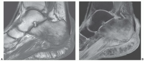
Figure 13.4 Sagittal T1- (A) and fat suppressed turbo spin-echo T2-weighted (B) images of the calcaneus demonstrating low signal intensity on T1- (A) and high signal intensity on the T2-weighted (B) images due to osteomyelitis in a diabetic. Routine radiographs were normal.

Infections typically involve the metaphyseal portion of long bones or areas near the physis in flat bones, such as the ilium. Radiologic changes are nonspecific in the early stages of infection. Localized swelling and distortion of the tissue planes may be the only findings.6,7 This may be followed with osteopenia in the area of marrow involvement or subtle lytic areas in the cortex. More defined bone destruction is usually not appreciated until 35% to 40% of the involved bone is destroyed, and thus is generally not evident for 10 to 14 days.10,12 Periosteal elevation may also be evident. The degree of involvement is often underestimated on routine radiographs. In this situation, computed tomography (CT) may be useful to more clearly define the extent of involvement. CT (Fig. 13.3) has been especially useful in determining the extent of disease prior to planning operative therapy and for detection of sequestra, cloaca (Table 13.1), and soft tissue changes.6,7,8,11,34,51

Radioisotope studies provide a sensitive tool for early detection of osteomyelitis. Technetium-99m, indium-111 labeled leukocytes, and technetium white cells or antigranulocyte antibodies provide sensitive and fairly specific methods for diagnosis of infection.2,6,7,13,27,38,52 However, the anatomic extent may be inaccurate, especially in the articular regions, and differentiation of soft tissue from bone involvement is not always possible.7 Also, in complex clinical settings such as previous surgery or instrumentation, neurotrophic changes or other conditions that create bone remodeling radionuclide imaging is less specific.16,53 This will be discussed in detail later in this chapter.

In recent years, PET has been effectively used to identify active infection.32,48,50


Acute Osteomyelitis

MRI is particularly suited to evaluate osteomyelitis due to superior soft tissue contrast and multiple image plane capabilities. Anatomic detail is superior to that provided by isotope studies. Subtle bone and soft tissue changes are more easily appreciated than on radiographs or CT examinations.3,4,51,52,53,54,55 In recent years, new pulse sequences, including diffusion-weighted imaging and the use of intravenous contrast have improved the utility of MRI for evaluating infection.53,55

As with other musculoskeletal pathology examination of patients with suspected infection requires at least T1-weighted and T2-weighted or STIR sequences, conventional spin-echo (SE) or fast spin-echo (FSE) sequences can be used.7,8,54 These sequences are needed to provide the necessary contrast between normal and abnormal tissues. T1-weighted sequences (SE or FSE) can be performed quickly and provide high spatial resolution.4,5,8,34 Infection is demonstrated as an area of decreased signal intensity compared to the high signal intensity of normal marrow fat (Fig. 13.4). Changes in cortical bone, periosteum, and muscle are less obvious on T1-weighted sequences (Fig. 13.4A). T2-weighted (SE or FSE) or STIR sequences demonstrate infection as areas of high signal intensity. We use fat suppression with FSE T-2 weighted sequences. Fat signal is reduced so areas of marrow inflammation are increased in signal intensity compared to normal marrow (Fig. 13.4B). In certain situations, more than two sequences may be required to improve tissue characterization. Short TI inversion recovery (STIR) sequences (Fig. 13.5) may provide valuable information about fat, water (cellular elements), or subtle inflammatory changes in bone marrow and soft tissues.4,7,8,34
We routinely add gadolinium-enhanced fat-suppressed T1-weighted images in patients with suspected osteomyelitis (0.1 mmol/kg b.w. intravenously) (Fig. 13.6).


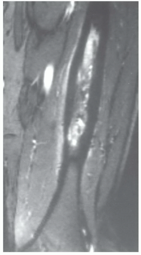
Figure 13.5 Osteomyelitis in the left femur. Coronal STIR sequence shows subtle thickening of the cortex with increased signal intensity in the marrow and adjacent soft tissues.


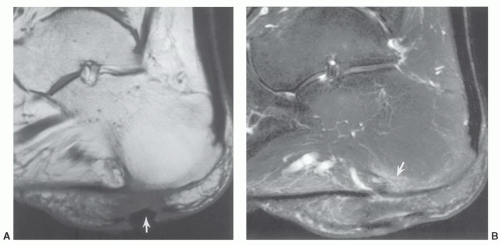
Figure 13.6 Diabetic with calcaneal osteomyelitis. A: Sagittal T1-weighted image demonstrates a soft tissue ulcer (arrow) and swelling. B: Contrast-enhanced fat-suppressed T1-weighted image demonstrates an area of enhancement (arrow) due to early osteomyelitis.

The anatomic extent of osteomyelitis can be clearly demonstrated by MRI.4,7,8,31 The extent of disease, including detection of skip areas, is easily established using multiple image planes. This information is particularly valuable when planning surgical debridement.


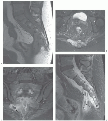
Figure 13.7 Patient with Crohn’s disease and sacral osteomyelitis with associated abscess formation. Sagittal T1- (A) and axial T2-weighted (B) images demonstrate perisacral soft tissue inflammation with an anterior soft tissue mass on the T1-weighted image and an area of soft tissue mass lateral to the sacrum on the T2-weighted image. Coronal (C) and sagittal (D) post-contrast fat suppressed T1-weighted images demonstrate the peripherally enhancing abscesses (arrows).

There have also been several studies comparing isotope studies, CT, and MRI for detection of early osteomyelitis.56,57,58 Chandnani et al.56 compared CT and MRI.MRIdemonstrateda94%sensitivity compared to 66%
for CT. Both techniques were equally specific for excluding osteomyelitis. Gallium-67 and indium-111 labeled white blood cells are more specific for infection than technetium scans.28,56 Beltran et al.57 compared technetium-99m methylene diphosphonate (MDP), gallium-67, and MRI for evaluating musculoskeletal infection. All techniques were equally effective in detecting osseous infection. However, MRI was more sensitive (100% compared to 69% for isotope scans) in identifying abscesses and distinguishing abscesses fromcellulitis (Fig. 13.7).57 Contrast enhancement and diffusion-weighted imaging add to the ease of detection of abscesses.4,31,55,59

The specificity of MRI for diagnosis of osteomyelitis needs to be more clearly defined. Increased signal intensity in marrow, cortical bone, periosteum, and soft tissue is noted on T2-weighted, STIR, and gradient-echo (T2*) sequences (Figs. 13.4,13.5,13.6,13.7).4,6,8,16,53


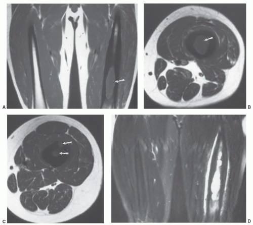
Figure 13.8 Osteomyelitis in the femur. Coronal (A) and axial (B and C) T1-weighted images demonstrate loss of normal fatty marrow in the femur (arrows). Signal intensity is increased on T2-weighted coronal image (D). Post-contrast fat suppressed T1-weighted images (E-H) demonstrate areas of peripheral enhancement in bone due to abscess formation. Juxtacortical soft tissue enhancement is also seen.

Morrison et al.53 evaluated fat-suppressed gadolinium-enhanced imaging in 51 patients with suspected osteomyelitis. These studies were compared with conventional T1- and T2-weighted MR images and technetium-99m MDP bone scintigrams. Focal enhancement with gadolinium was considered indicative of osteomyelitis (Fig. 13.6). In 73% of patients, the diagnosis was complicated by postoperative change, chronic osteomyelitis, or neurotrophic arthropathy. Despite this, enhanced fat-suppressed T1-weighted MR images demonstrated a sensitivity of 88% and specificity of 93% compared to a sensitivity of 79% and specificity 53% for non-gadolinium MR images (Fig. 13.8). Bone scans were 61% sensitive and 33% specific.53 Most
agree that sensitivity for detection of infection is improved by contrast-enhanced imaging. However, findings are not specific in differentiating infection from inflammation or neoplasms.47 Lack of bone enhancement effectively excludes infection.47,53,59


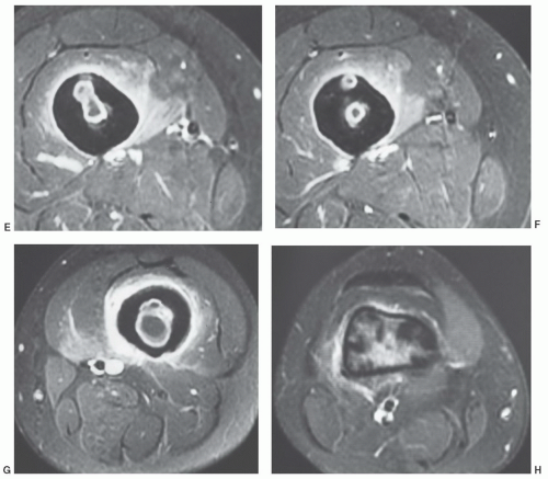
Figure 13.8 (continued)

More recently, Johnson et al.,60 studied the utility of signal abnormality on T1-weighted images for confirming osteomyelitis. Nineteen of twenty cases demonstrated confluent decreased marrow signal intensity (Fig. 13.9) for a 95% sensitivity and 91% specificity. More subtle changes on T1-weighted sequences were less specific.


Chronic Osteomyelitis

With acute infections, the clinical features combined with MR features are used to make the diagnosis. Biopsy may be necessary to confirm the organism. With subacute, chronic, or long-standing infections, image features may be more difficult to interpret. MR features combined with radionuclide scans or PET imaging may be required to define active infection. Chronic infection may be related to inadequate treatment, specific organisms, and other clinical features such as immunodeficiency.1,21,22,27,32,39,46,61,62,63

This section will consider low-grade and subtle infections such as tuberculosis, atypical mycobacterial infections, fungal infections, and specific conditions such as chronic relapsing multifocal osteomyelitis (CRMO) and Synovitis, Acme, Pustulosis, Hyperostosis, Osteitis (SAPHO) (Table 13.1).7,27,61,64,65,66,67,68

Diagnosis of chronic osteomyelitis is often delayed significantly due to nonspecific symptoms and radiographic features.25,27,38,61,64,65,66,67,68


Mycobacterium Tuberculosis

Tuberculosis continues to be a significant health problem in all countries, especially developing countries.69,70 Musculoskeletal involvement occurs in 1% to 3% of patients.70
Spinal involvement (accounts for 50%) of musculoskeletal involvement and tuberculous arthritis (accounts for 60%) respectively remain the most common sites of involvement (Fig. 13.10).64,70 Extraspinal osteomyelitis accounts for 19% of musculoskeletal tuberculosis. Pulmonary tuberculosis is evident in about 50% of patients with musculoskeletal involvement.70 Tuberculous osteomyelitis involves the metaphysis or epiphysis similar to pyogenic osteomyelitis. Periosteal reaction, bone sclerosis, and sequestra are less common than pyogenic infection.61,70 Changes may
resemble a benign bone tumor or, if more aggressive, round cell lesions such as Ewing sarcoma, lymphoma, or leukemia.7,31 CT, radionuclide scans, and MRI frequently add little to the specificity of the pathology.61,69 Therefore, biopsy and culture are required.


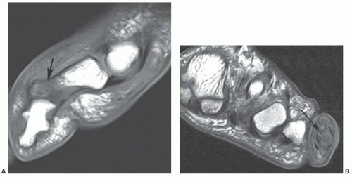
Figure 13.9 Sagittal (A) and axial (B) T1-weighted images of the fifth toe demonstrating decreased signal intensity in the distal aspect of the phalanx (arrow) in a confluent pattern due to osteomyelitis. The distal phalanges are congenitally fused. (From Berquist TH. Imaging of the Foot and Ankle. 3rd Ed. Philadelphia, Lippincott Williams & Wilkins, 2011.)


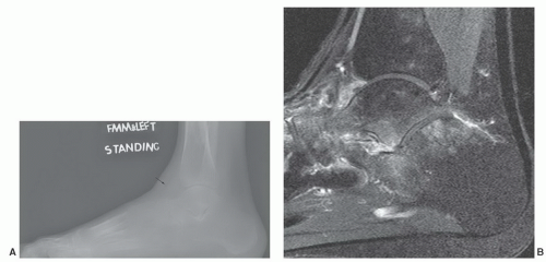
Figure 13.10 Multifocal tuberculosis. A: Lateral radiograph of the foot and ankle demonstrates marked osteopenia in the ankle, mid and hind foot. There is also an ankle effusion (arrow). Sagittal T2-weighted image (B) demonstrates periarticular marrow edema and axial (C) and sagittal (D) post-contrast fat-suppressed T1-weighted images demonstrate extensive bone and soft tissue involvement with synovial enhancement (arrow). Sagittal T1- (E) and T2-weighted (F) images of the spine demonstrate disc and osseous involvement at T12-L1 (arrow). (From Berquist TH. Imaging of the Foot and Ankle. 3rd Ed. Philadelphia, Lippincott Williams & Wilkins, 2011.)


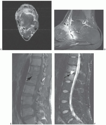
Figure 13.10 (continued)

In children, multiple lytic lesions may mimic fungal infection, pyogenic osteomyelitis, or neoplasms. The latter include eosinophilic granuloma, neuroblastoma, or lymphoma.38

Sharma61 described MR features of tuberculous osteomyelitis. Two types of lesions were described. Lesions might have predominantly low or intermediate signal intensity on T2-weighted sequences and low signal intensity on T1-weighted sequences. Features are related to central
caseating necrosis in tubercular granulomas. The second image feature described was low intensity with marginally higher intensity peripherally on T1-weighted images. Surrounding edema was common in this setting. Extraosseous soft tissue edema and abscess formulation were evident in about 80% of cases.61


Atypical Mycobacterial Osteomyelitis

Atypical mycobacteria (Table 13.3) are frequently drug-resistant and account for up to 30% of all mycobacterial infections.25 Themusculoskeletal system is involved in 1%to 10% of patients with mycobacterial infections.25,70,71 Infections may be caused by hematogenous spread or soft tissue contamination from soil, water, fish, birds, and other animals (Fig. 13.11, Table 13.3).17,25

Only gold members can continue reading. Log In or Register to continue

Stay updated, free articles. Join our Telegram channel

May 25, 2016 | Posted by in RHEUMATOLOGY | Comments Off on Musculoskeletal Infection

Full access? Get Clinical Tree

Get Clinical Tree app for offline access