1.1 Introduction to medical sciences
Chapter 1.1a Introduction to physiology: the systems of the body
The systems of the body
The physiological systems of the body are listed in Table 1.1a-I. Some of the terms may appear unfamiliar (the more commonly used descriptions follow in brackets). However, as they are fundamental to the organisation of most conventional medical textbooks, it is important that their names and functions are clearly recognised and understood.
Table 1.1a-I The 12 physiological functional systems of the body
The organs
 Information box 1.1 a-1 The organs: comments from a Chinese medicine perspective
The differences may be briefly summarised as follows:
| Physiological organs | Chinese medicine Organs |
| Solid physical structures | Terms to describe a collection of functions |
| The structure and function of the organs can be assessed by scientific means: dissection, physical examination, blood tests, ultrasound, x-ray imaging, etc. | The functions of the Chinese medicine Organs are assessed subjectively by checking the state of the Qi–using techniques such as pulse and tongue diagnosis |
| The function of the organ can be related to the structure of the organ. (e.g. the pumping action of the heart organ can be related to its muscular shape, electrical activity and valves) | The function of the Chinese medicine Organs is not related to any structure, although the Chinese medicine Organ may dominate the function of a physiological organ (e.g. the Qi of the Heart Organ, as interpreted in Chinese medicine, supports the heart organ) |
| For any one organ there is not necessarily a counterpart Chinese medicine Organ (e.g. pituitary) | For any one Chinese medicine Organ there is not necessarily a counterpart physiological organ (e.g. Triple Burner) |
Appendix I demonstrates in much more detail the nature of the differences between the functions of the physiological and Chinese medicine organs. The appendix gives a set of ‘Correspondence Tables’, each relating to a particular organ, and shows how the various functions of the physiological and Chinese medicine organs map onto one another (see Q1.1a-3).
The physiological levels
 Self-test 1.1a Introduction to physiology: the systems of the body
1. What is the definition of the term physiology?
2. Name two organs that form part of the following physiological (functional) systems:
3. List the physiological levels of organisation from least complex to most complex.
4. Describe one function of each of the following systems:
Answers
1. Physiology refers to the study of the function of all living organisms.
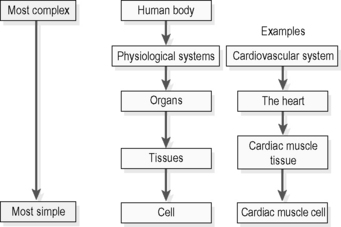
3. Physiological levels of organisation from least complex to most complex: the cell tissues, the organs, the systems, the body.
The physiological levels are summarised in Figure 1.1a-I.
Chapter 1.1b The cell: composition, respiration and division
The cell
Figures 1.1b-I to 1.1b-IV are diagrammatic representations of how cells in different tissues can appear under the light microscope. These diagrams indicate the wide variety of shapes of cells found in different tissues and how each cell within these tissues contains a large central structure, the nucleus. The nucleus also has a characteristic shape in cells of different tissues.
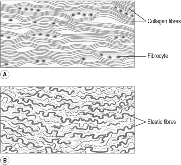
Figure 1.1b-I • Fibrous tissue, illustrating fibre-generating cells (fibrocytes) and collagen fibres.
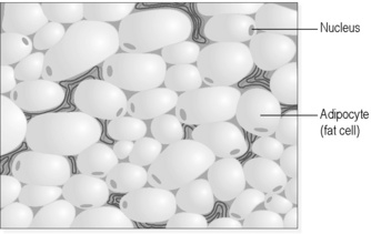
Figure 1.1b-II • Adipose (fat) tissue, illustrating fat-filled adipose cells and minimal connective fibres.
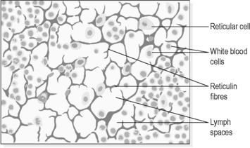
Figure 1.1b-III • Tissue from a lymph node, illustrating immune cells (white blood cells and reticular cells) and supportive reticulin fibres.
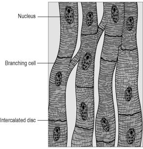
Figure 1.1b-IV • Cardiac muscle tissue, illustrating the fibre-like cardiac muscle cells linked by connecting ‘intercalating discs’.
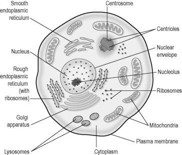
It can be helpful to use the idea of a generalised cell to study how all cells are made up and function. No single cell will be exactly like this generalised cell, but most cells will have the features portrayed in the generalised cell depicted in Figure 1.1b-V.
The structure of the cell
The plasma membrane acts as the link between the cell and the outside world. Large molecules, called proteins, in the membrane make the cell unique (they give the cell its ‘immunological identity’), and respond to various chemicals in the cell’s environment to bring about changes within the cytoplasm. In addition, the membrane allows the passage of nutrients into the cell and waste out of the cell (transport across the membrane) but is able to retain essential fluids and substances within the cell. This important feature of cell membranes, known as ‘semipermeability’ is described in Chapter 1.1c.
The essential information about the ‘generalised’ cell is listed in Table 1.1b-I.
Table 1.1b-I The important characteristics of the cell
Cell replication
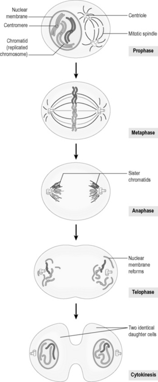
Mitosis and meiosis are illustrated in Figure 1.1b-VI. In this diagram, for the sake of clarity, only a single pair of the human cell’s complement of 23 chromosomal pairs is illustrated. The diagram shows how mitosis results in two identical daughter cells, whereas meiosis results in the production of four genetically unique daughter ‘half-cells’ or gametes.
Mutation
 Self-Test 1.1b The cell

 .
. .
.
 .
. Information Box 1.1b-I
 .
. .
.


