Diseases of the endocrine system

5.1 Diseases of the endocrine system


Chapter 5.1a The physiology of the endocrine system


Learning points


Introduction


The idea has already been introduced that the nervous and endocrine systems together can be considered as the foundation of that which makes the body an integrated whole. This is because they are both concerned with communication between one body part and another.


In the nervous system, the communication between one nerve and another takes place at a junction called the ‘synapse’. At the synapse, the release of a tiny amount of a chemical known as a ‘neurotransmitter’ enables a change that has taken place in one nerve cell to be transmitted to other nerve cells. In the endocrine system, the communication is also based on the release of chemicals. These chemicals, released by specialised endocrine cells, are known as ‘hormones’. Endocrine cells may be grouped together in organs known as ‘endocrine glands’, but also can be found loosely distributed within many of the organs and tissues of the body (see Q.5.1a I)image.


In contrast to neurotransmitters, hormones can stimulate changes in bodily cells only after travelling to local tissues via the tissue fluid, or to distant sites through the bloodstream. However, once hormones reach their target cells, the way in which they lead to internal changes within those cells is remarkably similar to the effect of neurotransmitters on target nerve cells. Both hormones and neurotransmitters connect with protein ‘receptors’ on the target cell membrane. Through this connection, these chemicals lead to a change in the internal physiology of the cell. As increasing numbers of these chemicals are discovered by scientists and their functions described, it is becoming clear that many of those which act as hormones in the body can also be found within the nervous system, where they function as neurotransmitters.


Through communication by means of hormones, the endocrine system encourages the state of homeostasis to be maintained within the body. In order to fulfil this role, the endocrine system has always to be responsive to changes in the internal and external environments of the body (see Q5.1a-2)image.


Cholecystokinin (CCK) (originating from cells in the duodenum), adrenaline (from the adrenal medulla), erythropoietin (EPO) (from the substance of the kidney) and testosterone (from the testes) are examples of hormones that have their impact on the digestive, cardiovascular, blood and reproductive systems, respectively. CCK and EPO are examples of hormones that are not released from specific endocrine organs. CCK is released from endocrine cells scattered within the wall of the duodenum, and EPO is released from endocrine cells within the tissue of the kidney. In contrast, adrenaline and testosterone are examples of hormones that originate from specialised endocrine organs, namely the adrenal glands and the testes, respectively.


The organs of the endocrine system


Figure 5.1a-I illustrates the anatomical location of the organs of the endocrine system. This chapter focuses on the most basic physiology of those endocrine organs that are affected in the important endocrine diseases. These are:


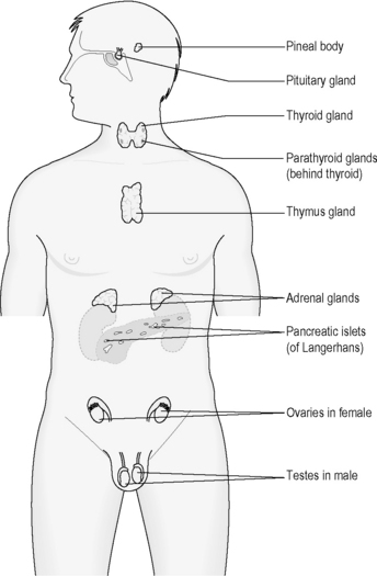
The role of the parathyroid glands in calcium homeostasis is described in Chapters 4.2a and 4.3a. The function of the ovaries and testes will be explored in Chapter 5.2a. The pineal gland and thymus gland will be not discussed further in this text.


The negative feedback loop


The negative feedback loop is the basic mechanism that underlies an important aspect of the action of these hormones. The negative feedback loop requires a detector to recognise a move away from balance, a control centre that recognises when the move has been so great that something has to be done about it, and an effector to bring about a change in the body to reverse the imbalance. A thermostat is a mechanical example of the negative feedback loop in action. The detector in this case is a thermometer. The control centre is the mechanism that is set to recognise when the temperature falls outside a desired range. The negative feedback loop is based on maximum desired temperature. When the temperature becomes too high, the effector, in this case the heating element, is switched off until the time when the temperature drops within the desired range once again.


These rather abstract concepts, which were introduced in Chapter 1.1c, can now be placed in more concrete terms. Endocrine cells play the role of both detector and control centre, while the release of hormone corresponds to the effector of the negative feedback loop. As a thermostat is designed to keep ambient temperature constant, the endocrine negative feedback loops work to keep bodily variables such as blood sugar or blood calcium at a steady level. This chapter offers many more examples of this negative feedback loop in action (for example see Table 5.1a-I), as each of the important endocrine organs is described in turn.


Table 5.1a-I The release of cholecystokinin (CCK): an example of the negative feedback loop in the regulation of hormones


.


The pituitary and the hypothalamus


The pituitary gland is a pea-sized organ that projects downwards from an area at the base of the brain called the ‘hypothalamus’. The site of the pituitary is deep within the head, approximately at the level of the bridge of the nose (Figure 5.1a-II). The extra acupoint Yintang, which is considered in Chinese medicine to have a profound influence over mental functions, overlies the region of the pituitary and hypothalamus.


A ‘stalk’ projects downwards from the hypothalamus through which nerve fibres pass into the pituitary. A delicate network of blood vessels connects the two areas through this stalk. These blood vessels carry hormones from the hypothalamus to the pituitary.


An alternative term for the pituitary gland is the ‘hypophysis’. The term ‘adenohypophysis’ refers to the anterior portion of the pituitary, which contains endocrine cells. The adenohypophysis is the source of six different hormones. The term ‘neurohypophysis’ refers to the posterior portion of the pituitary gland. This part receives nerve fibres from the hypothalamus, and is the source of two additional hormones, which are secreted directly into the bloodstream from the endings of these nerves.


Together, the hypothalamus and the pituitary perform a vital control function in the endocrine system. The hormones secreted by the hypothalamus and the pituitary affect the function of other endocrine organs, including the thyroid gland, the adrenal gland and the sex organs.


The hormones of the hypothalamus are either secreted into the general bloodstream by the nerve endings of the neurohypophysis, or carried within the blood vessels of the pituitary stalk to act on the adenohypophysis. In contrast, all the hormones released by the pituitary gland travel within the bloodstream to have effects on distant endocrine organs as well as other parts of the body.


There are at least 13 hormones that are released by the hypothalamus and the pituitary gland. All of these are commonly described by conventional practitioners by their initials rather than their full medical names, and include growth hormone (GH), thyroid stimulating hormone (TSH), adrenocorticotrophic hormone (ACTH) from the pituitary and growth-hormone-releasing hormone (GHRH), corticotrophin-releasing hormone (CRH) and gonadotrophin-releasing hormone (GnRH) from the hypothalamus.


It is helpful to understand that the hormones of the hypothalamus are the mechanism by which the hormones of the pituitary are controlled. For example, the release of growth hormone (GH) from the pituitary gland can be increased and decreased by two different hormones (GHRH and GHIH) from the hypothalamus. This is one example of the close connection between the nervous system (hypothalamus) and the endocrine system (pituitary gland).


The hypothalamus lies deep within the substance of the brain, and thus can be influenced by complex internal factors, including the stage of psychological development and the emotions. This might explain why such factors can have profound effects on hormone-controlled events such as childhood growth, timing of puberty and breastmilk production.


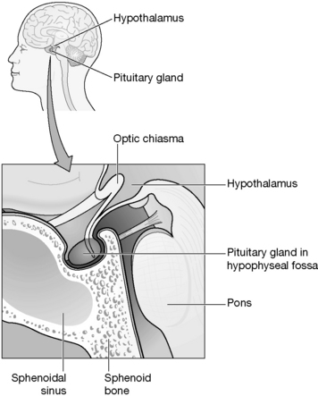
Table 5.1a-II lists the six hormones that are produced by the adenohypophysis.


Table 5.1a-II The hormones released from the anterior pituitary gland (adenohypophysis) and their functions


Hormone Function
Growth hormone (GH) Stimulates growth in many tissues
Thyroid-stimulating hormone (TSH) Stimulates the production of thyroid hormones
Adrenocorticotrophic hormone (ACTH) Stimulates the production of cortisol from the adrenal gland
Prolactin (PRL) Stimulates breastmilk production
Follicle-stimulating hormone (FSH) Stimulates the development of the ovarian follicle (the first half of the menstrual cycle)
Luteinising hormone (LH) Stimulates ovulation and the maturation of the ruptured follicle (corpus luteum)

The hormones oxytocin and antidiuretic hormone (ADH) are released from the neurohypophysis, but have their origins within nerve cells within the hypothalamus.


Growth hormone


Growth hormone is a hormone that stimulates the continued growth of the skeleton, muscles and soft tissues, including the major organs. This growth is not only essential during childhood, but is also important for the maintained health and liveliness of these tissues throughout adult life. Growth hormone affects the metabolism of the body, meaning that it alters the rate at which the complex chemical processes of the bodily tissues take place. Under the influence of growth hormone, the rate of protein and collagen synthesis increases, and the amount of sugar in the blood tends to rise to meet the body’s increased requirements for energy.


ADH, TSH, ACTH, FSH, LH and prolactin


The role of ADH was mentioned in Chapter 4.3a. This hormone is released in response to an increased concentration of the salts in the blood. It reduces the amount of water that is lost through the urine, and is important in the control of the homeostasis of the concentration of the blood. TSH and ACTH relate to the healthy function of the thyroid and adrenal glands, respectively. PRL, FSH and LH are all important in the physiology of the reproductive system. The function of these five pituitary hormones is described in more detail in Chapter 5.2a.


The thyroid gland


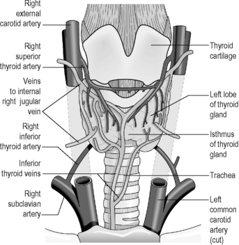
Figure 5.1a-III illustrates the thyroid gland, which is a bow-shaped organ situated just below the level of the laryngeal prominence (Adam’s apple) in the neck. The bow shape is a result of the gland being formed by two lobes separated by a narrow bridge of tissue called the ‘isthmus’. In health, the thyroid gland can be felt as a small region of vague softness below the voice box. This can be felt to rise and descend during swallowing.


The thyroid gland contains endocrine cells, which secrete two hormones, thyroxine (also known as T4) and triiodothyronine (T3). The thyroid gland draws upon iodine from the diet to manufacture sufficient quantities of these hormones. Iodine is found in seawater, and also in plant and animal products that originate from areas close to the sea. For this reason, iodine deficiency may occur in regions of the world that are distant from the sea, especially landlocked countries. However, in many developed countries, such as the UK, table salt contains additional iodine, meaning that iodine deficiency is rarely a problem.


The thyroid hormones T4 and T3 are both released in response to TSH (thyroid-stimulating hormone) from the pituitary. The release of TSH is primarily controlled by the release of TRH (thyroid-releasing hormone) from the hypothalamus. This is the mechanism whereby the activity of the brain affects thyroid function.


Like growth hormone, the thyroid hormones play an important role in physical growth and the rate at which the cells of the body convert nutrients into energy (the metabolic rate). They are also important in the healthy development of the growing nervous system, the appropriate function of the cardiovascular system and the smooth functioning of the gastrointestinal system. If in excess, the thyroid hormones can cause an increased use of bodily nutrients, leading to the production of heat, weight loss, feelings of nervousness, a rapid heart rate and increased peristalsis in the bowel.


The negative feedback loop of control of the thyroid hormones is affected by factors such as exercise, stress, low blood glucose and malnutrition. All these factors increase the release of TSH from the hypothalamus, and thus increase the release of T4 and T3. Conversely, raised levels of thyroid hormones in the bloodstream will inhibit the release of TRH from the hypothalamus and TSH from the pituitary gland (see Q5.1a-3)image.


The adrenal glands


The adrenal glands are situated on top of the kidneys, deep in the back of the abdomen, approximately at the level of the lumbar vertebrae L1 and L2. Each gland contains two distinct areas of endocrine tissue. The outer part of each gland is called the ‘cortex’, and this is the source of three types of steroid hormones, known as glucocorticoids, mineralocorticoids and androgens. The androgens secreted by the adrenal cortex are male sex hormones, but have a minimal effect in comparison to the effect of those secreted by the sex organs. The inner part of the adrenal gland is called the ‘medulla’. The adrenal medulla is the source of two hormones, adrenaline and noradrenaline, which play an important role in the sympathetic nervous system.


The pancreas


The pancreas plays an important exocrine role in the production of several digestive enzymes, which are released into the duodenum to break down the nutrients in food leaving the stomach. Studded within the exocrine tissue of the pancreas are little islands (islets) of endocrine cells, which secrete a range of hormones. These hormones are important for controlling the way in which the body utilises glucose after it has been absorbed into the bloodstream. In a way this can also be seen as an aspect of the digestive function.


The two most well-recognised hormones secreted by the islets are insulin and glucagon. These two hormones act in complementary ways to ensure that the cells of the body have access to a continuous supply of glucose, whatever the external conditions may be. This means that, whether the prevailing condition is one of feast or famine, the body tissues can be adequately nourished, at least in the short term.


Summary


This concludes the introduction to the organs of the endocrine system. Perhaps more so than for any other system, the delicate nature of the function of this system can only really become apparent after studying what happens when the system becomes out of balance.


image Self-test 5.1a The physiology of the endocrine system


Answers


1.


2.


3. To answer this question well you need first to make a distinction between the anterior pituitary (adenohypophysis) and the posterior pituitary (neurohypophysis). The neurohypophysis can be considered as a physical extension of the nerve cells of the hypothalamus. The two hormones released from the neurohypophysis are released in direct response to nervous messages reaching the hypothalamus. In this way they are similar in action to the hormone-releasing nerve cells of the hypothalamus itself.


In contrast, the adenohypophysis contains hormone-secreting endocrine cells. The hormones of the pituitary are released in response to hormones that have been first released from the hypothalamus, and so can be seen as in the control of the hypothalamus. This is the important difference between the pituitary and the hypothalamus.


Blood and urine tests


Simple blood tests can be used to assess the levels of the hormones in the blood. In some cases, the results alone may indicate whether or not endocrine disease should be suspected. For example, in suspected thyroid disease the most common first tests to be performed are serum tests to assess the levels of TSH, T4 and T3. Levels of these hormones that fall outside the normal range would be suggestive of thyroid disease. Such tests are easy for the doctor to perform, and not too inconvenient for the patient.


However, in other cases, because the level of the hormone varies dramatically throughout the day, and also according to individual need at the time, a single blood test cannot offer such conclusive results.


Sometimes a patient might be requested to collect all their urine over the course of 24 hours so that levels of hormone can be assessed from the urine. This has the advantage of overcoming the problem of varying levels throughout the day, but is well known for not being a very accurate method of assessment. Moreover, many patients find this an inconvenient and embarrassing test to perform.


Blood and urine tests may also be used to assess the effects on the body of the hormone in question. The most commonly performed test of this type is the assessment of the glucose (sugar) levels in the blood and the urine in cases of suspected diabetes mellitus. A raised blood glucose level is an indication of a lack of insulin, which is the underlying problem in diabetes mellitus. In both cases, a helpful result can be obtained by applying a drop of blood on a test card, or by dipping a test ‘stick’ into the urine.


The most accurate and clinically relevant form of assessing the blood glucose level is to take a specimen when the patient has been fasting for some hours. The least accurate means is to look for glucose in the urine. The latter may be performed to provide a very general overview of the problem, but the gold standard, the fasting blood sugar test, is obviously more inconvenient for the patient.


Stimulation and suppression tests


Stimulation and suppression tests are more complex forms of blood and urine tests. They usually involve assessing the hormonal response that takes place over a few hours following a certain ‘stimulus’ given to the body. For this reason, the patient may need to spend a day or more in hospital for such tests to be performed. The ‘stimulus’ is chosen to be something that would normally significantly increase (stimulation test) or decrease (suppression test) the level of the hormone in question.


In general, the stimulation test is used to investigate a disease resulting from insufficient hormone, and a suppression test is used to investigate a disease resulting from excessive secretion of a hormone. For example, suspected diabetes might be confirmed by using the glucose tolerance test. In this test, a patient who has been fasting from the previous night is given a glucose drink at a set time in the morning. Blood tests are taken at defined time intervals, including one just before the set time of the drink. The blood glucose can then be seen to rise and fall from the fasting period to a few hours after the drink. In health, the blood glucose never exceeds a predictable normal level, but in mild diabetes it may be seen to rise to above a healthy level, even though the level might then drop to within a healthy range after some time. Such an abnormality might not be picked up by a simple blood sugar test. The glucose tolerance test is an indirect form of a stimulation test. The glucose drink should, in health, lead to a release of insulin. The release of insulin in turn should prevent the rise in blood sugar from going too high. The fact that this fails to happen is an indication of insufficient release of insulin.


Another example is the test for a person with suspected disease of the adrenal cortex (Addison’s disease). In this case the patient is given an injection of a synthetic form of the pituitary hormone ACTH. In health, this hormone stimulates the release of cortisol and corticosterone from the adrenal cortex. In a patient with Addison’s disease, timed blood tests reveal a much lower rise in cortisol in the blood than would be expected. This is another example of a stimulation test that can reveal insufficient production of a hormone.


Stimulation and suppression tests are based on the complexities of endocrine physiology, a clear understanding of which is a challenge even for qualified doctors. From the perspective of a complementary medical practitioner, the important fact to appreciate about the investigation of endocrine disease is that a patient with suspected endocrine disease may be required to spend some time in hospital undergoing such tests.


Imaging tests


Very commonly, endocrine disease results from structural damage to an endocrine gland. In such cases the use of complex soft tissue imaging tests, such as the CT scan and MRI, can be helpful in defining the extent of the damage and whether or not the problem is operable.


For example, the pituitary gland can be clearly revealed by means of MRI. Figure 5.1b-I shows the sort of image that may be obtained. This image represents the upward growth of a pituitary tumour. Such growth can have marked endocrine consequences (as well as being very likely to impair vision by upwards pressure on the crossing of the optic nerve tracts) and also in the causation of headaches.
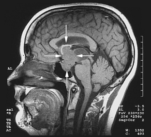


image Self-test 5.1b The investigation of the endocrine system


Enlargement of the thyroid gland: the causes of goitre


An enlarged thyroid gland is called a ‘goitre’. Large goitres are visible as a fullness of the neck, whereas smaller goitres can only be felt on examination. A goitre is considered to be present if it can be felt as a swelling that moves up and down with swallowing. This swelling is more than the vague softness that is usually felt in the region of the thyroid gland. Most goitres are small, and may not be noticed by the patient.


A goitre can become so large as to cause symptoms. The most common symptom is of visible swelling and the cosmetic problems that accompany this. Only in rare cases are there other symptoms. These include discomfort in the neck, difficulty swallowing and restricted breathing.


A goitre does not necessarily mean that there is a problem with the production of the thyroid hormones. The various causes of goitre are described below.


Stay updated, free articles. Join our Telegram channel

Oct 3, 2016 | Posted by in MANUAL THERAPIST | Comments Off on Diseases of the endocrine system

Full access? Get Clinical Tree

Get Clinical Tree app for offline access