Foot and Ankle Pain

49 Foot and Ankle Pain


Foot and ankle pain are independent risk factors for locomotor instability, impaired balance, and increased risk for falling, as well as compromised functional activities of daily living.15 Foot and ankle pain appear to affect approximately one in five middle-aged to older individuals. Interference with daily activities occurs in one-half to one-third of affected individuals but is rarely disabling outside the context of rheumatoid arthritis. Foot and ankle pain is significantly more common in women, a finding that has been attributed to gender-specific footwear.


Causes of Foot and Ankle Pain


The differential diagnosis of foot and ankle pain is vast and includes conditions of tendons, ligaments, muscle, bone, joints, periarticular structures, nerves, and vessels, as well as referred pain (Table 49-1).


Table 49-1 Differential Diagnosis of Foot and Ankle Pain


Tendon, Ligament, and Muscle

Bone

Joint

Periarticular Structures

Nerves

Vessels

Referred Pain


Courtesy Dr. George Raj, Non Surgical Spine and Joint Clinic PS, Bellingham, Wash.


The most common cause of pain of the foot and ankle is osteoarthritis (OA). Although OA is the most prevalent joint disease, its pathophysiology remains poorly understood. Research regarding foot and ankle OA in particular is limited by absence of a standard case definition. Ankle and foot OA results from damage and loss of the articular cartilage, which can cause inflammation, stiffness, pain, swelling, deformity, and limitation of function, such as walking or standing. Osteophyte formation can lead to impingement and further pain. In the foot, OA most commonly occurs in the big toe, the midfoot, and ankle. In the early stages, pain may occur only at the beginning and at the end of an activity, but as the condition progresses it can become constant, even at rest.


The ankle is a complex joint that is subjected to enormous forces during daily activities and in sports, especially running. It is also the joint most commonly injured in the human body. This combination of factors predisposes the ankle joint to degenerative changes, although the risk is lower than other weight-bearing joints, such as the hip and knee. The ankle also rarely develops arthritic changes without an identifiable cause. The most common cause of ankle OA is trauma and can develop following a fracture or repeated sprains. Other causes of OA are abnormal foot mechanics (flat and high-arched feet) and, rarely, systemic diseases such as hemochromatosos.


Foot and ankle pain is the presenting complaint in approximately 15% to 20% of newly diagnosed rheumatoid arthritis (RA) patients.6 Further, of those patients already diagnosed with RA, the prevalence of foot and ankle involvement has been estimated to be greater than 90%.7


Evaluation of the rheumatoid foot and ankle begins with a thorough history and physical examination. The location, timing, and duration of symptoms can help establish a specific diagnosis and help guide the subsequent course of treatment. Radiographs and advanced imaging modalities provide useful adjuncts in the evaluation of specific foot and ankle pathologies.


The treatment of the rheumatoid foot and ankle is aimed at both alleviating pain and preserving function (i.e., maintaining the ambulatory status of the patient). Initial nonoperative treatment includes medical management, physical therapy, shoewear modification, orthotics, and bracing. These measures provide substantial relief for many. For recalcitrant symptoms, surgical intervention may be necessary. Most surgical procedures fall into one of the following general categories: arthrodesis (joint fusion), arthroplasty (joint replacement), corrective osteotomy, ostectomy, and synovectomy (joint or tendon).


Functional Anatomy and Biomechanics


The ankle, or tibiotalar joint, is composed of the articulation between the foot (talus) and the lower leg (distal tibia and fibula). Its primary motion is plantar flexion and dorsiflexion in the sagittal plane. In addition, the articulation between the distal tibia and fibula allows a lesser amount of internal and external rotation to occur in the axial, or transverse, plane.


The foot may be loosely divided into three anatomic regions: forefoot, midfoot, and hindfoot. The forefoot consists of the toes and metatarsal bones, along with the metatarsophalangeal (MTP) and interphalangeal (IP) joints. The tarsometatarsal (TMT) joints connect the forefoot to the midfoot, which comprises the three cuneiform bones, the navicular, and the cuboid. Finally, the hindfoot, located below the ankle, consists of the talus and calcaneus. The joints of the hindfoot include the talocalcaneal (subtalar), talonavicular, and calcaneocuboid articulations.


Forefoot and midfoot motion is primarily plantarflexion and dorsiflexion in the sagittal plane, with some secondary pronation and supination in the coronal plane and abduction/adduction in the axial plane. Motion in the hindfoot is primarily composed of inversion/eversion in the coronal plane, with secondary internal/external rotation in the axial plane and plantarflexion/dorsiflexion in the sagittal plane.


Knowledge of these anatomic divisions is important because radiographs often demonstrate polyarticular disease in patients with RA. An intimate understanding of the local anatomy greatly aids in the establishment of an accurate diagnosis and formulation of an appropriate treatment plan.


Diagnostic Evaluation


Physical Examination


A thorough physical examination of the foot and ankle begins with gait analysis, even if simply observing the patient enter the examination room. Normal human gait is divided into two phases. The stance phase is the weight-bearing portion of the gait cycle and comprises roughly 60% of normal walking. It begins with heel-strike and then extends through foot-flat to toe-off. Meanwhile, the swing phase of gait extends from toe-off to heel-strike and comprises the remaining 40% of the gait cycle.


Patients with an “antalgic” gait pattern will have a shortened stance phase on the side of the affected limb, as they attempt to more quickly transfer their weight to the nonpainful limb. In addition to an antalgic gait, foot and ankle pain often results in the avoidance of ground contact with the painful part of the foot. A further problem noted in stance phase is dynamic collapse of the medial longitudinal arch, most apparent at foot-flat and toe-off.


During the swing phase of gait, a “steppage” gait may be noted. This is characterized by excessive hip and knee flexion to allow a patient’s foot to clear the ground in the setting of a footdrop. In patients with RA, it may be caused by attritional rupture of the anterior tibialis tendon, which is the main dorsiflexor of the ankle.


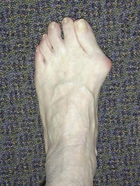
Following gait analysis, the foot and ankle are inspected, both with the patient sitting and standing. The location of swelling is usually well correlated with the joint(s) involved (e.g., ankle vs. talocalcaneal joint). Deformity should also be noted. Commonly seen deformities in patients with RA include hallux valgus, or bunion (Figure 49-1); hammertoes; and flatfoot deformity (characterized by hindfoot valgus/forefoot abduction). Callosities develop over regions of increased pressure and are associated with deformity and fat pad atrophy. Rheumatoid nodules can appear anywhere on the foot but are often found in areas of repetitive trauma (i.e., at the site of irritation from a tight shoe counter). Similarly, ulcerations appear in areas of repeated injury such as those found in tight-fitting shoes. Finally, wear patterns on shoes should also be noted. As Hoppenfeld observed8: “A deformed foot can deform any good shoe; in fact, in many cases the shoe is a literal showcase for certain disorders.”


Following inspection, the foot and ankle are thoroughly palpated. The dorsum of the foot and ankle has little overlying musculature. As such, many of the bones and tendons are immediately subcutaneous and a great deal of information can be gained from palpating these structures. It is helpful to palpate the foot and ankle by anatomic location (i.e., forefoot, midfoot, hindfoot, anterior and posterior ankle).


In the forefoot, the first metatarsal head and MTP joint can be palpated at the base of the hallux (great toe), at the medial aspect of the “ball” of the foot. Proceeding laterally, the lesser metatarsal heads and MTP joints can then be sequentially palpated. In patients with RA, such palpation often reveals tenderness, synovitis, and bursal swelling. In the second and third MTP joints, sagittal plane instability often results from attenuation of the plantar joint capsule. This can be appreciated by gently translating the second and third toes dorsally.


In the hindfoot the calcaneus is readily palpable, and its various parts can be palpated individually. A stress fracture should always be considered in patients with RA. Further, tenderness over the posterior aspect of the bone may indicate Achilles tendinitis while pain over the medial tubercle (palpable on the medial plantar surface) may indicate plantar fasciitis. Tenderness over the “sinus tarsi” of the hindfoot (located laterally, just anterior and distal to the tip of the fibula) indicates talocalcaneal joint pathology. Finally, posteromedial tenderness may be secondary to tenosynovitis, posterior tibial tendinosis, and tarsal tunnel syndrome (usually secondary to adjacent tenosynovitis).


In the ankle joint proper, tenderness over the anterior joint line usually correlates with ankle joint pathology including arthritis, synovitis, impingement, and osteochondral defect (OCD).


A more detailed description of these conditions and their correlation with anatomic location is provided later in the chapter and in Table 49-2.


Table 49-2 Anatomic Characteristics of Pain in the Foot and Ankle


Location Dysfunction
Forefoot
Midfoot Plantar fasciitis, arthritis, synovitis (rare)
Hindfoot
Anterior ankle Arthritis, synovitis, impingement, osteochondral defect
Posterior ankle
< div class='tao-gold-member'>

Stay updated, free articles. Join our Telegram channel

Jul 3, 2016 | Posted by in RHEUMATOLOGY | Comments Off on Foot and Ankle Pain

Full access? Get Clinical Tree

Get Clinical Tree app for offline access