Figure 11.1
Metatarsus adductus. Note the adducted hallux and forefoot and the slightly valgus hindfoot (Reproduced from Benson et al. Children’s Orthopaedics and Fractures, 2009, Springer)
The forefoot is adducted and often supinated.
Look at the lateral border of the foot for medial deviation.
Check for deep medial skin creases in severe deformity.
Check if the deformity is correctable.
Check the hindfoot, which will be in neutral or valgus, compared to the varus position in CTEV.
Examine all joints of the lower limb.
Look for DDH.
A rare severe form of metatarsus adductus is seen in the ‘serpentine (Z) foot’ or ‘severe skew foot’ (Fig. 11.2). This is characterised by a rigid adduction deformity, with lateral subluxation of the navicular and hindfoot valgus.
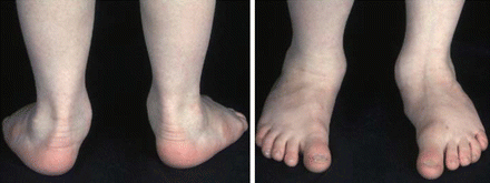
Figure 11.2
Serpentine deformity or Z-Foot (Reproduced from Benson et al. Children’s Orthopaedics and Fractures, 2009, Springer)
Treatment
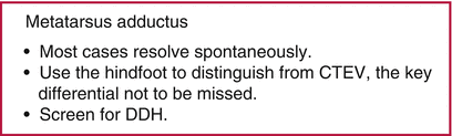
The majority of cases will resolve spontaneously. Any initial intervention should be in the form of stretches, moving to manipulation and serial casting if required. Length of treatment is variable but is often at least 10 weeks (Table 11.1). The goal is to achieve a straight lateral border of the foot.
Table 11.1
Initial treatment options in metatarsus adductus
Deformity correctable actively | No intervention required |
Deformity correctable passively | Stretching exercises by parents |
Rigid deformity or deep medial skin crease | Manipulation and serial casting |
Complication rates for surgical intervention are high and surgery is reserved for recurrent cases with severe deformity or late presentation. The fixed deformity seen in a Z foot often requires surgical intervention.
Surgical Intervention
Soft tissue release: abductor hallucis longus recession or release with or without tibialis posterior lengthening. Joint capsulotomies are more invasive.
Osteotomy: metatarsal open or closing wedge osteotomies can alter column length to correct fixed deformity where conservative therapy is not appropriate or has failed.
Talipes Calcaneovalgus
This deformity is more common in females and in first-born children (Fig. 11.3). It is often bilateral. Some cases are related to neurological conditions.
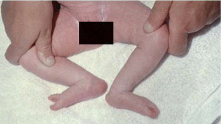
Figure 11.3
Calcaneovalgus feet in an infant (Reproduced from Benson et al. Children’s Orthopaedics and Fractures, 2009, Springer)
Symptoms and Signs
This condition presents shortly after birth. In contrast to CTEV, the hindfoot lies in a valgus position, with the foot dorsiflexed.
Look for skin creases anterior to the ankle.
Dorsiflexion can be marked, with the dorsal surface of the foot lying in contact with the tibia.
The deformity should be correctable passively.
Neurological examination is recommended as muscle imbalances caused by L5 spina bifida can cause this deformity. The condition is associated with DDH. Therefore, routine screening is recommended especially in unilateral cases. Other differential diagnoses include congenital postermedial tibial bowing and congenital vertical talus. Check if the deformity is fixed as expected in vertical talus.
Treatment
This deformity usually corrects spontaneously during the neonatal period. Severe deformity may require stretching exercises or serial casting.
Posteromedial Bowing of the Tibia
Posteromedial bowing of the tibia is a congenital disorder, which as its name suggests, leads to bowing of the tibia with the apex of the bow being posterior and medial. It normally affects the middle to distal portion of the tibia. The disorder is initially accompanied by a calcaneovalgus deformity which resolves spontaneously with time. The bowing itself normally resolves by the 4th birthday, although a residual leg length discrepancy is often apparent at skeletal maturity. The degree of leg discrepancy is thought to be independent of the initial degree of bowing and is typically about 4 cm.
Congenital Vertical Talus (CVT)
Congenital vertical talus or congenital convex pes valgus is a rare condition. The aetiology is unknown but family history is a known risk factor. Half of cases are bilateral and up to half are associated with an underlying genetic or neuromuscular disorder such as myelodysplasias, arthrogryposis or chromosomal abnormalities (Fig. 11.4).
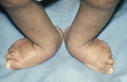
Figure 11.4
Bilateral CVT. Note the forefoot valgus and eversion. (Reproduced from Benson et al. Children’s Orthopaedics and Fractures, 2009, Springer)
Symptoms and Signs
This presents shortly after birth with a fixed deformity.
Examination findings reveal:
The foot is in dorsiflexion.
Medial arch is flat.
Hindfoot is often in a valgus position and potentially equinus.
The vertical talus position will result in a convex or “rocker-bottom” shape to the plantar surface of the midfoot.
There are four abnormalities that are always present in a true CVT:
Irreducible dorsal navicular dislocation.
Peroneus longus and tibialis posterior displacement such as they act as dorsiflexors.
Talo-calcaneal joint subluxation.
Fixed ankle equinus.
The most common differentials to consider are a calcaneo-valgus or plano-valgus foot, or an oblique talus. A true CVT is distinguished from the aforementioned conditions through the rigid, non-correctable position of the talus. Plantar flexion stress lateral radiographs (the Eyre-Brook view) help determine the position of the talus and it’s flexibility relative to the navicular and first ray (Fig. 11.5).
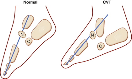
Figure 11.5
Diagram demonstrating the plantar-flexion stress lateral radiographs of normal and congenital vertical talus feet. A further key radiographic finding is the fixed dorsal dislocation of the navicular. The navicular is dotted as it is the last bone in the foot to ossify but its position can be determined by drawing a line through the first ray
Treatment
The aim of treatment is to restore the bones to their normal anatomical position, correcting the following deformities:
Talo-navicular dislocation.
Hindfoot equinus.
Forefoot eversion.
Non-operative
Non-operative treatment rarely succeeds in successfully treating CVT and so intervention is normally required.
Manipulations and serial casting: Manipulation into plantar-flexed and inverted positions form part of the ‘reverse Ponseti’ method. This method is successful, is the first-line of treatment and is increasing in popularity, although recurrence is an issue. A mini-open reduction of the talo-navicular joint is seen as part of the reverse Ponseti technique, along with any lengthening of involved dorsiflexors and evertors and an Achilles tenotomy, all performed at the end of the casting period.
Surgical Intervention
Surgery is normally performed between the ages of 6 months and 2 years and is usually preceded by serial ‘reverse Ponseti’ casting. A variety of techniques have been described which include soft tissue releases, lengthening of the dorsolateral tendons (Achilles, peroneals, extensors) and reconstruction of the calcaneonavicular (spring) ligament. Open or mini-open reduction techniques can be combined with soft tissue release to reduce the talus. This often requires maintaining reduction with a K-wire, later removed at 6 weeks. The recurrence rates of major open CVT surgery are often high (between 60 and 100%) and further revision procedures are often needed as child grows older.
Related posts:

Stay updated, free articles. Join our Telegram channel

Full access? Get Clinical Tree


