(1)
Temple University, Philadelphia, PA, USA
(2)
Department of Orthopedic Surgery, Sydney Kimmel School of Medicine at Thomas Jefferson University, Rothman Institute, Philadelphia, PA, USA
Keyword
Epidemiology of shoulder injury2.1 Introduction
Shoulder injuries, including both acute and chronic, are a frequent occurrence among overhead athletes [2]. Baseball pitchers, football quarterbacks, swimmers, volleyball players, and tennis players are included in this distinct group of athletes who consistently place an exquisite amount of stress on their shoulders. It is important to note that the overhead throw may produce a velocity greater than 7,000°/s and is the fastest movement recorded in humans [2, 3]. Because of this, the throwing athlete can be quite a challenging patient in sports medicine. To effectively perform a throw, there must be a delicate balance between shoulder laxity to accomplish extreme ranges of motion, so that velocity can be generated to the hand or racket and ultimately to the ball, and adequate stability to prevent subluxation or instability [8]. This is defined as “the thrower’s paradox.” Inherent stress is put on the stabilizing structures including the glenohumeral joint and the scapula and also soft tissue stabilizers such as the rotator cuff, scapulothoracic musculature, fibrocartilaginous labrum, and the joint capsule [2]. Awareness of the forces to which these structures are subjected to in the throwing motion is essential to the diagnosis and treatment of injuries [8]. It is also crucial for the overhead athlete to participate in a preventative stretching and strengthening program during the season and in the off-season to maximize performance and avoid injuries [7].
Unfortunately, not all shoulder injuries can be avoided and may occur due to muscle fatigue or weakness, poor mechanics and conditioning, and altered stability [4–6]. Also, although many shoulder injuries in the elite overhead athlete are common and predictable, there still may be some controversies as to the precise mechanisms by which these injuries occur [1]. In this chapter, we will review the biomechanics of a throw and the adaptive changes that occur in the dominant throwing shoulder. We will also discuss the evaluation and treatment of the many common pathologies observed in the shoulders of overhead athletes.
2.2 Shoulder Biomechanics and Pathoanatomy
2.2.1 Phases/Mechanics of a Throw
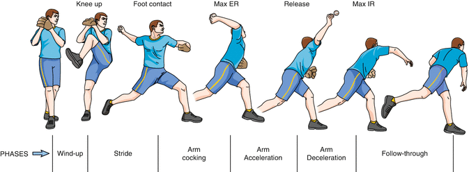
Overhead athletes most frequently subject their shoulders to the unstable position of maximal abduction and external rotation during a throw. It is important to obtain a thorough understanding of the biomechanical stresses placed on specific shoulder structures as they correlate to distinct phases of the throwing motion [8]. It is also necessary to take into account that the legs and trunk (core) play a significant role as force generators in the transfer of energy to the ball. The scapula aids in allowing for extreme ranges of motion by providing a stable platform for the humeral head to rotate. This is part of the “kinetic chain concept.” There are six phases included in the description of a throw, which are achieved in approximately 2 s, and have been described in Chap. 1 but are reviewed briefly in this chapter (Fig. 2.1).


Fig. 2.1
Elbow medial collateral ligament injuries (Ra’Kerry K. Rahman1, William N. Levine1 and Christopher S. Ahmad. Department of Orthopaedic Surgery, Center for Shoulder, Elbow and Sports Medicine, Columbia University Medical Center, 622 West 168th Street, PH 11th Floor, New York, NY 10032, USA)
Phase I is the wind-up or readying phase, in which there is the least amount of stress placed on the shoulder. Toward the end of this phase, the dominant shoulder is in slight internal rotation and abduction.
Phase II is the early cocking phase, which involves recruitment of the deltoid muscle early on and the supraspinatus, infraspinatus, and teres minor muscles later. The shoulder is moved into 90° of abduction with the elbow positioned slightly posterior to the plane of the body.
Phase III is the late cocking phase and involves planting of the lead striding leg with the shoulder culminating in its maximal external rotation to approximately 170–180°. The scapula retracts and aids in stabilization of the humeral head. Shoulder abduction remains at 90–100° and combined with the maximum external rotation, the humeral head, then translates anteriorly on the glenoid. The deltoid muscle firing decreases, and recruitment of the supraspinatus, infraspinatus, and teres minor muscles reaches its peak. Later in this phase, the subscapularis is fired as the torso rotates forward. The biceps, pectoralis major, latissimus dorsi, and serratus anterior muscles begin to fire at the end of this phase allowing maximum horizontal adduction.
Phase IV, acceleration, involves internal rotation of the shoulder 90° to the ball release point. In this phase, the scapula protracts and continues to provide a stable base for the humeral head as the body begins to move forward. This allows for a conversion of eccentric to concentric muscle function anteriorly and concentric to eccentric posteriorly. The triceps muscle is recruited early on in this phase, followed by the pectoralis major, latissimus dorsi, and serratus anterior later.
Phase V is known as deceleration and is the most violent phase of the throwing cycle due to the dissipation of the remaining kinetic energy to the ball. It is a reversal of the first three phases. Ball release occurs and the humeral rotation returns to 0°. Shoulder abduction is at 100° and horizontal adduction increases to 35°. Eccentric contraction of all of the above muscle groups occurs in order to slow down arm rotation.
Phase VI is last and is known as the follow-through. During this phase, the body moves forward along with the arm until motion stops. Muscle firing ceases to resting levels and the loads on the joint decrease.
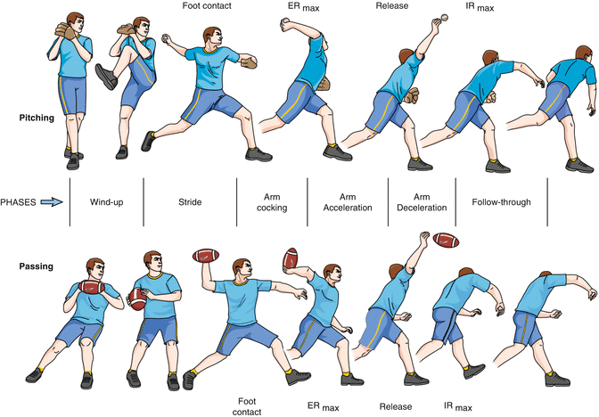
The phases of a throw described above are more descriptive of a baseball pitch. Although similar, there are some subtle differences when comparing the pitch with a throw in different sports. In football, the increased weight of the ball provides changes in mechanics (Fig. 2.2). It has been shown that quarterbacks rotate the shoulder sooner and assume maximal external rotation of the shoulder earlier on in the throw. This allows for more time in acceleration during internal rotation. Also, the shoulder horizontal adduction is increased and greater elbow flexion is achieved in the late cocking phase. This decreases the potential load on the shoulder. Arm velocity is decreased in the football throw because the quarterback must have a more erect position and a complete follow-through that is observed in the baseball pitch does not occur in football. Due to the decreased force generated when throwing the ball, shoulder injuries are not as common.
2.2.2 Adaptations of the Dominant Throwing Shoulder
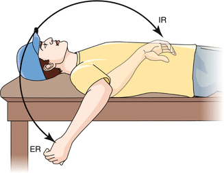
Due to the repetitive motion of the dominant upper extremity and the extreme stresses of the throw, adaptive changes occur that affect the stabilizing structures. In overhead athletes, the ability to externally rotate the humerus is very important to generate high velocities to the ball. This leads to an increase in humeral retroversion and a relative capsular laxity. It has been shown in many high-level throwing athletes that the dominant shoulder exhibits increased external rotation and decreased internal rotation in abduction. These changes occur due to laxity within the anterior inferior glenohumeral ligament (AIGHL), which normally functions as a restraint to anterior translation of the arm in abduction and external rotation. The coracohumeral ligament (CHL) is also a restraint to external rotation at the side and may develop laxity, especially in baseball pitchers, and as a result, an increased sulcus sign may be seen. Retroversion of the humeral head (an average of 10–20°) is also a finding in overhead athletes that may lead to increased external rotation of the dominant shoulder. The increase in external rotation can range from 9° to 16°. However, there are instances when the increase in external rotation does not make up for the loss of internal rotation. This typically occurs later in the athlete’s career and attributes to a loss of overall rotational motion of the dominant shoulder (Fig. 2.3). This is the general process and evolution of glenohumeral internal rotation deficit (GIRD).


Fig. 2.3
Muscular changes are also seen in the dominant arm of throwing athletes. Hypertrophy of the shoulder girdle muscles is common, while atrophy of the infraspinatus can be seen as well. External rotation strength, exhibited by the infraspinatus and teres minor muscles, has been found to be weaker in the dominant as compared to the nondominant shoulder. Conversely, muscles used in internal rotation, such as the subscapularis, latissimus dorsi, pectoralis major, and teres major, are typically stronger in the dominant throwing arm.
2.3 Clinical Presentation and Essential P/E
2.3.1 History
A very detailed history must be obtained for accurate diagnosis and treatment of shoulder injuries. Obtaining the mechanism of injury, duration, location, and timing of symptoms will give clues to the diagnosis. The age of the patient will also aid in possible differential diagnoses. For instance, young athletes will frequently have physeal injuries, labral pathology, or instability. On the other hand, older patients will more likely have pathology of the rotator cuff. Overhead athletes in the middle of their career may acquire laxity and rotator cuff pathology.
It is also important to inquire about specific history unique to overhead athletes, such as the phase of the throwing cycle that symptoms usually occur. Pain that occurs during the cocking phase may suggest labral pathology, internal impingement, or instability. During the late cocking or acceleration phases, anterior instability may be seen. In regard to posterior labral pathology or instability, this typically occurs with pain at the follow-through phase. Pain during deceleration or ball release may point toward a diagnosis of rotator cuff pathology. Timing of symptoms during the game, complaints of loss of throwing velocity, lack of command while throwing, and recent decrease in the number of pitch counts during games can also provide clues as to the possible diagnosis. It is also important to ask at what age the athlete began throwing and amount of rest in between seasons. Symptoms pertaining to the experience of numbness, tingling, or discoloration should raise the possible concern of neurologic or vascular pathology.
2.3.2 Physical Examination
2.3.2.1 Observation
Examination of the shoulder should begin with observation of positioning and symmetry, particularly of the scapula, as well as for any obvious gross deformities. Overdevelopment of the musculature of the dominant shoulder is common as discussed previously in this chapter. Atrophy of the supraspinatus and/or infraspinatus within their respective fossa, although usually subtle, may also be detected with inspection. It is also important to assess scapular positioning, which is usually in a protracted and depressed position when a shoulder injury is present.
2.3.2.2 Palpation and Range of Motion
All bony prominences and joints should be palpated for tenderness, including the glenohumeral joint, acromioclavicular joint, coracoid process, bicipital groove, acromion, scapular, cervical spine, and along the clavicle. Range of motion should be assessed both at the glenohumeral and scapulothoracic joints. The examiner should look for asymmetry or winging of the scapula (scapular dyskinesis). This may be secondary to weakness of the periscapular musculature or, although rare, possible nerve injury. Throwers should be assessed particularly in external and internal rotation in both the seated and supine positions. In the abducted shoulder, decreased motion in internal rotation and increased in external rotation may be due to posterior capsular tightness, and this may lead to an overall loss of motion. Discrepancies in passive and active range of motion may suggest muscle dysfunction or restriction secondary to pain.
2.3.2.3 Strength Testing
It is also important to test the strength of all rotator cuff muscles, scapular muscles, and the deltoid during physical examination. Testing of the subscapularis includes the lift off and the belly press. During the lift-off test, the patient places the dorsum of the hand on his or her buttock. With a subscapularis dysfunction, the patient will not be able to lift their hand off the buttock in this position. The belly press test will also elicit a limitation in internal rotation strength. As the patient firmly presses his or her hand into the lower abdomen with the elbow kept forward, they will not be able to maintain this position with a subscapularis dysfunction. The infraspinatus and teres minor can be evaluated for their strength in external rotation by having the patient resists external rotation with his or her arm by the side and abducted to 90°. It is common for an overhead athlete to have an increased strength in internal rotation in the dominant arm as compared to the nondominant arm. This may also coincide with a slight decrease in strength in external rotation and abduction.
Stay updated, free articles. Join our Telegram channel

Full access? Get Clinical Tree


