Diseases of the reproductive system

5.2 Diseases of the reproductive system


Chapter 5.2a The physiology of the reproductive system


Learning points


Introduction


The function of the reproductive system in both sexes is to ensure the reproduction of the human species. To do this, first, the reproductive system of each sex has to be able to produce healthy sex cells (gametes). These are the ova (singular ovum) from the female, and the spermatozoa (sperm) from the male. A second function of the reproductive system of each sex is to enable the meeting of these gametes (fertilisation) during the act of sexual intercourse.


The gametes originate from specialised cells called ‘germ cells’, which are formed within the ovaries in females and the testes in males. The gametes are the result of the particular form of cell division known as ‘meiosis’ (see Q.5.2a-1)image.


After fertilisation, it is the role of the female reproductive system to provide a suitable environment for the implantation and nourishment of the fertilised egg (zygote). This role continues for the 40 weeks of pregnancy, as the zygote divides and enlarges through the stages of embryo and fetus in the womb. The female reproductive tract then has to ensure the safe expulsion of the fetus during labour. Finally, it has to undergo all the changes of the postnatal period, which allow the woman to return, as much as possible, to her pre-pregnant state.


Section 5.2 focuses on the male reproductive system and the non-pregnant female reproductive system. The study of the male reproductive system is conventionally termed ‘andrology’, and the study of the non-pregnant female reproductive system is termed ‘gynaecology’ (derived from the Greek words for man and woman, respectively). Section 5.3 is dedicated to the conditions of pregnancy, labour and the postnatal period. The study of these conditions is conventionally referred to as ‘obstetrics’. The present chapter considers the physiology of the male and the female reproductive system in turn, and concludes with a brief discussion about the physiology of sexual intercourse.


The physiology of the male reproductive system


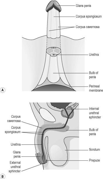
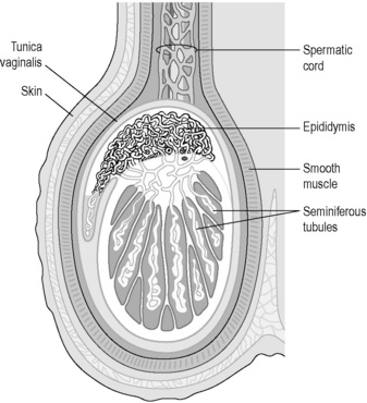
The male reproductive system consists of the testes, the epididymis, the urethra, the penis and associated glands, including the prostate gland. Figure 5.2a-I illustrates the anatomy of this system and Figure 5.2a-II shows the structure of the testis and scrotum in more detail.


The testes are the paired male sex glands, or gonads, and sit suspended from the perineum within the sacs formed by the scrotum. The two main products of the testes are spermatozoa (originating from the germinal, or germ, cells of the seminiferous tubules) and the male sex hormone testosterone (originating from the interstitial cells of Leydig).


The pituitary gland produces the two hormones follicle-stimulating hormone (FSH) and luteinising hormone (LH), which play an important role in the function of the testes. FSH stimulates the production of spermatozoa from the germ cells. LH stimulates the production of testosterone from the testes. The levels of both these hormones surge at puberty (see Q5.2a-2)image.


A mature sperm cell has a head, a body and a motile tail, which propels the sperm cell forwards by means of a whip-like motion. The formation of sperm is most efficient if it can take place at about 3°C below body temperature, which is aided by the fact that the testes sit in the scrotum. Spermatozoa are secreted from the germinal cells of the testis into the epididymis. The epididymis is a complex of very fine tubules, which store and direct spermatozoa towards the vas deferens. Testosterone is secreted from the cells of Leydig directly into the bloodstream. Thus the testis is, strictly speaking, both an exocrine and an endocrine gland.


The spermatic cords emerge from a gap in the musculature above the inguinal ligament and provide the testes with a blood and nerve supply. They also contain the vas deferens. The purpose of the vas deferens is to carry the spermatozoa away from the testes and the epididymis, and then to branch towards the urethra, from which the spermatozoa can be ejaculated. It is this tube that is severed and tied off in the procedure of vasectomy (see Q5.2a-3)image.


The prostate gland and seminal vesicles are exocrine glands that produce nutrient-rich lubricating fluid to maximise the survival of the spermatozoa and also to promote comfortable sexual intercourse. The seminal vesicles open out into the distal portion of the two vas deferens, and the prostate gland is situated at the base of the bladder at the point at which the vas deferens ducts enter the urethra. The prostate hugs the most proximal region of the urethra, which is why it is described as the ‘prostatic urethra’. The fluids produced by these glands mix with the spermatozoa in the urethra during ejaculation to form semen (see Q5.2a-4-Q5.2a-6)image.


The female reproductive system


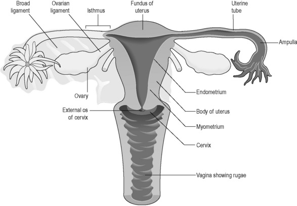
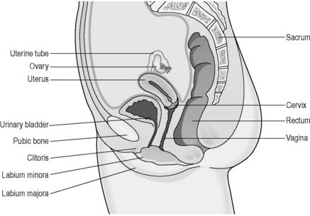
The female reproductive system lies at a deeper level in the body than in the male (Figures 5.2a-III and 5.2a-IV). The ovaries sit deep in the basin formed by the pelvis, and lie either side of the womb (uterus). The ovaries lie so that they are close to the openings of two tubes (the fallopian tubes) that extend sideways from the uterus. The uterus is a pear-sized organ sitting just behind and above the bladder, protected in front by the bridge formed by the pubic bone. The neck of the uterus (the cervix) opens into the bodily space called the ‘vagina’. The entrance of the vagina, the vulva, is surrounded by the fleshy lips of the labia minora and labia majora in the perineum.


The ovary


The ovary is the female sex gland, or gonad. The ovary originates from exactly the same embryonic tissue as the testis. During development of the male fetus, this embryonic tissue, originally positioned in the abdomen, develops into the testis and migrates into the scrotum under the influence of male sex hormones. In the female fetus, in the absence of these male hormones, the tissue remains where it is and develops into the ovary.


The ovary is the site for the maturation of the ova, and this takes place during the course of the first half of the menstrual cycle, during which usually only a single ovarian follicle enlarges. Ovarian follicles each consist of a single ovum and are lined by ovarian cells. By the time the female baby is born, her ovaries contain all the ova, each within its own ovarian follicle, that will be released during her childbearing years.


During the first half of the menstrual cycle, a single ovarian follicle begins the process of maturation (Figure 5.2a-V). This means that, over the course of approximately 14 days, the follicle enlarges and develops a fluid-filled cavity. This so called Graafian follicle then merges with the outer part of the ovary and ruptures to release the single ovum into the space surrounding the ovary. This process of rupture and release is what is termed ‘ovulation’. The whole process is stimulated firstly by FSH from the pituitary gland, and this explains its name. FSH is the same hormone that in men stimulates the development of spermatozoa from germinal cells.
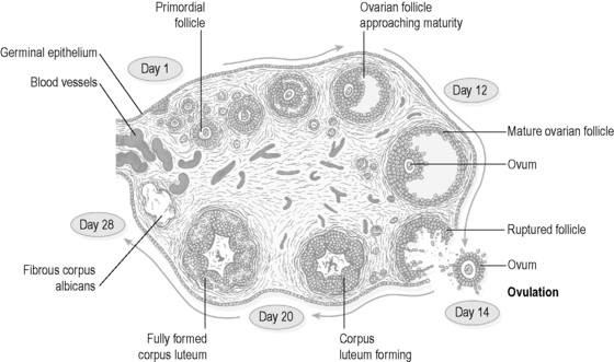


During the first 14 days of the menstrual cycle, the multiplying follicle cells secrete the female sex hormone oestrogen, under the influence of FSH. The rising oestrogen levels have a suppressive (negative feedback) effect on the secretion of FSH. As The FSH levels start to drop, the pituitary then releases a surge of a second hormone, LH. It is this hormone that stimulates ovulation. After ovulation, the remaining follicle develops into a body known as the ‘corpus luteum’ (the origin of the term ‘luteinising hormone’). The corpus luteum no longer produces oestrogen, and instead releases the other female sex hormone, progesterone.


The corpus luteum matures and then degenerates over the course of the next 14 days. As the corpus luteum degenerates, it ceases to produce progesterone. However, if fertilisation occurs, this degeneration does not happen, and instead the corpus luteum continues to develop. This occurs because, as the zygote multiplies and grows, it secretes the hormone human chorionic gonadotrophin (HCG) which stimulates the corpus luteum and prevents its degeneration. Thus, in early pregnancy the progesterone levels continue to rise.


To summarise; after puberty, the ovarian follicle is the source of a cyclical release of the female sex hormones. Oestrogen is released in the first half of the menstrual cycle, and progesterone in the second half of the menstrual cycle. This cycle is controlled by the cyclical release of the pituitary hormones FSH and LH (see Q5.2a-8)image.


The onset of puberty in the girl


As is the case for boys, in girls the onset of puberty is also stimulated by a rise in the production of FSH and LH. This usually occurs between the ages of 10 and 14 years.


Two to three years prior to this surge in FSH and LH in both boys and girls there is an increase in the release of GH from the pituitary gland. The release of GH stimulates the growth spurt that occurs in children just before puberty. In girls, the rise in FSH and LH stimulates the production of increasing amounts of oestrogen from the follicle cells of the ovaries. Initially, these pituitary hormones are not present in sufficient quantities to stimulate ovulation. However, the oestrogen produced affects diverse body tissues to bring about the early changes of puberty, including breast development and the growth of pubic hair. As puberty approaches, the levels of FSH and LH increase until ovulation is stimulated.


At approximately the same time as the onset of ovulation, menstruation begins. The onset of menstruation is termed the ‘menarche’. It is recognised that ovulation may occur for a few cycles before the first menstrual bleed. This means that there is a possibility of pregnancy even before menstruation has started.


The onset of puberty in both girls and boys in westernised cultures is occurring at an increasingly early age. The average age of onset of menstruation is nearly 3 years earlier now than it was a century ago. This suggests that it is not purely biological ‘programming’ that stimulates the dramatic increase in the release of FSH and LH that initiates puberty. It is now believed that both nutritional and psychological factors also play a part in this hormonal change. These are believed to influence the physiology of the pituitary via the production of specific hormones from the hypothalamus.


A child who is less well nourished is more likely to experience a delayed onset of puberty. Conversely, a child who achieves a degree of psychological maturity, particularly with regard to understanding or experience of sexual matters, appears to experience an earlier onset of puberty. Both these factors tend to promote the earlier onset of puberty in girls who grow up in ‘modern’ cultures.


Menstruation


The changes that take place in the ovary after the onset of ovulation have a cycle of approximately 28 days, although any interval between 22 and 35 days is considered conventionally to be within the normal range. These changes are mirrored by cyclical changes in the uterus and vagina as well as many other tissues in the body. These changes are the result of the rise of oestrogen in the first half of the cycle and in progesterone in the second half of the cycle.


Oestrogen can be considered as a hormone that stimulates fleshiness. In the first half of the cycle, it continues to promote the healthy development of the breasts and pubic hair. It also stimulates and maintains the changes to the skin, body fat and bony skeleton (in particular the hips and pelvis) which are characteristic of the female body. In the uterus, it stimulates a thickening of the endometrium, with the development of deep mucus-forming glands and a rich supply of blood vessels. About 3 days before ovulation a thick sticky mucus is secreted from the cervix. This mucus encourages the passage of spermatozoa at the time when the woman is likely to be most fertile. This phase of the menstrual cycle is termed the ‘proliferative phase’, a term which refers to the changes taking place in the endometrium at that time.


Oestrogen promotes fleshiness of the tissues of the vagina and the labia majora and minor. It also promotes the vaginal secretions, which are particularly important for comfortable sexual intercourse.


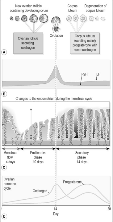
Progesterone can be considered to be the hormone that prepares the body for pregnancy and promotes the first 3 months of pregnancy. After ovulation, the rising levels of progesterone stimulate a watery swelling of the endometrium and the production of a thick mucus. This mucus is believed to encourage the passage of the spermatozoa into the fallopian tubes, where fertilisation can take place. The changes in the endometrium provide a suitable environment for the zygote. If implantation does not occur, at about 6–8 days after ovulation the corpus luteum starts to degenerate, and progesterone levels drop. It is this drop that causes the lining of the endometrium to start to break down, culminating in menstruation at 14 days after ovulation. There are thus two distinct phases to this second half of the menstrual cycle, termed the ‘secretory phase’ and the ‘premenstrual phase’ (Figure 5.2a-VI).


Normal menstruation takes place over the course of 3–7 days. As blood, endometrial lining cells and tissue fluid are shed, chemicals are released that stimulate the muscle wall of the uterus to contract, so that bleeding is minimised, and the products are effectively cleared from the uterus. This contraction is the cause of the cramping pain of menstruation. A degree of pain is therefore considered to be normal from a conventional perspective. It is important to understand that menstrual blood is not pure blood. A ‘heavy’ period may not necessarily indicate a large loss of blood cells, as much of the volume of what is lost may be tissue fluid.


While menstruation is occurring, the release of FSH initiates the next cycle, with the stimulation of the maturation of another ovarian follicle. This is why the first day of the menstrual bleed is conventionally considered to be the first day of the whole menstrual cycle.


The menopause


The ‘menopause’ is the term commonly used to describe the time in life, usually between the ages of 45 and 55 years, when menstruation ceases. Strictly speaking, the conventional use of the term menopause is to describe the precise time of cessation of menstruation. The term ‘climacteric’ is used to describe the period of approximately 10 years that precedes and follows this event (see Q5.2a-9)image.


The ageing ovaries are the cause of the onset of the climacteric. Gradually, the ovaries become less and less responsive to the pituitary hormones. This means that more and more of the menstrual cycles fail to lead to ovulation, and the production of oestrogen drops. The rate of drop in oestrogen levels varies widely between women, and this might explain why some women experience more profound symptoms than others during the climacteric.


Other features of a lack of oestrogen include a reduction in the strength of the pelvic floor muscles, with increased risk of prolapse of the vagina and uterus. Urinary problems, including urinary infections and stress incontinence, can occur because of a combination of reduced pelvic floor tone and changes in the lining of the skin around the opening of the urethra.


The notorious ‘hot flushes’ are a consequence of a rapid drop in oestrogen levels. The symptoms of these include bouts of profuse sweating and heat intolerance, often occurring at night. These can be accompanied by a range of bodily and psychological symptoms, which are not conventionally considered necessarily to be related to a lack of oestrogen.


A lack of oestrogen also promotes loss of calcium from the bones. This can occur very rapidly during the climacteric and can result in the symptoms of osteoporosis.


As the ovaries become less responsive to the control of the pituitary hormones, menstrual cycles become increasingly irregular. Some women experience increased frequency of periods, sometimes with excessive bleeding, for months to years, before the menopause. Usually, just before the menopause, the periods become lighter and more infrequent.


As the climacteric approaches, women become increasingly infertile as fewer and fewer cycles lead to ovulation. However, onset of the menopause does not necessarily mean that ovulation has ceased. In some women ovulation may still occur for a short time. This explains why it is that, in very rare circumstances, pregnancy can still happen after a woman’s periods have ceased. Women who reach the menopause before the age of 50 years are counselled to continue to use contraception for 2 years after the menopause, and those over 50 years old are advised to continue using contraception for 1 year.


The physiology of sexual intercourse


Sexual intercourse is necessary for natural conception to occur. It enables the meeting of the spermatozoa and the ovum in the fallopian tubes. It is, of course, also a very important aspect of bonding in human relationships.


Despite the overt differences, the physiological processes that take place during sexual intercourse are very similar in men and women. The sexual act, for both men and women, has been described as taking place in five phases, although these phases tend to merge into one another. These phases are:


Sexual desire: the emotional/mental response to the thought, sight, touch or smell of another person.


Sexual arousal: following sexual desire, physical changes take place in the sex organs (genitalia). The penis and clitoris become erect and the vagina becomes lubricated. The nipples can become erect. The heart rate can increase. There is a strong desire for sexual intercourse. This phase is mediated by the parasympathetic aspect of the nervous system.


Plateau: sexual pleasure is intensified and sustained before and after penile penetration has taken place. This phase is also mediated by the parasympathetic nervous system.


Orgasm: orgasm usually results in ejaculation of semen in men. In penile/vaginal intercourse, the semen is directed deep into the vagina, close to the entrance of the cervical canal. 90% of women are able to achieve orgasm, although only 25% will be able to reach orgasm through penile penetration into the vagina alone. There are rhythmic contractions of the perineal muscles of the women at orgasm. These contractions encourage the upward movement of the semen towards the cervix. The orgasmic phase is mediated by the sympathetic aspect of the nervous system.


Resolution: after orgasm, both partners are relaxed, and the body gradually returns to a non-aroused state. In this phase the parasympathetic nervous system is dominant. The resolution state encourages rest, and for the woman this is the ideal state for the continued passage of the spermatozoa into the cervical canal and thence into the uterus.


image Information Box 5.2.a-II The female reproductive system: comments from a Chinese medicine perspective


From a Chinese medicine perspective, the physiology of the female reproductive system is complex. The Extra Yang Organ, the Uterus, is the central organ, but the Spleen, Stomach, Liver, Heart and Kidneys all have important roles. The four Extraordinary Vessels, the Ren Mai, the Du Mai, the Chong Mai and the Dai Mai, are important regulatory channels. Additional channels, the Bao Mai and the Bao Luo, which link the Uterus with the Heart and the Kidneys, respectively, are also important for the regulation of the female reproductive system.


Blood and Kidney Essence (Jing) are the important substances for a healthy female reproductive system. It is Heart Blood that fills and nourishes the uterus, and so the Spleen and the Liver Organs are important for the manufacture and the smooth movement of this Blood. However, Jing is required in addition to Heart Blood for menstruation to occur, as it is the basis of the menstrual Blood. In women, Jing is believed to flow in 7-year cycles. The Spleen and Jing may be the foundation of the action of oestrogen, as together they give rise to fleshy characteristics, fertility and strong bones.


The deep origin of all three of the Extraordinary Vessels (Ren Mai, Du Mai and Chong Mai) is the region of the Kidneys. These then all flow through the Uterus before reaching their more superficial pathways.


The Chong Mai, as the Sea of Blood, has a strong influence on menstruation. The Chong Mai is linked to the Stomach Channel via St 30 (Qichong). This explains how nausea can be a problem in conditions such as dysmenorrhoea and morning sickness.


The Ren Mai governs the cervix, vagina and the vulva. It also helps to provide the other Yin substances important in female reproductive physiology (Yin, Essence and Fluids), as it is the Sea of the Yin Channels. Therefore, it is important in the regulation of fertility, puberty, pregnancy and the menopause.


Because of its complex pathway, the Du Mai also influences the vagina and the vulva. The Du Mai together and the Ren Mai together can be seen as two branches of one circuit that interlinks the Uterus with the other Organs. The Dai Mai encircles the waist, and so crosses the leg Channels. In this way it influences the smooth flow of Qi in the Liver Channel and harmonises The Kidneys and the Spleen.


The relative strength of the organs and substances in the four menstrual phases correspond to the physiological phases understood in conventional terms. These are (with conventional terms in parentheses):


At the climacteric, there is a decline in the Kidney Essence and in the Qi and Blood of the Ren Mai and the Chong Mai. There is thus a general drying up of body fluids (Yin Deficiency). This culminates in the cessation of menstruation.


image Self-test 5.2a The physiology of the reproductive system


Answers


1. Semen is composed of the male sex cells, the spermatozoa, and fluid. The spermatozoa originate from germ cells within the testes. The fluid contains nutrients for the spermatozoa, and provides an environment in which the spermatozoa can swim freely. The fluid originates from the seminal vesicles and the prostate gland.


2.


All these effects begin at puberty and the changes will start to regress if testosterone and oestrogen levels drop.


3. Oestrogen is produced in increasing amounts in the first (proliferative) half of the menstrual cycle. Progesterone is produced in the first part of the second half of the menstrual cycle (secretory phase), although levels decline after about 7 days. It is the drop in progesterone that is the stimulus for menstruation.


4. The symptoms of the menopause are attributed to falling levels of oestrogen. Sudden drops in oestrogen are considered to induce more profound symptoms than a more gradual drop in oestrogen levels.


The investigation of the female reproductive system


The investigation of the female reproductive system includes


Imaging tests


Because the uterus and ovaries are deep organs, imaging tests are very often performed to provide more information.


The pelvic ultrasound scan is a non-invasive examination which can provide very useful information. The test usually involves the use of a probe placed on the skin of suprapubic part of the abdomen. In some cases a vaginally inserted probe can give more information. Tumours and cysts of the ovaries and uterus can be visualised by means of an ultrasound examination. The formation of the Graafian follicle can be seen, and so ultrasound can give an indication of healthy ovulation.


Endoscopy of the vagina (colposcopy) is a test that requires the insertion of a vaginal speculum. A colposcope (a form of endoscope) is used to provide detailed images of the surface of the cervix, and permits the removal of cervical biopsies and treatment of the cervix. This investigation is most commonly used to provide more information and treatment after an abnormal cervical smear test.


Endoscopy of the uterus (hysteroscopy) requires the insertion of a fine endoscope into the uterus via the cervical canal. This is usually performed in an outpatient setting in a conscious patient. The test can be uncomfortable, as the cervix is very sensitive. Hysteroscopy is used in the investigation of menstrual disorders, and is the means by which samples of the endometrium may be obtained for microscopic examination.


Laparoscopy is a procedure in which a fine endoscope is inserted into the peritoneal cavity which surrounds the pelvic organs. The endoscope is inserted via a small incision made adjacent to the umbilicus. Laparoscopy can reveal the details of the external anatomy of the ovaries and the uterus. It can visualise ovarian cysts and the changes that occur in endometriosis. Laparoscopy is also the means by which some important gynaecological procedures can be performed. These include sterilisation, removal of ovarian cysts and removal of eggs for assisted-fertilisation techniques.


Hysterosalpingography is a test to examine the interior anatomy of the fallopian tubes. A ‘dye’ that is opaque to x-rays is injected into the cervical canal, and x-ray images taken of the pelvic area. The dye can reveal the interior shape of the uterus and fallopian tubes, and thereby whether or not the fallopian tubes are blocked.


Endometrial biopsy is performed by means of hysteroscopy. A sample of the lining of the womb is removed from a conscious patient by means of a spoon-shaped instrument called a ‘curette’. The patient is likely to experience cramping pain and some bleeding after this procedure.


A less invasive method of obtaining endometrial samples is by means of a fine-ended suction instrument called a ‘Pipelle curette’. This samples far less endometrium but still has a greater than 90% detection rate for endometrial cancer.


The investigation of breast diseases


The investigation of breast diseases includes


The investigation of the male reproductive system


The investigation of the male reproductive system includes:


The investigation of sexually transmitted diseases


The investigation of sexually transmitted diseases includes:


Blood tests


Blood tests can provide evidence of a sexually transmitted infection. These include the test for human immunodeficiency virus (HIV), in which antibodies to HIV are indicative of infection. Blood tests can also reveal evidence of infection by hepatitis B and C and syphilis.


GU clinics offer the HIV test on a strictly confidential basis. Because of the consequences of being tested as HIV-positive, patients will be offered pre-test and post-test counselling, irrespective of whether the test is positive or negative (see Q5.2b-1).


Amenorrhoea and oligomenorrhoea


Amenorrhoea is defined as total lack of menstruation. It may be primary or secondary. Primary amenorrhoea means that there has never been an onset of menstruation. This diagnosis is made if the menarche has not occurred by the age of 16 years.


Secondary amenorrhoea indicates a cessation of menstruation. This diagnosis is made when menstruation has ceased for more than 70 days. Oligomenorrhoea indicates infrequent periods with a cycle length of greater than 35 days. In general, the causes of oligomenorrhoea are the same as those of secondary amenorrhoea (see Q5.2c-1)image.


Primary amenorrhoea


Secondary amenorrhoea and oligomenorrhoea


The most common cause of secondary amenorrhoea is pregnancy. This common cause may actually be a possibility that has been overlooked by the patient, but needs always to be considered in any case of absence of menstrual bleeding.


Polycystic ovarian syndrome


In polycystic ovarian syndrome (PCOS) the ovarian follicles do not develop properly. The ovary becomes enlarged, with four or more unruptured follicles (cysts). The condition is associated with weight gain, oligomenorrhoea or amenorrhoea, excessive body hair in a male distribution and infertility. It is believed that insulin resistance and oversecretion of insulin might be the fundamental problem in PCOS.


This hormonal imbalance, which is more common in obese people, is believed to affect the LH surge from the pituitary gland (so preventing rupture of the ovarian follicles and causing infertility) as well as leading to excessive formation of male sex hormones (which cause excessive body hair and also acne). PCOS is associated with a greater than average risk of non-insulin-dependent diabetes mellitus, high blood pressure and cardiovascular disease. There is also a small increased risk of cancer of the womb and of the ovaries.


PCOS is very common. Ultrasound studies have shown that up to 20% of women have evidence of PCOS. However, only a few will exhibit the more severe manifestations of the syndrome.


In PCOS, ultrasound examination of the ovaries shows unruptured ovarian follicles and a blood test reveals an imbalance in the levels of FSH and LH, and in some cases raised levels of testosterone and prolactin.


In PCOS women are advised to lose weight, stop smoking and to increase participation in gentle exercise, as these changes can normalise insulin levels. The antidiabetes drug metformin may be prescribed, as it reduces insulin resistance, particularly in anyone who is classified as overweight according to their body mass index (BMI > 25).


If amenorrhoea is the primary problem, the combined oral contraceptive pill is prescribed to induce regular menstruation and protect the woman from endometrial cancer.


The drug clomiphene may be prescribed to induce ovulation in PCOS. If this fails to help with conception, surgical drilling of the ovaries can reduce the production of unnecessary sex steroid hormones and improve fertility rates.


Excessive male-pattern body hair in PCOS can be treated by means of medication. The oral contraceptive may be effective in reducing the hairiness. Alternatively, the drug cyproterone acetate may be prescribed. This has the effect of opposing the action of the male sex hormones.


Menorrhagia


Menorrhagia is defined as menstrual bleeding that is too heavy, and specifically relates to a menstrual loss of more than 80 ml/month. However, in practice the degree of menstrual loss is very difficult to assess. Usually loss is expressed in terms of the number of tampons or towels required, and the numbers of episodes of ‘flooding’ (see Q5.2c-2)image.


The main health consequence of menorrhagia is blood loss. This can, in severe cases, lead to iron-deficiency anaemia, as the loss of iron in the menstrual blood is too great to be replaced by the diet alone. Very rarely, the blood loss is so profuse as to require emergency treatment with transfusion.


However, for most people, the main problem with menorrhagia is the inconvenience of having a prolonged and heavy period. Although many women feel tired and uncomfortable during a heavy period, they may never lose sufficient blood to become clinically anaemic.


Dysfunctional uterine bleeding


The most important and common cause of menorrhagia is functional. This means that there is no structural or readily measurable cause for the bleeding. The term ‘dysfunctional uterine bleeding’ (DUB) is applied to a range of syndromes, all of which involve heavy bleeding. In DUB the bleeding is usually regular. If the pattern of bleeding is chaotic, and with no predictable pattern, then the term ‘metrorrhagia’ is applied.


DUB is most common just after the menarche and in the years leading up to the menopause. Usually, the underlying problem is due to a defect in the maturation of the ovarian follicle or the formation of the corpus luteum. The reasons for this are usually unclear, although the root lies partly in the control of the whole process by the pituitary gland. This, of course, explains why emotional and mental factors can play an important role in the development of heavy periods.


The defective follicular development may cause the levels of oestrogen and progesterone to become subtly unbalanced, and this is the cause of the heavy bleeding. However, most women with DUB demonstrate no measurable abnormalities in the release of pituitary or sex hormones that might support this theory.


The pattern of problematic bleeding varies according to the precise underlying problem. In some cases the spotting and blood loss occurs for some days before the onset of menstruation. This bleeding may start at the time of ovulation (mid-cycle bleeding). In other cases there is slight bleeding or spotting for some days after the period.


In less common cases the underlying problem is that ovulation does not occur (anovulatory cycles), and the lining of the womb becomes thickened by excessive oestrogen production. This form of DUB is common just before the menopause. The time between periods becomes longer (up to 6–8 weeks), but when the period comes it is very heavy, and can be associated with flooding.


Another problem that can occur in the few years before the menopause is a result of fluctuating levels of oestrogen from the failing ovary. The result is more frequent periods, which may also be heavy, and flooding.


Investigation of persistent DUB is important to eliminate more serious causes (see later). If no physical cause is found, treatment is primarily directed at controlling bleeding. In women who have developed anaemia, iron tablets may be prescribed in the short term. Very rarely, a blood transfusion may be necessary to replace blood that has been lost rapidly.


Simple medications that may be used to reduce the volume of heavy bleeds include non-steroidal anti-inflammatory drugs (NSAIDs) such as mefenamic acid. Tranexamic acid is a drug that inhibits the breakdown of blood clots, and this may be used to halt a heavy bleed, but must be avoided if the patient has any risk factors for thrombosis.


DUB can also be treated by means of hormonal drugs. Contraceptive methods can be helpful for many women. The oral contraceptive pill, the progesterone-containing intrauterine device, progesterone implants or injections will all control heavy bleeding. These treatments will of course also prevent pregnancy. In some women, withdrawal from these treatments after a few months may result in normalisation of the periods.


Norethisterone is a synthetic progesterone (progestogen) which is commonly prescribed in cases of very severe bleeding to control premature breakdown of the womb lining. This is taken daily from day 5 to day 25 of the cycle. In most women this will keep a bleed at bay until the tablets are stopped. Withdrawal of the tablets on day 26 leads to a ‘withdrawal bleed’. This treatment is continued for 4–6 menstrual cycles, and may be sufficient in some women to normalise their periods.


In resistant cases, surgical treatments may be offered. Dilatation and curettage (D&C) is the surgical procedure in which the lining of the womb is scraped away with a curette. It is a low-risk procedure and can bring about a temporary (4–6 months) relief from heavy bleeding.


A more permanent approach is to remove the lining of the uterus by heat or laser via a hysteroscope. This procedure, which requires a 1–2 day stay in hospital, is called ‘endometrial ablation’. This procedure causes sterility, but is less invasive than a hysterectomy. In about 80% of women who have this procedure there is either total loss of or a relative reduction in periods.


Hysterectomy is now performed less commonly for DUB because the recent developments of drug treatments and endometrial ablation are effective and are much less invasive alternatives.


Premenstrual syndrome


Premenstrual syndrome (PMS) is a functional condition that involves low mood and uncomfortable physical symptoms. PMS occurs from any time in the second half of the menstrual cycle (between ovulation and the first few days of menstruation). PMS is more common between the ages of 30 and 40 years. At the onset of menstruation, or during the period, the symptoms disappear and the woman may even feel euphoric for a few days. The symptoms of low mood and physical discomfort start again after ovulation. It is estimated that in 15% of women the mood and physical disturbances of PMS are so severe as to disturb day-to-day living and personal relationships.


The perimenstrual syndrome is a particular pattern of PMS which is described as occurring in the 2–3 days prior to menstruation.


The emotional and mental symptoms of PMS include irritability, anxiety, depression, tearfulness and confusion. Sweet cravings may be prominent in some people. Physical symptoms include abdominal bloating, breast tenderness, swelling of the ankles, headache, nausea and clumsiness.


In perimenstrual syndrome, the most common symptoms are tiredness and headache in the 2 days prior to menstruation. Migraine can occur during this time in some women. There may also be bloating and abdominal pain.


The cause of PMS is not clearly understood. Theories that propose a hormonal cause have not been consistently supported by experimental measurement of hormone fluctuations in women with PMS. A recent hypothesis is that it is how progesterone is metabolised in the brain and interacts with the GABA-A neurotransmitter receptor that determines sensitivity to the premenstrual phase.


Treatment of PMS ideally should begin with lifestyle advice. Dealing with stress is probably the most important, but in many cases this is the most difficult lifestyle factor to deal with. In some women, normalisation of diet and adoption of whole foods can be very helpful. Regular exercise can also be very beneficial for some people.


Vitamin B6 (pyridoxine) and oil of evening primrose are two relatively ‘natural’ prescribed medications which can be of help for some people. The drug bromocriptine may also be useful in reducing symptoms.


For some women the regular cycle induced by the oral contraceptive pill can be helpful, presumably because it provides a regular level of (synthetic) oestrogen and progesterone.


Selective serotonin reuptake inhibitor (SSRI) antidepressants and the anxiolytic drug alprazolam (which augments GABA-A function) may also be useful in some women.


Agnus castus fruit extract is a herbal remedy which has good scientific evidence supporting the fact that it can be of help in up to 50% of people.


If the problem is very severe, some women are offered surgical treatment. The most invasive approach is to remove the womb and both ovaries and then treat with HRT. This drastic treatment approach reportedly has a 96% satisfaction rate.


Dysmenorrhoea


Dysmenorrhoea is painful menstruation. This can be classified into two broad types, which may overlap. The first type, primary dysmenorrhoea, classically starts shortly after the menarche and peaks between the ages of 15 and 25 years. In primary dysmenorrhoea there are severe crampy pains, which begin after the onset of menstruation and usually last for less than 24 hours. They may be so severe as to lead to vomiting. Up to three-quarters of young women experience some form of primary dysmenorrhoea. The problem usually improves as the woman gets older, and may be markedly better after childbirth.


Secondary dysmenorrhoea, by definition, has an underlying physical cause. The most common causes are endometriosis or pelvic inflammatory disease. Fibroids may also be a cause of secondary dysmenorrhoea. Secondary dysmenorrhoea tends to begin a few days before the onset of menstruation and can continue through to late in the period. It tends to peak towards the end of the period. However, in practice, it may be difficult to distinguish primary from secondary dysmenorrhoea on the basis of symptoms alone.


Drug treatment of dysmenorrhoea involves two main approaches. Suppression of ovulation by means of an oral contraceptive may be preferred by many young women who also desire an effective contraceptive method. Alternatively, NSAIDs may be very effective in reducing the intensity of the pain. Commonly prescribed NSAIDs include mefanemic acid, and ibuprofen.


Problems during the menopause


The menopause is a natural physiological event, and like the menarche is a time of transition from one stage of life to another. Although it is a time in life when the body functions associated with fertility are ceasing to be active, a healthy climacteric should not be dominated by negative symptoms. Studies indicate that up to 25% of women have no symptoms during the menopause. However, 35% have mild to moderate symptoms and 40% suffer from severe symptoms.


Hormone replacement therapy


HRT used to be commonly prescribed to counteract the major menopausal symptoms, which include hot flushes and vaginal dryness, and to prevent the long-term complication of osteoporosis. These symptoms and complications result from the declining levels of oestrogen from the failing ovaries, and by providing oestrogen HRT counteracts this decline.


However, recent research into the effects of HRT in large groups of women exposed the fact that oestrogen replacement carries no cardiovascular benefits, and nor does it protect against dementia. Moreover, long-term HRT slightly increases the incidence of stroke, deep vein thrombosis and breast cancer. For this reason, HRT is no longer prescribed as a first-line treatment for the prevention of the development of postmenopausal osteoporosis. HRT may be considered as treatment for disabling hot flushes. Topical forms of HRT, in the form of oestrogen creams, are used to counteract the problems that may result from vaginal atrophy.


HRT consists of a synthetic or naturally derived form of oestrogen. As it is not healthy for the uterus to be exposed to oestrogen alone (as unopposed oestrogen can promote the development of endometrial cancer), most preparations also contain a synthetic progesterone-like compound known as a ‘progestogen’. This additional progestogen is only necessary for those women who have an intact uterus. Those who have had a hysterectomy can be prescribed oestrogen-only HRT.


HRT comes in the form of tablets, skin patches or implants. In women with an intact uterus, the treatment is prescribed so that a ‘period’ is induced every 28 days. This bleed happens because the treatment consists of a regular daily dose of oestrogen, and a dose of progestogen to be taken on only 12 of the days of the ‘cycle’. The ‘period’ is in fact a ‘withdrawal bleed’ which follows the start of the 16 days when the progestogen is not taken.


Before prescription of HRT the doctor needs to ensure that the woman is in good health. In particular, HRT is not advisable if there is any evidence of breast or endometrial cancer, liver disease or a history of recent thromboembolism. The blood pressure is checked, because in some women HRT may cause a rise in blood pressure.

Stay updated, free articles. Join our Telegram channel

Oct 3, 2016 | Posted by in MANUAL THERAPIST | Comments Off on Diseases of the reproductive system

Full access? Get Clinical Tree

Get Clinical Tree app for offline access