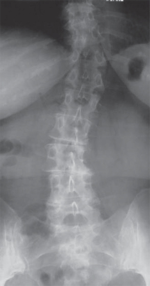
32 Degenerative or de novo scoliosis presents in adults, in contrast to idiopathic scoliosis of adolescence. Degenerative adult spinal deformity is brought on by asymmetric disk collapse with facet arthropathy and incompetence, leading to unbalancing of the spine into coronal, sagittal, and rotational deformity. Osteoporotic fractures may contribute to the degenerative deformity. Degenerative scoliosis is present in 6% of the population over 50 years of age, presenting with mechanical back pain from uncompensated sagittal or coronal imbalance and, more commonly, radicular leg pain from stenosis. Original classifications of degenerative scoliosis consist of primary or secondary type. Primary degenerative curves have a predominance of degenerative features. Secondary degenerative curves are those superimposed upon other conditions, such as spondylolysis, congenital anomalies, idiopathic scoliosis, trauma, or infection. A Scoliosis Research Society (SRS) classification system for adult spinal deformity has evolved based upon curve type, degenerative modifiers, and global balance. Detailed classification schemes allow comparison of factors that predict need for surgery. A patient’s history of complaints should be carefully defined, distinguishing leg pain from back pain. Degenerative scoliosis patients tend to have a predominance of radicular leg symptoms from stenosis. Adult deformity patients with preexisting adolescent idiopathic deformities tend to have a predominance of mechanical back pain. Patients often complain of asymmetry and loss of the waistline with early satiety. Pelvic obliquity results in one pant leg longer than the other. Pain in the region of the buttocks or gluteal region referred from the lumbar spine is often described by the patient as “hip” pain. Patients relate increased standing and walking intolerance with back pain from sagittal imbalance, which results in muscle fatigue from the attempt to compensate. In contrast to isolated spinal stenosis, radicular complaints associated with degenerative scoliosis tend to be static and unrelieved by sitting, rather than intermittent and relieved by spinal flexion, as seen in classic neurogenic claudication. The physical examination begins with observation of the standing spine. Generally, a loss of lumbar lordosis is noted that may be partially compensated for by hip and knee flexion. Observed gait may list to one side of an uncompensated coronal deformity. Coronal imbalance is assessed with a plumb line from the base of the cervical spine to the gluteal crease. Pain on palpation may be present over an inflamed facet joint or along the 12th rib as a result of impingement against the iliac crest. An examination includes evaluation of the lower extremities for leg length discrepancy, pelvic obliquity, contractures, and arthritic deformity of the hips or knees. Pain, numbness, paresthesias, muscle weakness, and atrophy of the lower extremities result from chronic nerve root compression. Anterior thigh pain with femoral stretch testing or lower leg calf pain with straight leg raise may distinguish symptoms emanating from the upper or lower lumbar nerve roots, respectively. Neoplasm and vascular disease are considered in the elderly patient with recent onset of back pain or activity-related leg pain. Diagnostic testing begins with plain radiographs from thoracic to lumbar regions in the anteroposterior (AP) and lateral plane. Degenerative curves are most commonly in the lumbar spine and centered on L3 (Fig. 32.1). Full-length radiographs of the spine assess spinal balance and compensatory thoracic curvatures. Loss of lumbar lordosis is observed on lateral films and presents prior to coronal plane deformities (Fig. 32.2). Bending radiographs demonstrate segmental instability in the sagittal plane and curve flexibility in the coronal plane. Magnetic resonance imaging (MRI) provides excellent visualization of the degenerative state of the intervertebral disk and degree of stenosis and nerve impingement. Computed tomography (CT) with 3D reconstructed images or myelography is also useful for assessment of deformity and nerve root foraminal stenosis. Diskography may or may not be helpful. Selective nerve root or facet blocks may be more useful in distinguishing symptomatic levels. Electrodiagnostic studies provide information on the levels of nerve compression or underlying peripheral neuropathy. Bone density tests are an important adjunct for treatment of associated osteoporosis. Fig. 32.1 AP radiograph of degenerative lumbar scoliosis with compensatory thoracic curve.
Degenerative Scoliosis
![]() Classification
Classification
![]() Workup
Workup
History
Physical Examination
Spinal Imaging
Related posts:

Stay updated, free articles. Join our Telegram channel

Full access? Get Clinical Tree


