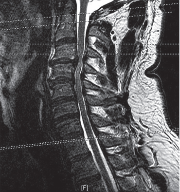
20 Cervical spondylotic myelopathy is the most common cause of spinal cord dysfunction in older individuals. The onset of symptoms, however, can be insidious. The stepwise progression of the disease is slow and may be difficult to predict. Since the natural history of the disease is variable, management can be challenging. Surgical intervention has been shown to prevent neurologic deterioration and to provide superior outcomes as compared with conservative management.1 Because surgery can reliably halt the progression of disease, but typically not reliably reverse symptoms or reliably restore pre-disease function, there is significant interest in determining the factors that would predict which patients are likely to have progressive neurologic decline. Thus, current research efforts are focused on elucidating the anatomic, physiologic, and genetic features that influence the development of progressive myelopathy in patients who have spinal cord compression. The Nurick classification, a 6-point scale from 0 (root signs but no cord involvement) to V (chair-bound or bedridden), is the most commonly utilized system for categorizing the degree of myelopathy.2 A modified Nurick classification system has also been utilized in which grade 0 identifies patients with no symptoms and grades I through VI correspond to the classic grading system.3 The Japanese Orthopedic Association has also developed a scoring system for cervical myelopathy, termed the JOA score, based on degree of motor and sensory dysfunction in the extremities and urinary dysfunction. This system is more complex and specific than the Nurick classification and consequently is less commonly utilized. A careful history is imperative in the evaluation of myelopathy, as the symptoms of cord compression are often vague and can relate to difficulty with ambulation and extremity dysfunction, which patients may not necessarily relate to spinal disease. For instance, loss of upper-extremity dexterity and decreased fine motor skills will manifest as difficulty manipulating small objects, as in buttoning shirts, or difficulty with handwriting or typing. Patients may also note difficulty with balance or bowel/bladder dysfunction. When taking a history, family members may be helpful in corroborating subtle changes in gait or dexterity of which patients themselves may be unaware. The physical examination of patients with myelopathy is paramount for both diagnosis and surgical decision making. Clinical symptomatology and the severity of myelopathy are widely variable among patients with identical imaging studies. Furthermore, clinical manifestations, rather than imaging findings, dictate the need for surgical intervention. Patients with cervical spondylotic myelopathy demonstrate upper motor neuron findings in the lower extremities and mixed upper and lower motor neuron findings in the upper extremities. For instance, C5–C6 pathology may cause flaccid weakness of the deltoid and biceps with concomitant spasticity of the triceps, wrist flexors, and lower extremities. Patients who demonstrate lower motor neuron signs in the lower extremities should be evaluated for lumbar spine pathology, as lumbar and cervical stenosis are associated and can occur in conjunction. The most common initial signs and symptoms of cervical myelopathy are: (1) gait abnormalities, (2) upper-extremity clumsiness, (3) spasticity of the extremities (lower more frequent than upper), (4) multisegmental sensory involvement, (5) Hoffmann sign, and (6) Lhermitte sign. Gait dysfunction is most often characterized by a broad-based, shuffling gait resultant from lower extremity spasticity and proprioceptive deficits. Upper-extremity examination is likely to reveal a mixed picture of flaccid and spastic weakness, whereas lower-extremity examination is often characterized by spasticity, hyperreflexia, positive Babinski sign, and clonus. The initial diagnostic imaging evaluation of cervical myelopathy should consist of plain radiographs of the cervical spine, including anteroposterior (AP), lateral, and flexion-extension films. Cervical myelopathy can result from both primary congenital cervical stenosis and acquired cervical stenosis, each of which has unique radiographic findings. Congenital cervical stenosis is relatively rare and results from abnormal development of the vertebral column. Patients with congenital stenosis are predisposed to spinal cord injury from minor trauma. Acquired cervical stenosis, or cervical spondylosis, refers to the more common pathology of age-related changes to the cervical spine. The AP diameter of the spinal canal can be assessed on lateral radiographs and Pavlov’s ratio (AP diameter of the canal over that of the vertebral body) calculated. A value less than 0.8 is associated with neurologic changes and worse outcome. In the case of congenital stenosis, decreased canal diameters occur at the level of the mid-vertebral body (developmental segmental sagittal diameter), whereas in cervical spondylosis, decreased canal diameters are seen near the endplates secondary to encroachment of the intervertebral disk and osteophytes into the spinal canal (spondylotic segmental sagittal diameter). Flexion-extension radiographs are helpful for assessing dynamic changes, such as spondylolisthesis, which can result in positional symptomatology and demonstrate potential dynamic instability. This is of particular importance, as these patients demonstrate particularly good outcomes with surgical decompression and fusion.4 The current gold standard imaging technique for evaluation of patients with cervical spondylosis is magnetic resonance imaging (MRI), as it provides multiplanar images that visualize neural elements, intervertebral disks, and ligamentum flavum, accurately identifying the location(s) of cord compression. Furthermore, MRI can detect the presence of signal change (myelomalacia) within the spinal cord, which may be associated with more severe cord compression and may have prognostic significance. A sagittal MRI demonstrating multilevel spondylosis and associated myelomalacia is demonstrated in Fig. 20.1. Ossification of the posterior longitudinal ligament (OPLL), an entity that can result in cervical myelopathy independent of other spondylotic changes, is best seen on computed tomography (CT) studies, but can also be characterized on MRI in many cases. CT and CT myelography are useful in assessing bony structures, including the neural foramen, though they are less commonly utilized with the availability of MRI. Fig. 20.1 Sagittal T2 MRI demonstrating multilevel cervical stenosis and associated myelomalacia.
Cervical Myelopathy: Posterior Approach
![]() Classification
Classification
![]() Workup
Workup
Spinal Imaging

Stay updated, free articles. Join our Telegram channel

Full access? Get Clinical Tree


