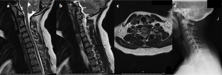
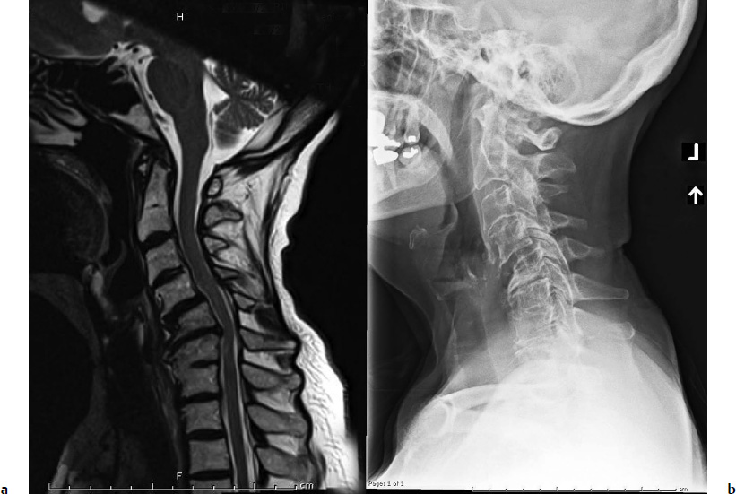
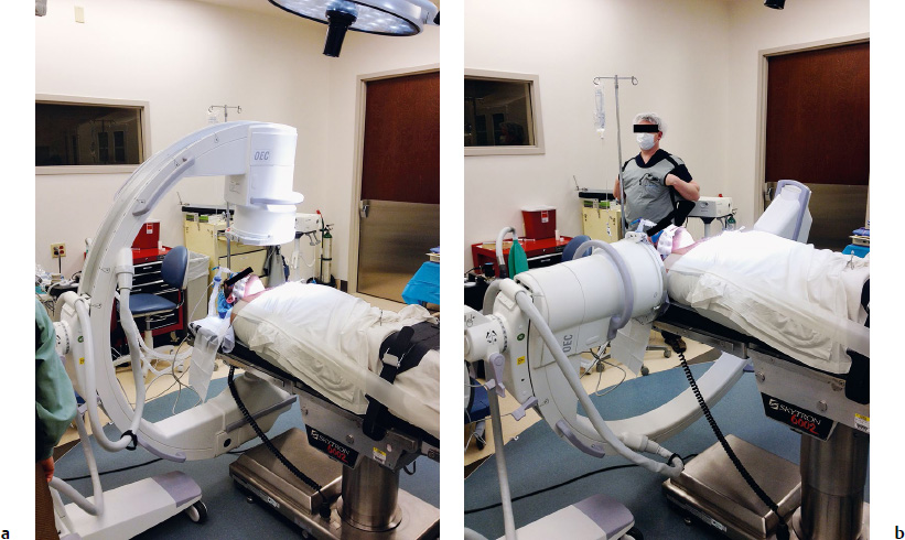
7 Cervical disk arthroplasty is meant to serve several key purposes. The primary goal is restoration of normal disk height and segmental motion, and removal of degenerative pathology causing dysfunction. Secondary goals include preservation of global spine mechanics/alignment, functional motion in the remainder of the cervical region, and theoretic reduction in adjacent level pathology. Anterior cervical diskectomy and fusion (ACDF) is a well-known treatment for patients with cervical degenerative changes resulting in radiculopathy or myelopathy and currently considered the gold standard.1 Although quite successful, ACDF is not without complications and potentially negative long-term effects.1–5 In recent years, alternative treatment options have been aggressively pursued and studied in an effort to avoid pitfalls associated with ACDF procedures. As with fusion, identification of the pathology, alignment of the cervical spine, adjacent level changes, and recovery of neurologic function all remain important. New concerns that arise with disk arthroplasty include proper patient selection, kinematics, motion quality, wear properties, and fixation of the implant. There are numerous arthroplasty designs that are currently in use worldwide, all with different insertion techniques and designs, attempting to address the aforementioned goals. Anterior cervical diskectomy and fusion is the current gold standard for treatment of cervical radiculopathy and myelopathy related to disk-level pathology, though it is not without potential pitfalls, which have been well described in the literature and include donor-site morbidity, pseudarthrosis, loss of normal motion, postoperative immobilization, and adjacent segment pathology. The most commonly cited morbidity associated with harvesting of the iliac crest is donorsite pain, which can be as high as 31% at 2 years following the index procedure.5 Pseudarthrosis rates are reported anywhere from 3 to 27% depending on the study, and increase with the number of fusion levels.1,4 Clinical adjacent-level pathology occurs at a annual rate of 2.9% in patients who have undergone ACDF.2 However, radiographic adjacent-level pathology has been reported as high as 92% within 5 years following ACDF.3 Disk arthroplasty is a viable alternative that avoids some of the obvious pitfalls of ACDF. There is no need to harvest graft material or risk pseudarthrosis. Immobilization is not necessary postoperatively and patients have been shown to return to activities, including work, sooner when compared with fusion.6 There is still much debate regarding the true cause-and-effect relationship regarding adjacent segment pathology, and no consensus exists despite reports in the literature. Numerous studies clearly show that significant changes occur at adjacent levels following cervical fusion compared with disk arthroplasty.7–10 Theoretically, these abnormal changes could accelerate the natural progression of degenerative changes. Further study and longer-term follow-up data are still needed regarding the effects of fusion and arthroplasty on adjacent-level pathology. Patient selection plays an important role in identifying who may be a candidate for disk arthroplasty. Healthy patients with single, disk-level pathology including herniation/spondylotic disease causing radiculopathy or myelopathy are ideal (Fig. 7.1). Patients who present with sagittal plane abnormalities, multilevel spondylolysis, segmental instability, significant medical comorbidities, or active infections should be excluded (Fig. 7.2). Patients with a history of previous cervical surgery, metabolic bone disease, progressive neuromuscular disease, or significant osteoporosis, or those on corticosteroid therapy, are poor candidates. Patients with radiographically confirmed ankylosis, ossification of the posterior longitudinal ligament (OPLL), kyphosis, end-plate abnormalities, or facet joint arthroses should also be excluded. Treatment of cervical myelopathy secondary to factors other than single-level pathology should be avoided. Preservation of motion is a key premise in the use of disk arthroplasty and one of the most important theoretic advantages over fusion procedures. The goal is functional motion preservation at the surgical level as well as adjacent levels. Unfortunately, due to several factors including implant design, methods of measurement, and variable definitions of motion, this becomes extremely difficult to truly assess and compare. Long-term data are again lacking, further limiting conclusive information at this point. Another important factor to consider is not simply maintenance of motion, but the actual quality of motion that is preserved. Defining normal motion of the cervical spine is a challenging task that has been poorly described in the literature. This is likely because of the complex nature of the functional spinal unit in the cervical region. The majority of studies analyzing disk arthroplasty and fusionin fact do not define “normal” motion but instead refer to what should be called functional motion. Comparative studies looking at pre- and postoperative differences among arthroplasty versus fusion patients are not beginning with normal motion segments. They are segments that are functional, yet many have degenerative changes present. It is also important to take into consideration other properties of a disk that can affect overall kinematics including viscoelastic properties, center of rotation (COR), and coupled motions. These also have proven difficult to quantify, and the lack of measurement standards limit the ability to compare data among studies. Fig. 7.1a–d Cervical magnetic resonance imaging (MRI) (a,b,c) and plain film (d) of a young otherwise healthy patient displaying single-level pathology at the C6/7 level on the left. Fig. 7.2a,b Cervical MRI (a) and plain film (b) displaying sagittal deformity with multilevel degenerative changes. Despite many of these limitations, studies have attempted to evaluate cervical kinematics and quality of motion following arthroplasty.9,11–14 In general, functional motion appears to be well preserved at the index level when arthroplasty is used regardless of device.9,15 Results of adjacent level changes are more difficult to interpret, however. Biomechanical in vitro studies have clearly shown significant kinematic abnormalities at adjacent levels following fusions compared with arthroplasty.7,8 Recent systematic review and meta-analysis studies looking specifically at kinematics of adjacent segments following disk arthroplasty versus fusion have shown no significant difference in range of motion statistically or clinically.16,17 Maintenance of normal sagittal alignment in the cervical spine is an important goal of arthroplasty as well. Many factors can influence surgical level alignment, the most important being surgeon technique. Although alignment may vary at the implant level or adjacent levels, global spine alignment does not appear to be significantly affected and is well maintained in patients with arthroplasty as well as fusion.16 Data are limited regarding the in vivo wear properties of current artificial disks. Explanted arthroplasty devices from both human and nonhuman subjects have been studied. Both polymeric and metal ions or debris have been identified in adjacent tissues depending on the implant used. Inflammatory reactions were found in response to soft tissue debris, though no evidence thus far of adjacent osteolysis has been reported.18 Concerns exist regarding the effects of metal hypersensitivity and formation of pseudotumors, which have been reported.19 It is difficult to make any significant claims regarding in vivo wear properties due to the limited number of devices studied and the fact that there are only short-term data currently available. At 2 years, arthroplasty compared with ACDF has shown significant differences in favor of arthroplasty. These outcomes appear to be maintained at 4 years with significantly higher rates of overall success, and improvements in Neck Disability Index (NDI), neck and arm pain scores, and Short Form (SF-36) Health Survey physical component scores.20 Longer-term data are needed to further evaluate maintenance of superiority over ACDF. Unfortunately, these data become more and more difficult to obtain with time. Follow-up rates decrease with time, and a moderate decrease can already be seen at the 4-year time point. This leads to potential limitations of data interpretation. Arthroplasty in general is technically more demanding than a standard ACDF procedure. Additional steps are required, and more attention to detail is needed to ensure accurate insertion of the device. Patient positioning is important prior to the procedure to ensure that the cervical spine is in the neutral sagittal alignment. This is in contrast to a fusion, in which the ideal positioning is slightly lordotic. A typical shoulder bump is not needed for positioning. A small towel roll, however, can be placed behind the cervical spine to help maintain neutral alignment. Verification of alignment and adequate visualization of the operative level is performed with anteroposterior (AP) and lateral fluoroscopy (Fig. 7.3). If the patient’s spine is placed in lordosis, there is a tendency toward asymmetric end-plate preparation, leading to device malpositioning and cervical spine kyphosis due to excessive removal of the posterior end plates. If adequate visualization of the operative level cannot be accomplished, the arthroplasty procedure should be abandoned for ACDF. The remaining steps in patient positioning are similar to a standard fusion. Surgical approach remains the same for cervical arthroplasty as fusion. A standard Smith-Robinson approach is performed to expose the affected level. Extreme care is taken to preserve and identify the normal anatomy as this allows for correct referencing in preparation for the arthroplasty. If this basic step is overlooked from the beginning, all work proceeding forward is jeopardized. One of the key technical points during preparation of the disk space is ensuring that you are directly in the midline. There are several anatomic reference points that can be used to locate the center of the vertebral body, including (1) exposure of the longus colli musculature bilaterally or (2) performing the diskectomy and exposing the uncovertebral joints bilaterally. Once the center of the vertebral body is determined, that point is marked to ensure accurate referencing moving forward. Whether using a keeled device, screw fixation, or a bone ingrowth implant, centering of the arthroplasty is important. The entire disk is removed similar to a fusion procedure. It is imperative, however, that all neural elements are fully decompressed including the spinal cord and bilateral neural foramen. If the neural elements are not adequately decompressed, continued motion can lead to recurrent or new symptoms. Distraction across the disk space should be sufficient only to return the disk height to normal and enable acceptance of the arthroplasty. Difficulty can often arise with insertion of the arthroplasty trials. This should not be mistaken for lack of disk distraction. The majority of the time difficulty stems from narrowing in the medial-lateral plane at the uncovertebral joints. Careful and methodical widening of the disk space is performed with the combination of a high-speed bur, curette, and Kerrison punch until the implant trials are accepted. Awareness of the path of the vertebral artery is also important during this portion of the procedure to avoid a potentially devastating vascular injury by taking the decompression too far lateral. If these points are not understood, preparation of the disk space can be both challenging and frustrating, and the potential arises for improper disk space preparation or iatrogenic complications.
Cervical Disk Arthroplasty
 Introduction
 Arthroplasty Rationale
 Patient Selection
Kinematics
Alignment
Wear Properties
 Surgical Outcomes
Surgical Related Issues
Positioning
Approach
Disk Preparation
< div class='tao-gold-member'>
Related posts:
 Posterior Cervical Minimally Invasive Microendoscopic Foraminotomy
Anterior Cervical Decompression and Fusion Techniques: Anterior Cervical Decompression and Fusion, Corpectomy, and Foraminotomy
Indications and Techniques of Cervical Pedicle Screws C3-7 for Degenerative Conditions
Cervical Sagittal Balance: What Is Normal and What Is the Effect on Reconstruction Outcomes
Complications of Anterior Surgery: Vertebral Artery Injury, Esophageal Perforation, and Dysphagia
Navigation for Cervical Reconstruction Surgery
Posterior Cervical Minimally Invasive Microendoscopic Foraminotomy
Anterior Cervical Decompression and Fusion Techniques: Anterior Cervical Decompression and Fusion, Corpectomy, and Foraminotomy
Indications and Techniques of Cervical Pedicle Screws C3-7 for Degenerative Conditions
Cervical Sagittal Balance: What Is Normal and What Is the Effect on Reconstruction Outcomes
Complications of Anterior Surgery: Vertebral Artery Injury, Esophageal Perforation, and Dysphagia
Navigation for Cervical Reconstruction Surgery
![]()
Stay updated, free articles. Join our Telegram channel

Full access? Get Clinical Tree


