Fig. 31.1
Schematic transverse section through the thorax and arm demonstrating the classification of muscles included in the shoulder girdle, depending on their locations and the courses of the nerves. Muscles in d1 of Table 31.1 are demonstrated as green. Muscles in d2, v1, and v2 are demonstrated as blue, orange, and pink, respectively. Muscles in each group (d1, d2, v1, and v2) are shown in Table 31.1. CP coracoid process
Table 31.1
Location of shoulder girdle muscles
Location | Muscle | Nerve | Roots | |
|---|---|---|---|---|
Dorsal (d) | d1 | Levator scapulae | Dorsal scapular | C5 |
Rhomboids | C5 | |||
Anterior serratus | Long thoracic | C567 | ||
d2 | Subscapularis | Subscapular | C56 | |
Teres major | C56 | |||
Latissimus dorsi | Thoracodorsal | C678 | ||
Ventral (v) | v1 | Subclavius | Subclavian | C56 |
v2 | Pectoralis major | Med and lat pectoral | C567 | |
Pectoralis minor | Medial pectoral | C8T1 |
31.1.2 Morphological Consideration of the Brachial Plexus
During the development of the location of muscles, the relationship between each muscle and the innervating nerve has been described to be phylogenetically preserved. Therefore, morphological consideration of muscles of the shoulder girdle may give a clue to the understanding of the complex structure of the brachial plexus.
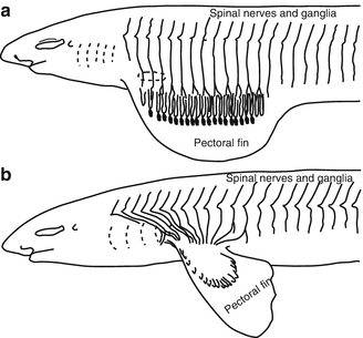
A spinal nerve firstly separates into the anterior and posterior branch to respectively innervate the ventral and dorsal trunk muscles of the same segment. The brachial plexus should be composed of the anterior branch, because the upper extremity emerges in the ventral side of the body trunk. The consideration of the development of the shark’s fin makes it easier to understand why anterior branches at the several segmental levels complicatedly form a plexus (Fig. 31.2). If the development of the fin slowly progressed while the base of the fin kept opening to the body trunk, the segmental order of the innervation should be preserved and the concentration should not take place. Yet, while the base of the fin is actually closed during the rapid development, myotomes are mixed and innervating nerves make anastomosis to form a plexus. During these processes, the segmental orders of nerves are significantly destroyed [1].


Fig. 31.2
Schemes of the upper part of adult sharks showing the nerve-supply of the fin. (a) The pectoral fin is expanded, and its nervous, muscular, and skeletal elements are distributed. (b) The base of the fin is closed, and concentration of myotomes innervating nerves take place
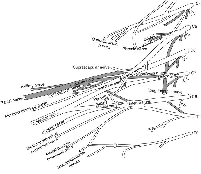
However, following two rules of innervating nerves have been approximately preserved during the development of the brachial plexus. Firstly, branches of the brachial plexus are separated into a ventral and dorsal layer based on locations of innervated muscles. In other words, the dorsal muscles (d) are innervated by nerves of the dorsal layer, and the ventral muscles (v) are by nerves of the ventral layer. Second, the closer the location of the muscle is to the body trunk, the more proximal the origin of the innervating nerve from the brachial plexus is. To be specific, the dorsal scapular and long thoracic nerve (d1 in Table 31.1), and subclavian nerve (v1) arise near the intervertebral foramina, while the subscapular and thoracodorsal nerve (d2), and pectoral nerve (V2) arise at even the infraclavicular area (Figs. 31.1 and 31.3).


