(1)
Shoulder Center of Kentucky, Lexington Clinic, 1221 South Broadway, Lexington, KY 40504, USA
Keywords
Winging of the scapula Scapula dyskinesis Sick scapula 29.1 Introduction
Injury to the scapula and its musculature is not uncommon. Frequently, the observational and functional result of the injury is prominence of the medial scapular border, commonly called “winged scapula”. Clinical presentations of scapular winging include neurologically based scapular winging, scapular muscle detachment, snapping scapula , and kinetic chain or muscle inhibition based scapular dyskinesis. These can all be organized as manifestations of scapular dyskinesis (abnormal scapular motion during arm movement) which can be seen as causes or results of various types of shoulder injury.
29.2 Neurologically Based Scapular Winging
The most common perception of the “winged” scapula, with prominence of the medial scapular border, is that it occurs as a result of damage to the nerves supplying the scapular stabilizing muscles. The exact frequency of neurologically based winging is poorly defined. Injury may be traumatic, iatrogenic, or idiopathic and the clinical interpretation requires careful history and examination. The three nerves most often involved include the long thoracic, spinal accessory, and dorsal scapular nerves.
The long thoracic nerve innervates the serratus anterior muscle and originates from the ventral rami of C5, C6, and C7. It is a purely motor nerve. The course of the nerve is clinically relevant as it predisposes the nerve to frequent injury. After passing through the middle scalene, the nerve crosses under the clavicle and remains superficial along the lateral chest wall. Here, it can be subjected to blunt trauma, diverse athletic injuries, or traction [1, 2]. Numerous reports of compressive neuropathy exist from lateral positioning or prolonged convalescence. Iatrogenic injury should be considered in patients’ status recovering from invasive lateral thoracic surgery such as radical mastectomy or axillary lymph node dissection. Finally, Parsonage–Turner type neuritis may be the underlying cause in patients experiencing significant pain or with recent viral infection. Recovery from idiopathic causes is usually apparent by 1 year but has been reported up to 2.
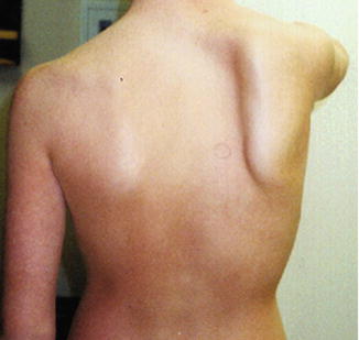
Long thoracic nerve injury creates loss of the serratus anterior muscle function which results in translation of the scapula superiorly and medially and disruption of normal scapulohumeral kinematics. Clinically, this rotation causes the inferior angle to become notably prominent in both static and dynamic examination (Fig. 29.1). The diagnosis can be confirmed on EMG around 6 weeks following initial trauma. Initial treatment measures include observation with supportive care, rehabilitation, and possibly follow-up EMG at 3-month intervals. Rehabilitation should focus upon preservation of glenohumeral motion and scapular stabilization by activation of the rhomboids and lower trapezius. Strengthening of the surrounding scapular stabilizers can be challenging; however, clinicians should avoid the use of long-lever exercises which can be over taxing and exacerbate symptoms. Rhomboid and lower trapezius strengthening is best achieved through the use of short-lever maneuvers which encourage scapular retraction and depression.


Fig. 29.1
Example of long thoracic nerve palsy creating scapular winging
Surgery for long thoracic nerve palsy may be indicated for patients with 1 year of symptoms, functional deficits, and no signs of recovery. Scapulothoracic fusion has been considered and may result in satisfactory results. However, due to the morbidity of the procedure it is generally reserved for high demand laborers and salvage situations. Fasciodesis procedures (sling) have also been utilized but can result in high rates of attritional loosening and poor function. Therefore, muscle transfers can be performed in an effort to restore a semblance of dynamic kinematics. Transfer of the sternocostal head of the pectoralis major has found the greatest measure of success [3, 4]. The selected portion of the tendon is reflected from its insertion on the humerus, tunneled ventral to the scapula and attached to the inferior angle of the scapula by various techniques. The tendon length generally requires augmentation with fascia latae or other graft for length. Published results have generally been favorable with an increase in variety of postsurgical outcomes scores [3, 5, 6].
Scapular winging can also be the product of injury to the spinal accessory nerve (cranial nerve 12) [4]. Upon loss of the trapezius activation, the scapula assumes a more inferior and lateral, or “drooping” posture. Winging is often less prominent than with serratus anterior palsy; however, the atrophy in the upper trapezius, loss of muscle tone and inability to shrug are easily discernible. Inability of the lower trapezius to achieve and maintain the retracted “functional” position of the scapula leads patients to report pain and weakness with forward elevation and abduction. Compensatory muscle spasm in the rhomboids and levator scapula are common. The diagnosis can be corroborated by EMG. Interpretation of EMG findings must be carefully performed as a false-negative report can occur due to placement of the recording electrode in the normal underlying rhomboids instead of the thin, atrophic overlying trapezius. For idiopathic causes or neuritis, supportive management and observation are recommended up to 1 year. In cases of iatrogenic transection or penetrating trauma, surgery is often advocated.
Scapulothoracic fusion may provide static stability and relieve pain but the loss of motion and complications may be unacceptable. The Eden-Lange Transfer was developed to provide a dynamic medial and superior restraint [3, 4]. In this procedure, the levator scapula and rhomboids are transferred approximately 5 cm laterally and secured via drill holes into the scapular body to improve mechanical advantage and substitute for trapezius function. Both short-term and long-term positive outcomes have been previously reported following this procedure [5, 7].
Less commonly, weakness of the major and minor rhomboid muscles is encountered in deficits of the dorsal scapular nerve. The nerve is a branch of the C5 nerve root and may be involved in radiculopathy. With loss of the rhomboids, unopposed pull of the serratus anterior results in lateral rotation of the inferior angle of the scapula. Atrophy of the rhomboids may be observed. EMG and supportive observation as outlined earlier are recommended. Fasciodesis procedures have met with limited success.
29.3 Scapular Muscle Detachment
This problem is not well known or well categorized with limited results reported [8]. As a result, these patients have experienced symptoms for months and years. The pathoanatomy appears to be an anatomic or physiologic detachment of the lower trapezius and rhomboids from the spine and medial border of the scapula. The large majority of cases present after an acute traumatic tensile load, half involving seat belt restrained motor vehicle accidents but there are multiple other causes such as throwing, catching, or lifting a heavy object with the arm at full extension, pulling against a heavy object, hanging on the rim after dunking a basketball, and electrical shock such as electrocution or cardioversion. The presenting symptom cluster is very uniform with early posttraumatic onset of localized pain along the medial scapular border. The pain increases in intensity as the condition evolves and averages 8/10 numeric pain rating. There are major limitations of arm use away from the body in forward flexion or overhead positions. Increased upper trapezius activity and spasm, resulting from lack of lower trapezius activity, creates migraine-like headaches. Neck and shoulder joint symptoms may be present due to dyskinesis and will often become the focus of treatment, including surgery with infrequent positive results.
The physical exam also exhibits a consistent cluster of findings including the localized tenderness, often a noticeable and palpable soft tissue defect, either due to the detachment or the muscle atrophy, altered scapular resting position as well as dynamic dyskinesis including snapping scapula , shoulder impingement and weakness in forward flexion, and clinical decrease or relief of symptoms with scapular corrective maneuvers. MRI and CT imaging are of minimal benefit. The cuts may not be in the correct plane, the detachment scar is not easily demonstrated, the lower trapezius is detached from the spine but then lays back over in the supine retracted imaging position, or the chronic changes are too subtle for the static imaging processes. Consistency of the history and physical exam findings allows for a reliable clinical diagnosis.
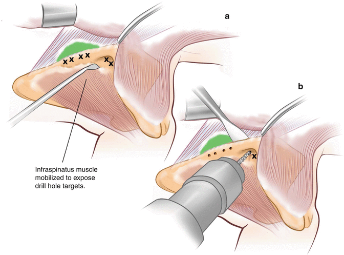
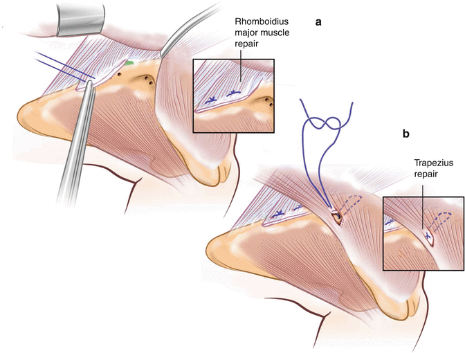
Most of these patients have had workups to rule out neurologic or bony causation, and have had varying types of treatment, including local or distant surgery and various rehabilitation protocols. If they have failed an appropriate scapular rehabilitation program and do not demonstrate other anatomic defects, surgical reattachment is indicated. This is accomplished by direct reattachment through pairs of drill holes in the medial scapular border and scapular spine (Fig. 29.2) [3, 8]. The detached and scarred rhomboids are mobilized and reattached onto the dorsal aspect of the scapula about 1 cm from the medial edge (Fig. 29.3). The lower trapezius is mobilized and reattached along the proximal scapular spine.


Fig. 29.2
Illustration of drill hole placement when performing the scapular muscle reattachment procedure. Mobilization of the infraspinatus away from the medial border of the scapula (a) in order for placement of drill holes for muscle reattachment (b)

Fig. 29.3
Illustration of the reattachment of the rhomboid and lower trapezius muscles. Reattachment of the rhomboid muscle is performed initially (a) followed by reattachment of the lower trapezius muscle (b)
Postoperatively, the arm is protected in neutral rotation for 4 weeks but gentle scapular retraction is encouraged immediately. At 4 weeks, closed chain activation up to 90° abduction with the hand stabilized is started. By 6–8 weeks, as the repair has healed and early strength is gained, motion over 90° is allowed and the patient is started on the standard scapular strengthening program. Maximum strength is not regained for about 6–9 months, probably reflecting the chronic muscle disuse and atrophy. Results from a 2-year follow-up of a small cohort show that pain scores decreased from 8/10 to 2/10, and ASES scores improved from 38/100 to 68/100 [8]. These results are durable at 2-year follow-up.
29.4 Snapping Scapula
The diagnosis and management of snapping scapula can be clinically challenging. The condition has been estimated to be present in up to 30 % of asymptomatic individuals yet can cause crippling pain. Snapping scapula most frequently represents a disruption of the smooth gliding of the scapula over the thoracic cage and periscapular muscles [9]. A thorough understanding of the three-dimensional kinematics and anatomy is required to appreciate the varied etiologies. The normal coupled scapular motions of posterior tilt and external rotation are decreased as the arm elevates often due to tissue tightness, muscle weakness, and in some instances compensatory movement patterns following injury. Consequently, the normal movement of the instant center of rotation of the scapula from the superior medial border to the AC joint is disrupted, causing the scapula to rotate around the medial border, creating excessive pressure and leading to symptoms [10].
Stay updated, free articles. Join our Telegram channel

Full access? Get Clinical Tree


