Fig. 6.1
3D CT scapulae reconstruction with a schematic representation of secondary acromial ossifications centres
6.2 Acromion Anatomy
6.2.1 Description
The acromion is a scapular structure projected anteriorly from the lateral border of the scapular spine, which represents continuity. It presents superior and inferior surfaces, and medial and lateral borders. The inferior lip of the crest of the spine is continued with the lateral acromion border, which is thick and irregular. On the other hand, the medial border corresponds to a prolongation of the scapular crest of the spine upper lip. It is short and is occupied anteriorly by an oval-shaped articular facet, directed medially and upwards, to join with the lateral end of the clavicle. Both acromial borders join anteriorly, forming a triangle called acromial angle [7, 8].
6.2.2 Muscles and Ligaments Insertions
The mid portion of the deltoid muscle originates along the lateral border of the acromion, including the most anterior portion of the acromial process [8]. The deltoid insertion in the acromion has an average thickness of 5.4 mm, corresponding to approximately 74 % of the anterior acromion thickness [9]. These anatomic features must be considered when performing an acromioplasty, because a large resection could affect the deltoid origin.
On the other hand, the coracoacromial ligament is a triangular fibrous lamina, its apex is attached to the acromion and its base to the lateral border of the coracoid process (Fig. 6.2). The upper surface of the ligament is related to the surface of the deep deltoid muscle, while the inferior surface is oriented towards the glenohumeral joint and the periarticular muscles, from which it is separated by a synovial pouch (subacromial or subdeltoid pouch) [8]. The ligament has two bands: lateral (stronger and thicker) and medial (with a variable insertion in the acromion) [10].
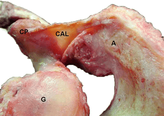

Fig. 6.2
Anatomic dissection of coracoacromial ligament (CAL), which is located between the lateral border of coracoid process (CP) and acromion (A). Glenoid fossa (G) is also shown (From Morphology Department, Universidad de los Andes, Santiago, Chile)
6.2.3 Function
The acromion, together with the coracoacromial ligament and the coracoid process, form the coracoacromial arch (Figs. 6.3 and 6.4), which is a curved structure meant to protect the glenohumeral joint. Specifically, the acromion and the coracoacromial ligament limit the upper translation of the glenohumeral joint [11].
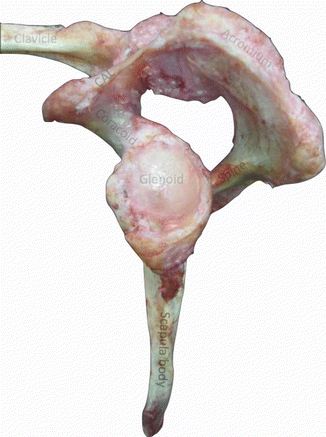
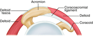

Fig. 6.3
Anatomic dissection of coracoacromial arch, formed by the coracoid process (CP), coracoacromial ligament (CAL) and acromion (A), establishing a curved structure over the glenoid process (G) (From Morphology Department, Universidad de los Andes, Santiago, Chile)

Fig. 6.4
Diagram of the acromion and coracoacromial ligament (Modified with permission, Arthroscopic Rotator Cuff Surgery: A Practical Approach to Management, Chapter 7, 2008, K. Yamaguchi, R. Tashjian Copyright Springer Science + Business Media)
6.3 Anatomic Variations and Changes with Age
Bigliani et al. (1986) described three types of acromion according to its inferior surface morphology and classified it as type I (flat acromion), type II (curved acromion) and type III (hooked acromion) [3] (Fig. 6.5). The prevalence described in the literature for each type depends upon the analysed population, but in general the evidence shows a higher frequency of type II acromion (near 50–55 %), followed by type III (25–30 %) and type I (15–20 %) [12]. In a healthy population, the incidence of type I acromion decreases with age, as type III increases [13]; therefore, it is considered that aging leads to secondary changes in the coracoacromial arch [14]. It has also been observed that with age the acromion presents degenerative changes characterized by an anterior spur which appears. In subjects below 50 years of age, the prevalence is considered to be 7 %, while above this age the prevalence rises to 30 % [15].
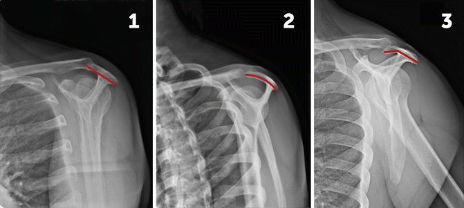

Fig. 6.5
Outlet views of shoulder radiographies with the three types of acromion described by Bigliani (1986): type 1 (flat acromion), type 2 (curved acromion) and type 3 (hooked acromion)
6.4 Acromial Morphology and Rotator Cuff Injury
Intrinsic and extrinsic factors intervene in rotator cuff injury. The evidence between acromial morphology and its association with rotator cuff injuries remains controversial. Neer (1983) established that 95 % of patients with a rotator cuff tear present a space conflict at the coracoacromial arch. Equally, Bigliani et al. (1986) showed that the incidence of type III acromion was 70 % in patients with rotator cuff injury, compared to 38 % in the general population [3, 16]. However, studies performed have not been able to show such association [17, 18], assuming that also age could be a confusion factor in this topic, especially taking into account the higher prevalence of type III acromion as years go by [13], even more when age is considered an independent risk factor for rotator cuff injury [19]. Therefore, acromial morphology would represent a degenerative process with aging rather than an innate anatomical feature [18]. Gill et al. (2002) found no significant association between acromial morphology and rotator cuff injury in patients over 50 years of age [18].
6.5 Pathology: Os Acromiale
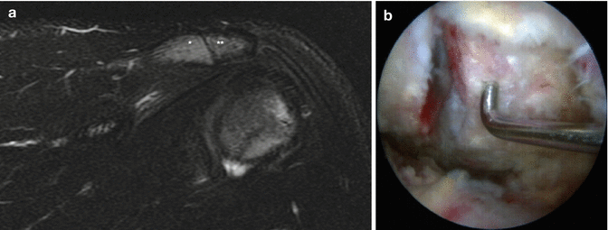
Os acromiale (OA ) is an incomplete fusion of the secondary acromial ossification centres, thus resulting in an epiphyseal fragment which is separated from the rest of the bone structure [20]. The most usual location is between the meso-acromion and meta-acromion, named “common os acromiale” (approximately 76 % of the cases) (Fig. 6.6), while the separation between the pre-acromion and meso-acromion centres is less frequent (“terminal osacromiale”, 15 % of the cases). The location between meta-acromion and basi-acromion is exceptional (1.8 %) [21, 22]. The degree of fusion varies from a fibrous union (complete or partial) up to a diartrodial joint [23].
