A common pitfall when interpreting foot and ankle radiographs, made by both student and practitioner alike, is mistaking normal anatomic shadows for pathology. To truly appreciate abnormalities of position, form, density, and architecture, the interpreter must have a working knowledge and appreciation of normal radiographic anatomy. In the foot and ankle, this requires correlation of normal three-dimensional osteology (the bone specimen) to two-dimensional radiographic anatomy (the radiographic image). The reader is, therefore, encouraged to compare the radiographic image to a foot skeleton when interpreting radiographic studies.
The following collection of illustrations, tracings of “rectus” foot images, and their respective radiographs should be used as a general reference guide. Slight variations in the appearance of each bone occur depending on the position of the foot relative to the image receptor. For example, a pronated or supinated foot affects the overall radiographic appearance of the entire foot as well as each individual bone (Figure 5-1). Therefore, it is important to concentrate on the correlation of gross anatomy to the radiograph, not to memorize the appearance of each bone per se.
The illustrations that follow include a variety of radiographic views (Figures 5-2 to 5-12).
Sarrafian’s Anatomy of the Foot and Ankle1 was used as the reference for the terminology in the labels. Occasionally, however, I used the best generic term to label a radiographic “shadow” that Sarrafian had not specifically identified. The label key is given in Box 5-1.
I have spent much of my career studying foot and ankle bone specimens (normal three-dimensional osteology) and correlating them to two-dimensional radiographs.2–8 The original manuscript for this chapter included a complete reference guide to radiographic anatomy of the foot and ankle. But, because of its large size and numerous images, it was not included in this textbook. The manuscript, however, has been accepted for publication in five parts and should be in print by the time this second edition becomes available.9–13 The project correlates detailed radiographic anatomy of the entire adult foot and ankle (two-dimensional) to osteology (three-dimensional). Images of each foot and distal leg bone (“front” and “back” perspectives) are presented alongside a corresponding radiographic image for comparison. It should serve as a baseline (“normal”) that future researchers can use as well as a reference that both students and practitioners can use for comparison when interpreting radiographs and distinguishing abnormal findings from normal.
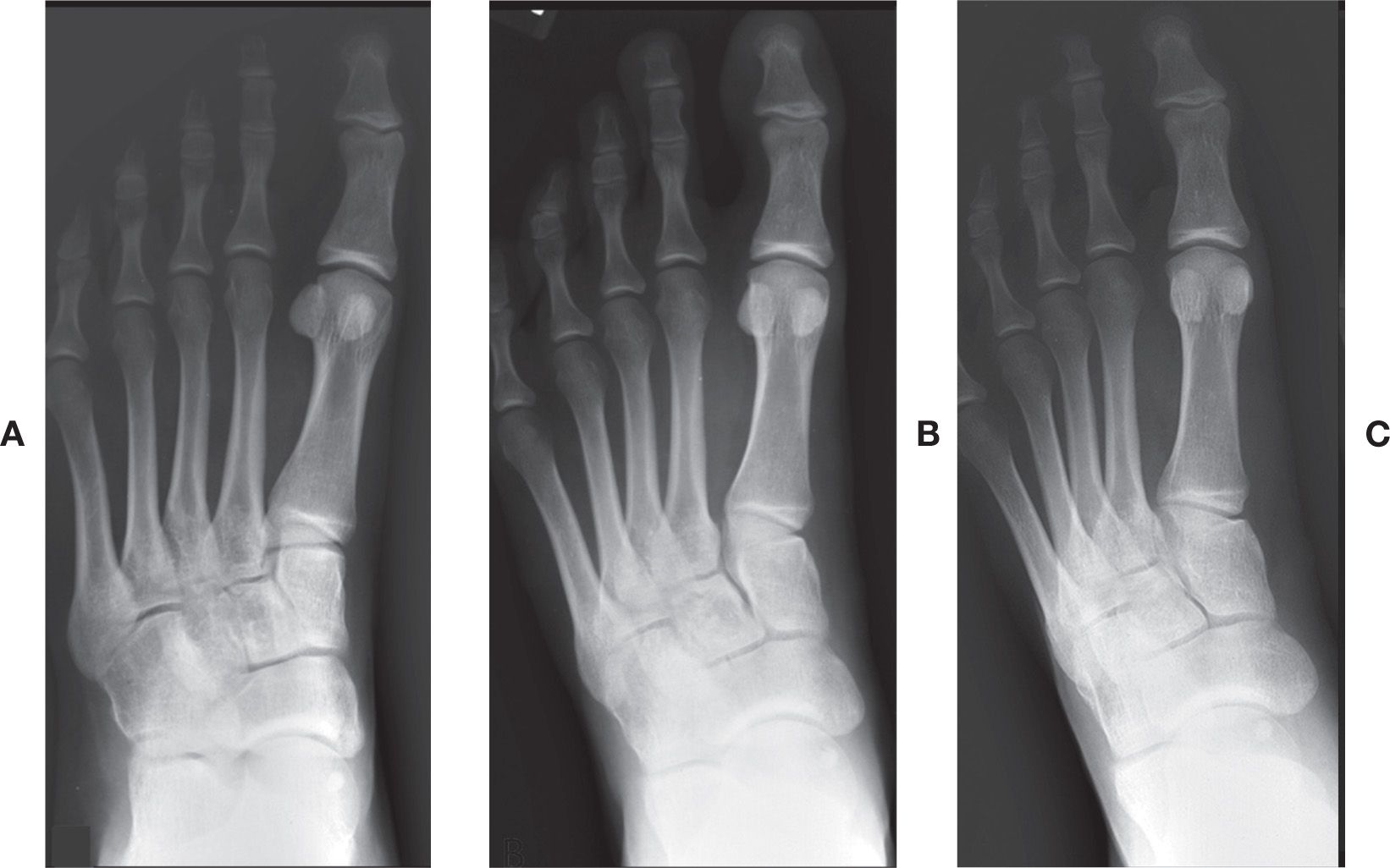
FIGURE 5-1. The effect of pronation and supination on the position and form of bones (all images of the same foot): A: Pronated; B: Rectus; C: Supinated.
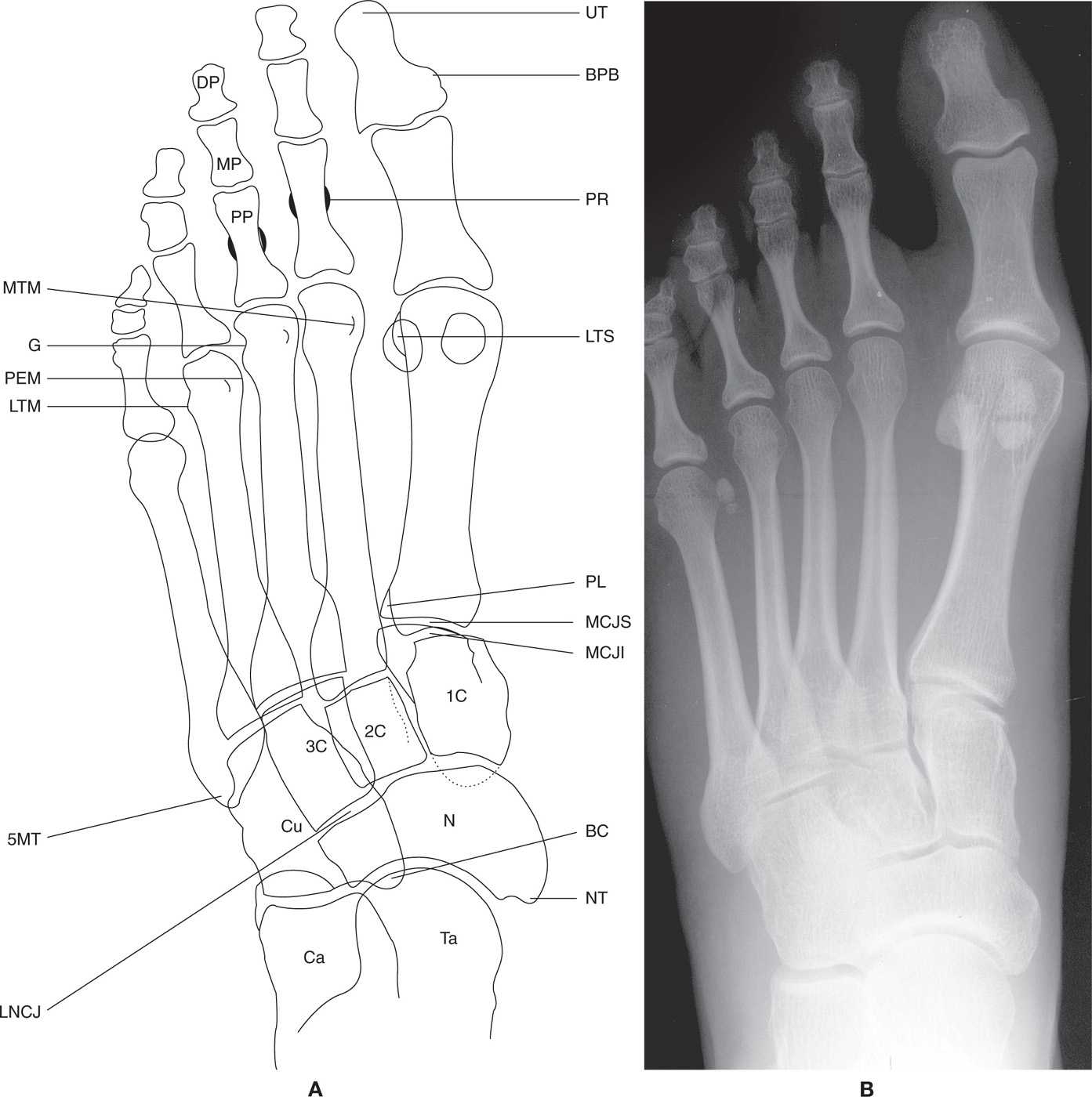
FIGURE 5-2. A and B: Dorsoplantar (DP) foot view.
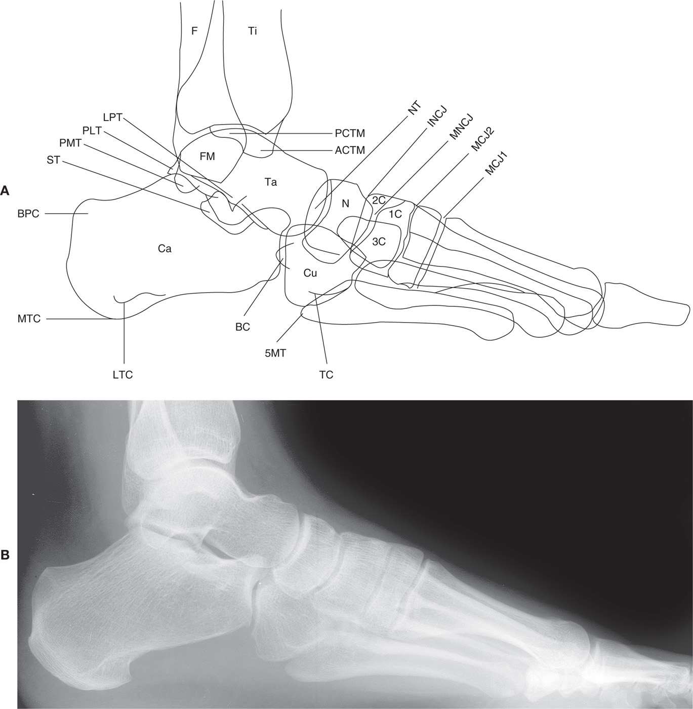
FIGURE 5-3. A and B: Lateral foot view.
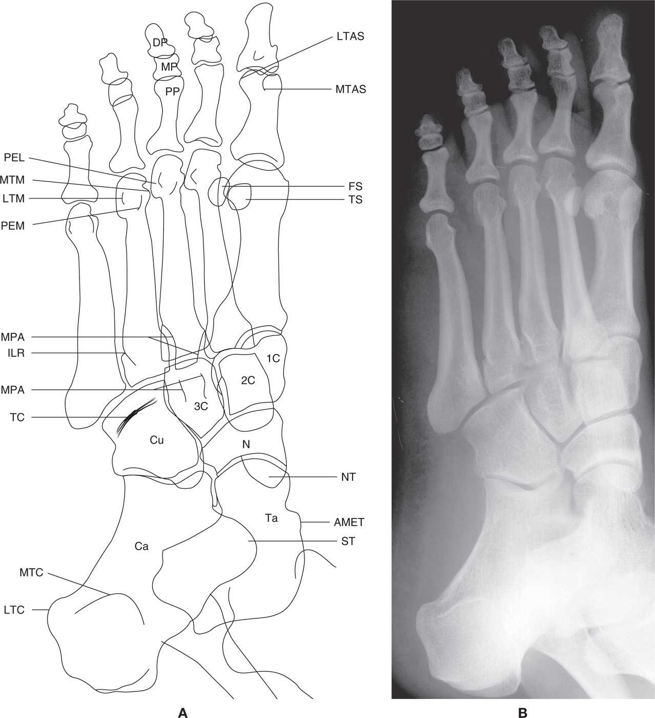
FIGURE 5-4. A and B: Medial oblique foot view.
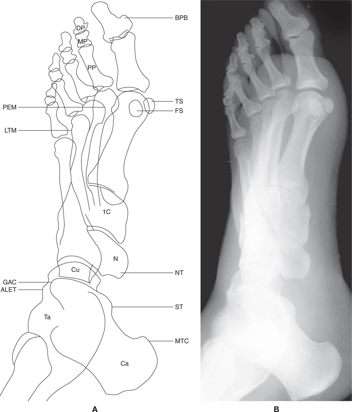
FIGURE 5-5. A and B: Lateral oblique foot view.
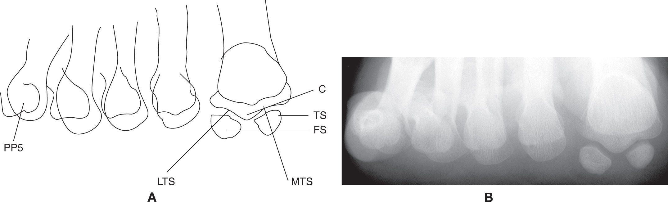
FIGURE 5-6. A and B: Sesamoid axial view.
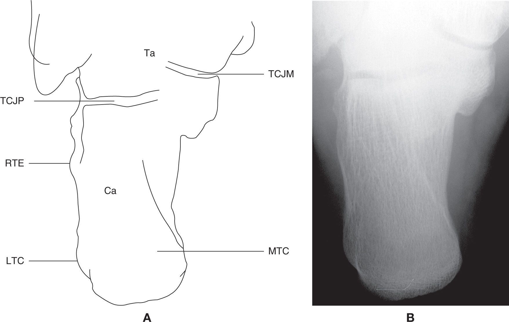
FIGURE 5-7. A and B: Calcaneal axial view.
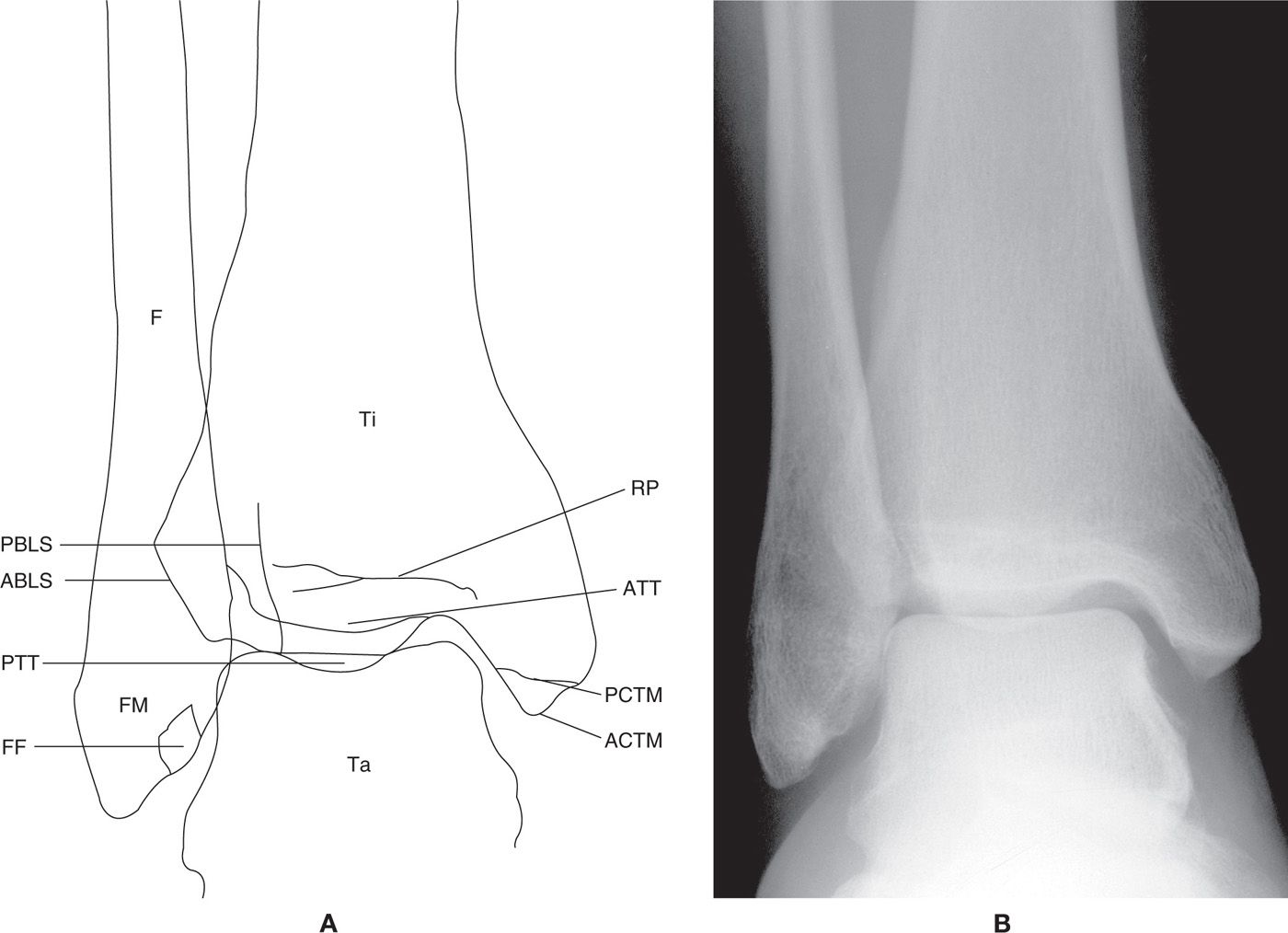
FIGURE 5-8. A and B: Anteroposterior (AP) ankle view.
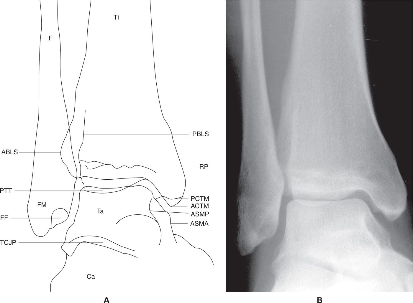
FIGURE 5-9. A and B: Mortise ankle view.
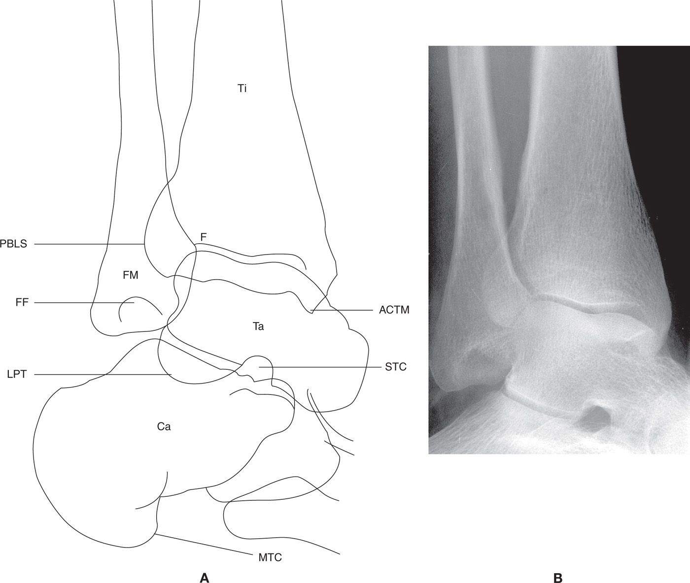
FIGURE 5-10. A and B: Medial oblique ankle view.
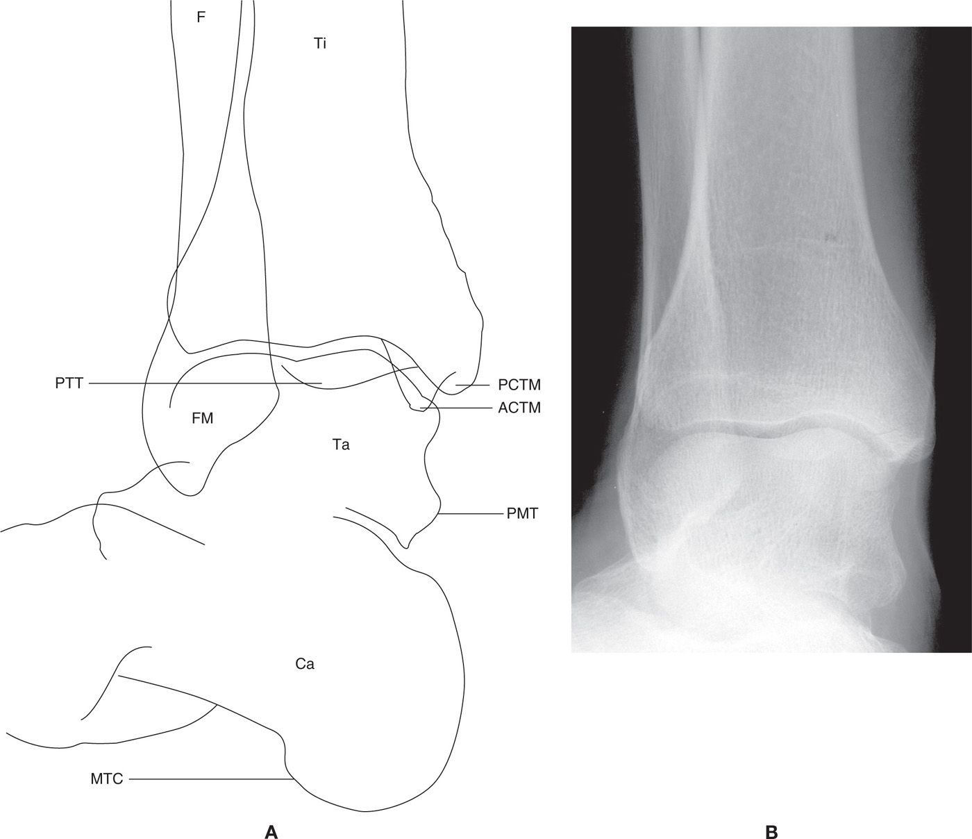
FIGURE 5-11. A and B: Lateral oblique ankle view.
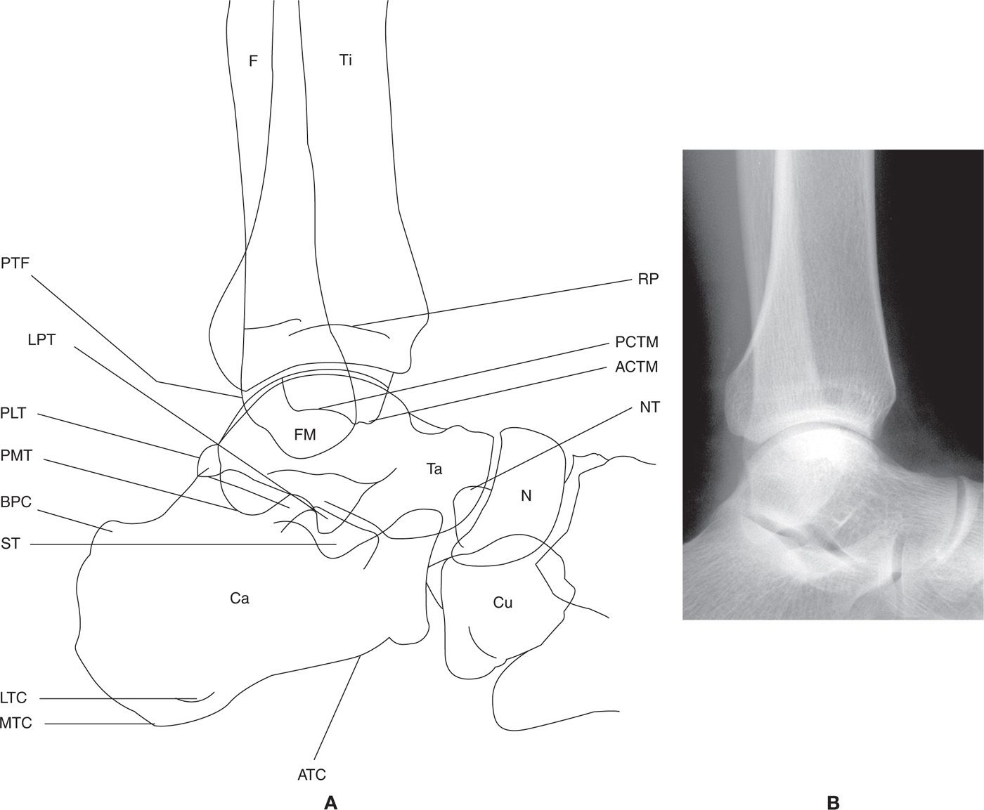
FIGURE 5-12. A and B: Lateral ankle view.
BOX 5-1 Label Key
1C | First cuneiform (medial cuneiform) |
2C | Second cuneiform (intermediate cuneiform) |
3C | Third cuneiform (lateral cuneiform) |
5MT | Tuberosity of fifth metatarsal base |
ABLS | Anterior border of distal tibial lateral surface |
ACTM | Anterior colliculus (of tibial malleolus) |
ALET | Anterolateral extension of trochlear surface (talus) |
AMET | Anteromedial extension of trochlear surface (talus) |
ASMA | Articular surface for medial malleolus, anterior margin |
ASMP | Articular surface for medial malleolus, posterior margin |
ATC | Anterior tuberosity (anterior tubercle) |
ATT | Anterior tibial tubercle |
BC | Beak of cuboid |
BPB | Bony projection, distal phalanx base (varies in size) |
BPC | Bursal projection, posterior calcaneus |
C | Crista (crest of metatarsal head) |
Ca | Calcaneus |
Cu | Cuboid |
DP | Distal phalanx |
F | Fibula |
FF | Fibular (digital) fossa |
FM | Fibular malleolus |
FS | Fibular sesamoid |
G | Groove separating tubercle and articular surface |
GAC | Great apophysis (anterior process of calcaneus) |
INCJ | Intermediate naviculocuneiform joint |
ILR | Interosseous ligament rugosity |
LNCJ | Lateral naviculocuneiform joint |
LPT | Lateral process (talus) |
LTAS | Lateral trochlear articular surface (hallux proximal phalanx) |
LTC | Lateral tuberosity (lateral tubercle) of calcaneus |
LTM | Lateral tubercle (for metatarsophalangeal ligaments) |
LTS | Lateral trochlear surface (first metatarsal) |
MCJ1 | First metatarsocuneiform joint |
MCJ2 | Second metatarsocuneiform joint |
MCJI | Inferior aspect of first metatarsocuneiform joint |
MCJS | Superior aspect of first metatarsocuneiform joint |
MNCJ | Medial naviculocuneiform joint |
MP | Middle phalanx |
MPA | Medial and lateral margins of plantar apex of bone |
MTAS | Medial trochlear articular surface (hallux proximal phalanx) |
MTC | Medial tuberosity (medial tubercle) of calcaneus |
MTM | Medial tubercle (for metatarsophalangeal ligaments) |
MTS | Medial trochlear surface (first met) |
N | Navicular |
NT | Tuberosity of navicular |
PBLS | Posterior border of distal tibial lateral surface |
PCTM | Posterior colliculus (of tibial malleolus) |
PEL | Proximal extension of metatarsal head articular surface, laterally |
PEM | Proximal extension of metatarsal head articular surface, medially |
PL | Tubercle for insertion of peroneus longus tendon |
PLT | Posterolateral tubercle (talus) (trigonal process) |
PMT | Posteromedial tubercle (talus) |
PP | Proximal phalanx |
PR | Phalangeal ridge |
PTF | Posterior tubercle (fibula) |
PTT | Posterior tibial tubercle |
RP | Remnant of physis |
RTE | Retrotrochlear eminence |
ST | Sustentaculum tali |
STC | Sinus tarsi/tarsal canal |
Ta | Talus |
TC | Tuberosity of cuboid |
TCJA | Talocalcaneal joint, anterior |
TCJM | Talocalcaneal joint, middle |
TCJP | Talocalcaneal joint, posterior |
Ti | Tibia |
TS | Tibial sesamoid |
UT | Ungual tuberosity (tuft of distal phalanx) |
Related posts:

Stay updated, free articles. Join our Telegram channel

Full access? Get Clinical Tree


