Postoperative Evaluation and Complications
ANDREW J. MEYR AND CORINE L. CREECH
INTRODUCTION
Radiographic evaluation forms an essential component of the preoperative assessment and surgical decision making for the majority of foot and ankle reconstructive procedures, so it should be expected that radiographs would also be utilized postoperatively in the assessment of these interventions. In an admittedly philosophic manner of speaking, however, postoperative radiographs often represent a self-fulfilling prophecy in this respect. If, for example, we preoperatively define a hallux abductovalgus deformity radiographically based on an increased first intermetatarsal angle, and subsequently perform a metatarsal osteotomy specifically designed to decrease the first intermetatarsal angle, then we have “fixed” the deformity as we have defined it, regardless of patient complaint.
It is sometimes difficult to remember in clinical practice that imaging studies and radiographic angles are only tools that surgeons utilize for medical decision making, and although they may be useful, they are not the only pieces of information that we have available for our patient evaluation. A “Cartesian” way of thinking is overreliance on radiographs in the perioperative period.1,2 Rene Descartes (1596–1650) was a French scientist whose philosophy proposed that all of science could be explained by the concept of “matter in motion.” In other words, the body is nothing more than a series of measureable working parts, and pain or pathology is an objective deviation of this system. Ergo, if a surgeon can physically put the parts back in working positions, then the machine should function again asymptomatically. Anyone working in a clinical setting knows from experience that this is not always the case.
This brings to mind the common adage within the field of reconstructive foot and ankle surgery to “treat the patient, not the x-rays.” Although many layers of meaning may be ascribed to this expression, the most basic connotation cautions surgeons against a completely radiographic-focused perioperative patient evaluation. Using the previous example, it would be unusual for a patient to present specifically complaining of a “painfully increased first intermetatarsal angle,” so we should not necessarily expect the patient to be completely without complaint just because our surgical intervention has created a radiographically rectus joint. Although perioperative radiographs are important, patient complaints often go beyond our subjective and objective radiographic findings.
With that said, however, postoperative radiographs following reconstructive foot and ankle surgery are considered the standard of care and have many potential useful applications for patient-care decision making during the postoperative period. This chapter intends to serve as an introduction for several of these applications including (1) assessment of deformity correction, (2) serial assessment of osseous healing, and (3) verification of hardware.
ASSESSMENT OF DEFORMITY CORRECTION
As it relates to reconstructive foot and ankle surgery, deformity correction can be either elective (structural deformities such as a bunion or flatfoot) or nonelective (as in fracture malalignment) in nature. Regardless, each requires knowledge of normal alignment measures and radiographic anatomy.
Hallux Abductovalgus
Elective deformity correction is usually accompanied by a unique set of angles and measurements for the given anatomic location. As a clear example of this, Chapter 13 (Foot Segmental Relationships and Bone Morphology) details the numerous objective radiographic measurements that can be utilized for evaluation of the first metatarsal–phalangeal joint and hallux abductovalgus deformity. Surgeons will often base their specific surgical plan on whether each of these measurements are normal versus abnormal, and then recommend specific surgical procedures that will have an effect on that specific subset of measurements. If a given radiographic measurement is identified as abnormal preoperatively, then it is reasonable to conclude that the surgical intervention should correct for this abnormality (Figure 23-1).
Although first metatarsal osteotomies are one of the most common surgical procedures performed within foot surgery, postoperative measurement can represent a clinical challenge and controversy because of the morphologic changes created within the bone by the surgery itself. For example, the anatomic landmarks used to define the distal metaphyseal–diaphyseal junction for the long axis of the first metatarsal can be disrupted by performance of the osteotomy at this anatomic location. In these situations it has been suggested to instead utilize a “center-of-head” technique for calculation of the distal reference point instead of the metaphyseal–diaphyseal junction (Figure 23-2).3
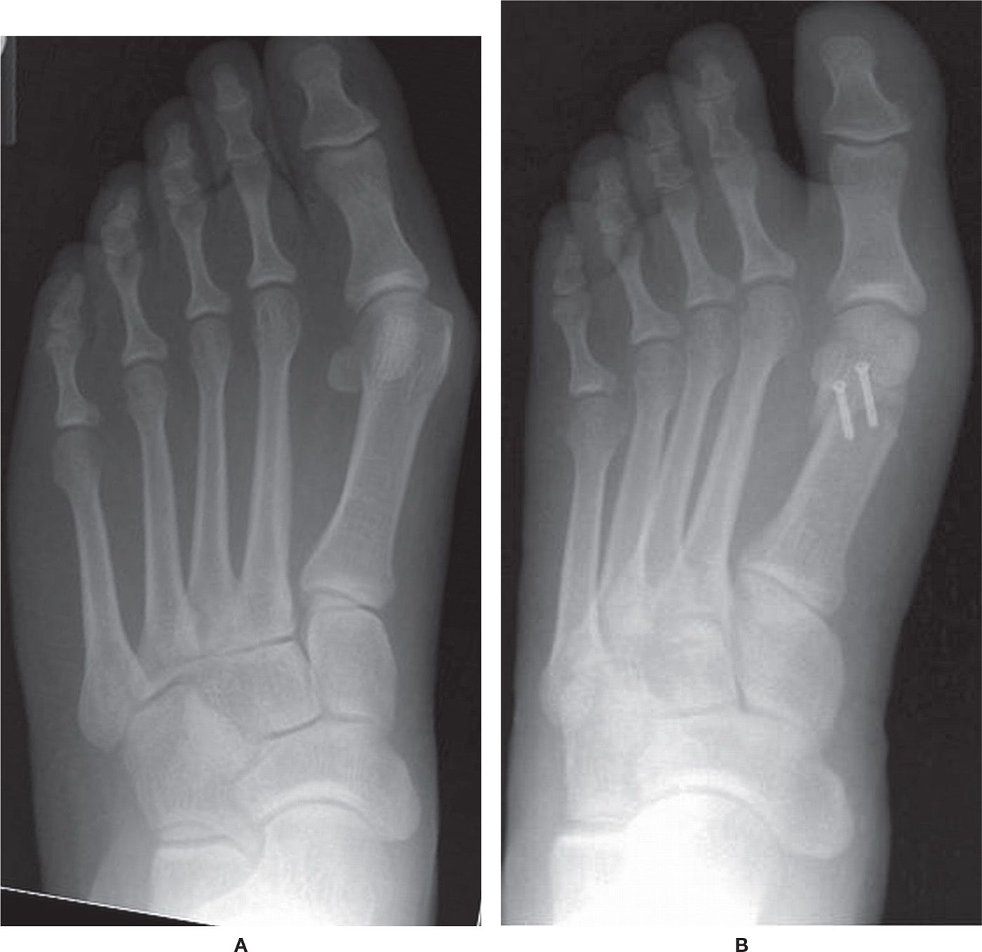
FIGURE 23-1. Preoperative (A) and postoperative (B) dorsoplantar radiographs of hallux abductovalgus deformity demonstrating change in the measurement of angular deformity. A: The first intermetatarsal angle (12°), hallux abductus angle (30°), and a tibial sesamoid position of 5 are all abnormally increased. These abnormalities can be addressed with the selected surgical procedure, in this case, a distal first metatarsal osteotomy. B: Postoperatively, all preoperative angular abnormalities have been corrected to within their normal ranges: the first intermetatarsal angle is decreased to 4°, the hallux abductus angle is decreased to 12°, and the tibial sesamoid position is decreased to a position 2.
Another consideration to keep in mind is that structural deformity is primarily assessed with weight-bearing radiographs taken in the angle and base of gait, while postoperative radiographs are primarily performed non–weight bearing and often with the patient in a cast or other bandaging materials. It may realistically take several weeks or even months to obtain a true weight-bearing postoperative radiograph in the angle and base of gait. Surgeons should exercise caution to not “over read” structural positional changes postoperatively with non–weight-bearing radiographs, as these views are known to have at least some effect on angular measurements (Figures 23-3 and 23-4).4–6
Other Structural Deformities
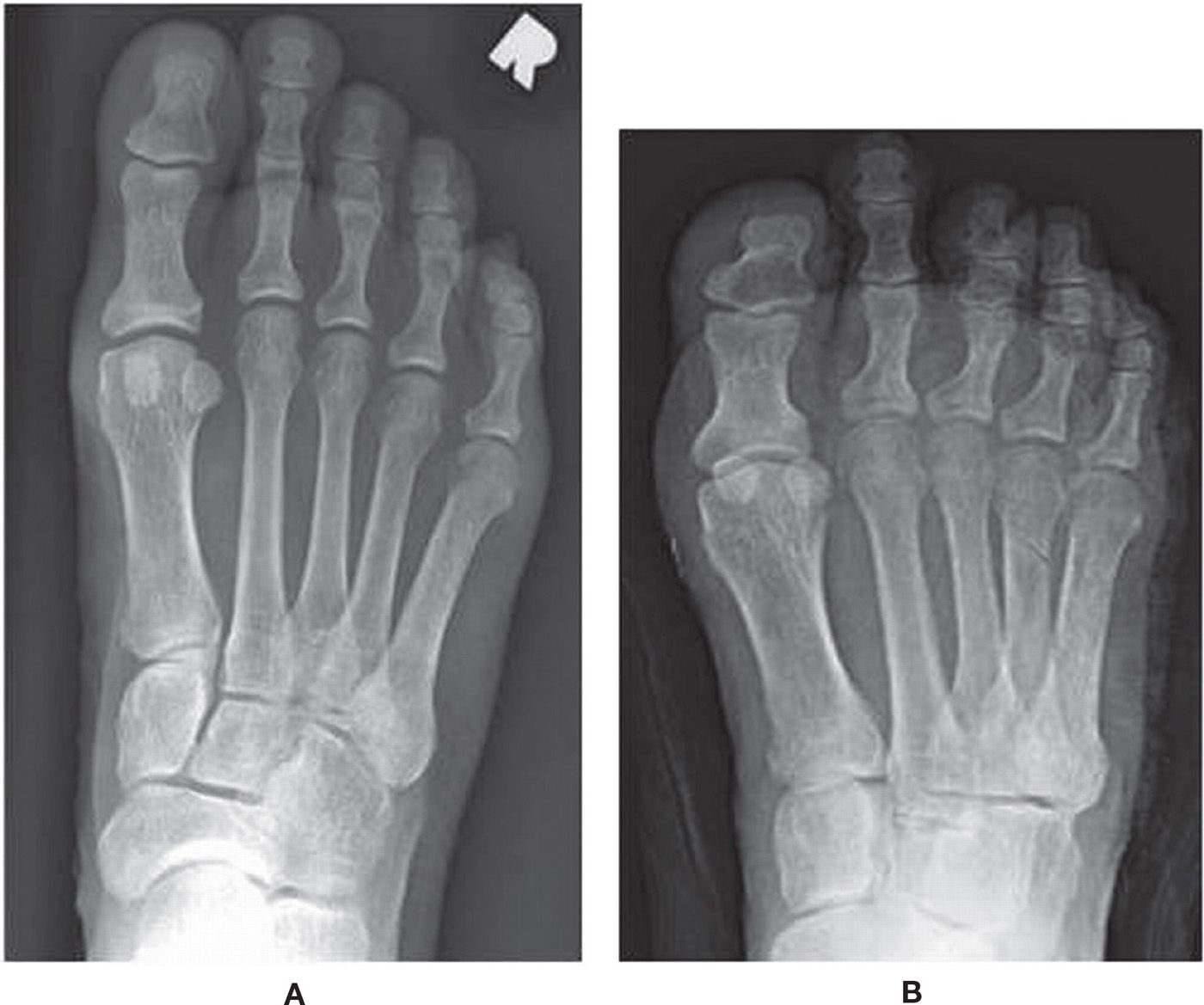
The assessment of postoperative correction for other lower-extremity pathologies occurs in much the same way, although each structural abnormality often has a distinct set of measurements used to objectify both deformity and correction. Examples include so-called “shortening” lesser metatarsal osteotomies which aim to restore the metatarsal parabola (Figure 23-5), dorsiflexory first metatarsal osteotomies which restore the lateral talar–first metatarsal angle into a parallel relationship for cavus foot deformity (Figure 23-6), and lateral calcaneal lengthening procedures which decrease the cuboid-abduction angle and increase talar head coverage for a flatfoot deformity (Figure 23-7). If radiographs and specific angular measurements are utilized preoperatively to objectively define deformity, then the same views and measurements should be reevaluated in the postoperative period to ascertain correction and return to normally accepted measurement ranges.
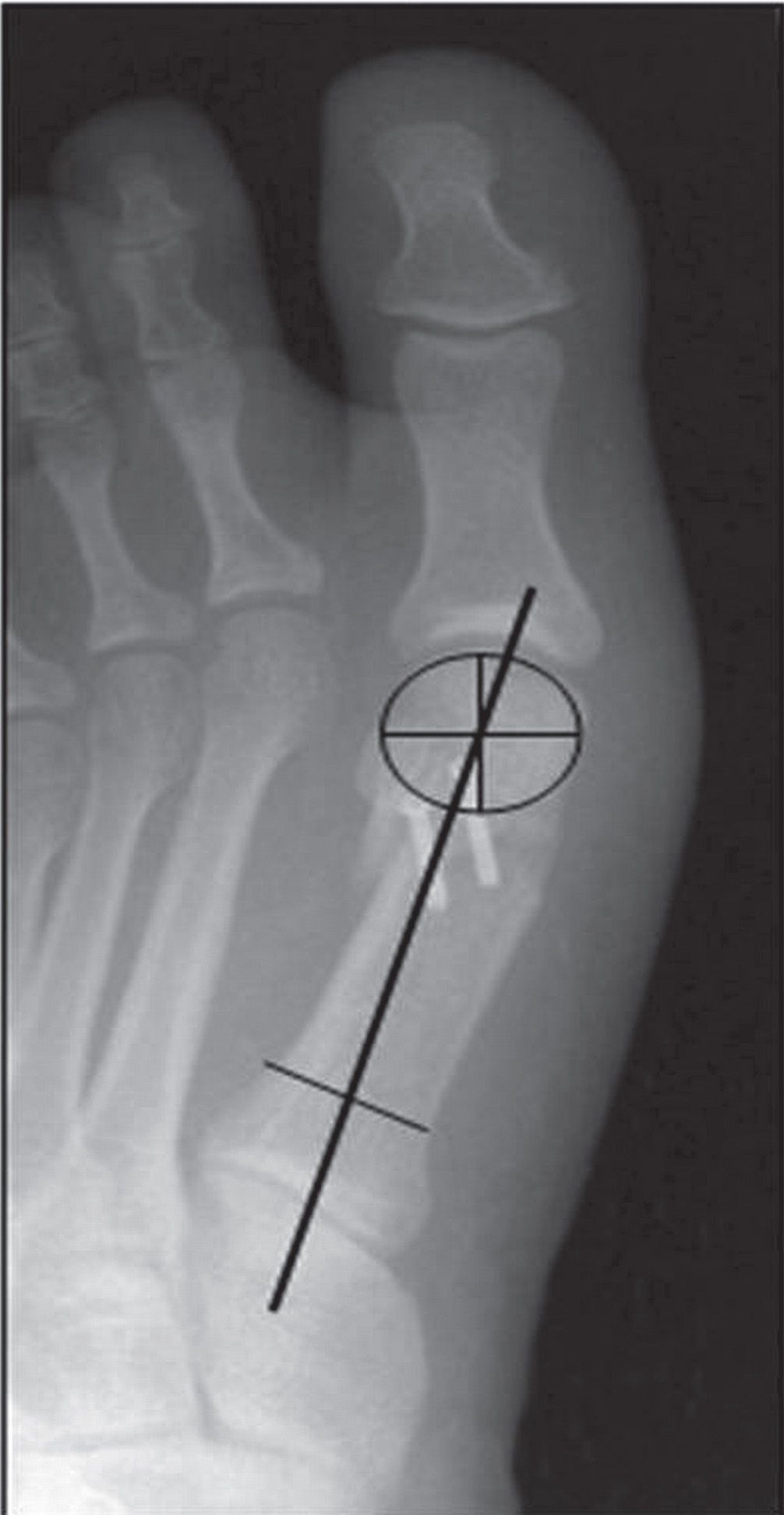
FIGURE 23-2. Measurement of a radiographic angle with disrupted anatomical landmark. The head of the first metatarsal is converted into a circle, and the long axis of the first metatarsal passes through the center of this circle (in addition to a bisection of the proximal metaphyseal–diaphyseal junction).
One radiographic view that requires specific mention with respect to deformity correction is the calcaneal axial. This view should be considered in the perioperative period for assessment of frontal plane deformities of the rearfoot and ankle.7 It is difficult to assess for varus and valgus deformities of the calcaneus, subtalar joint, and ankle joint without this view (Figure 23-8).
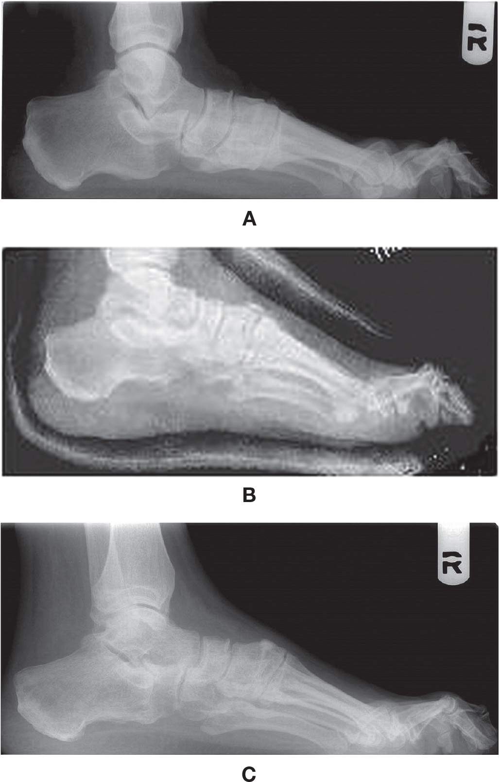
FIGURE 23-3. The effect of weight bearing on the lateral radiographic view. A: The preoperative radiograph demonstrates sagittal plane malalignment with a decreased calcaneal inclination angle, increased talar declination angle, and abnormal lateral talar–first metatarsal angle. B: The immediate postoperative radiograph appears to demonstrate improvement of these same measurements; however, one can appreciate the overlying cast and that this view was performed non–weight bearing. C: True assessment of postoperative change often cannot be performed until several weeks to months later with a true weight-bearing view. The radiograph now demonstrates improvement of these measurements compared to the preoperative radiograph (A), although not to the same degree as the immediate non–weight-bearing radiograph (B).
Traumatic Reconstructions
Acute trauma presents another situation in which the goal of surgical intervention is to return an osseous relationship from abnormal to normal. Surgical correction, however, is usually not aimed at returning deformity to within a normally accepted range, but is instead aimed at returning the anatomy to exactly where it was before the injury. As a common rule of thumb, foot and ankle surgeons use 2 mm as a guideline for “exact” anatomic reduction. Any resulting displacement or deformity greater than 2 mm is generally considered to be a less than ideal outcome for the foot and ankle (Figure 23-9). This is an objective measurement that can be calculated relatively easily with the advent of digital radiographic software. As previously, most specific anatomic locations have unique criteria and measurements used to define “normal.” However, there are two locations in the lower extremity that warrant specific mention with respect to traumatic postoperative radiographic assessment.
Related posts:

Stay updated, free articles. Join our Telegram channel

Full access? Get Clinical Tree


