45
Volar Dislocations of the Proximal Interphalangeal Joint
Lisa L. Lattanza and Steven Z. Glickel
History and Clinical Presentation
A 20-year-old woman sustained an injury to her right ring finger during a tackle while playing rugby. She was not able to describe the exact mechanism, as it happened in a “pile-up.” She was seen in the emergency department that day.
Physical Examination
A swollen and tender proximal interphalangeal (PIP) joint of the right ring finger was noted, with slight rotational deformity of the digit. The patient was unable to actively extend the PIP joint. There was brisk capillary refill of the digit and the skin was intact.
Diagnostic Studies
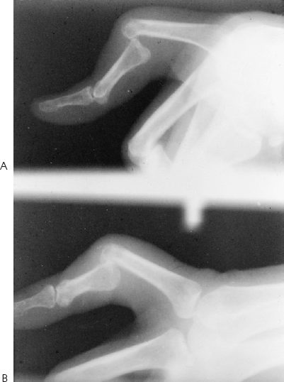
Anteroposterior and lateral radiographs of the right ring finger showed a volar PIP joint dislocation without fracture (Fig. 45-1).
PEARLS
- Flex the PIP and MP joints when reducing the dislocation.
- Determine if the dislocation is straight volar or volar rotatory.
- Mobilize early if stable.
PITFALLS
- Most volar dislocations do not require aggressive operative intervention.
- Beware of late PIP joint stiffness or boutonniere deformity.
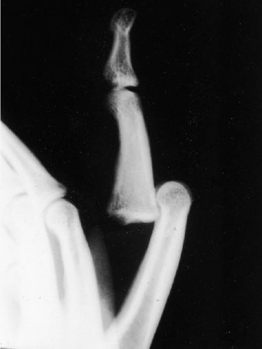
Figure 45-1. Volar proximal interphalangeal (PIP) joint dislocation.
Differential Diagnosis
PIP joint sprain
Dorsal PIP joint dislocation
Volar PIP joint dislocation
Middle phalanx fracture
Proximal phalanx fracture
PIP joint fracture/dislocation
Diagnosis
Volar PIP Dislocation
Unlike dorsal PIP joint dislocations, volar dislocations are rare. It is important to understand the mechanism of injury and the resultant damage to the underlying structures to devise a sensible treatment plan. It is also necessary to differentiate the straight volar dislocation from the volar rotatory dislocation as the structures damaged, and therefore the treatment, are different.
Nonsurgical Management
There are two types of volar PIP joint dislocations: a straight volar dislocation and the volar rotatory dislocation. To fully understand the management of these two distinct entities, it is essential to understand the different injuries.
Straight volar dislocations occur with a volarly directed force on a semiflexed digit. The primary injury is to the central slip of the extensor mechanism. These injuries are generally easily treated with closed reduction and splinting in extension to allow the central slip to heal. This is the same treatment as would be indicated for a closed acute boutonniere deformity. The distal interphalangeal (DIP) joint should be left free to move. However, prior to applying the splint, it should be determined that the joint is stable after reduction. With the digit anesthetized, the collateral ligaments of the PIP joint should be tested for stability with lateral stress applied in both full extension and 30 degrees of flexion. The patient should also be asked to fully extend the PIP joint after reduction to assess the extent of injury to the central slip. In general, it is easier to reduce a volar dislocation when the central slip has been completely ruptured than one where the disruption is partial. Hence, there is probably greater damage to the extensor mechanism of a volar PIP joint dislocation that is easier to reduce than in one that is more difficult to reduce. There is some controversy in the literature about whether the central slip should be repaired if it is completely ruptured. Spinner and Choi recommended repair of all disrupted extensor mechanisms, but Peimer et al showed that patients had a better functional outcome if treated closed. Postreduction radiographs should be taken to confirm that joint congruency has been maintained.
Volar rotatory dislocations result from a volarly directed force on a semiflexed interphalangeal (IP) joint with a concomitant varus or valgus stress. It can be differentiated from a straight volar dislocation on radiographs (Fig. 45-2). With this type of injury there is damage to one collateral ligament as well as varying degrees of injury to the volar plate and central slip. The dislocation cannot be reduced with longitudinal traction, as this causes a “noose” effect, trapping the condyle in the tear between the central slip and lateral band. The joint can usually be reduced atraumatically if the PIP and MP joints are first placed in flexion before applying traction to allow the volarly displaced lateral bands to relax. If the joint does not reduce with this maneuver, there is soft tissue interposition of a collateral ligament or central slip.