60
Extraarticular Distal Radius Fractures
Kydee K. Sheetz and Matthew D. Putman
History and Clinical Presentation
A 73-year-old woman tripped over a piece of fencing and landed on her extended left hand. She noted immediate pain and deformity, and presented to the emergency room shortly after injury. On presentation, she was noted to have gross swelling and angulation of the left wrist, and radiographs were obtained (Fig. 60–1). She complained of localized wrist pain.
Physical Examination
No nerve or vascular compression or injury was noted. The fracture was reduced and splinted using finger-trap traction (Fig. 60–2). There was concern regarding the extent of comminution, shortening, and the residual angulation deformity, and the patient was referred for immediate follow-up to the clinic. She presented 2 days later in her volar-dorsal splint with the reduction alignment unchanged.
PEARLS
- Stability of the DRUJ is essential.
- Begin forearm rotation as soon as possible. At minimum, support forearm in supination.
- Carefully analyze degree of comminution.
- Do not expose tendons or nerves to ensure fixation.
PITFALLS
- Unrecognized subluxation of the DRUJ
- Pronation “contracture”
- Failure of fixation secondary to comminution.
- RSD tendon rupture
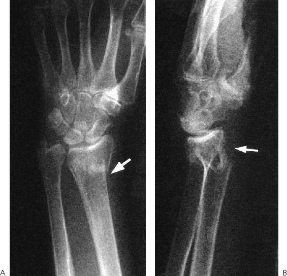
Figure 60–1. Anteroposterior (AP) (A) and lateral (B) radiographs of the patient’s wrist immediately after injury, demonstrating displacement, angulation, shortening, and comminution.
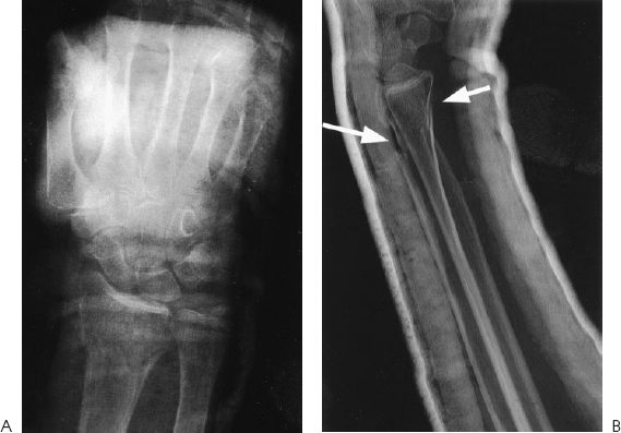
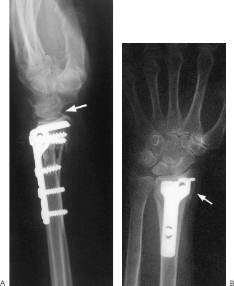
Figure 60–3. AP (A) and fossa lateral (B) radiographs of the patient’s wrist postoperatively, demonstrating restored alignment and appropriate length.
Diagnostic Studies
Prereduction radiographs (Fig. 60–1) show approximately 50% dorsal displacement, greater than 20 degrees of dorsal angulation, and volar, dorsal, and radial column comminution. Reduction improved the dorsal displacement and decreased the dorsal angulation to less than 20 degrees. However, volar and dorsal comminution is still evident, as demonstrated by the double cortical shadows on the anteroposterior (AP) and lateral postreduction view (Fig. 60–2). In addition, there is visible radial shortening. Radiographs should be repeated postoperatively to verify that alignment and length have been restored (Fig. 60–3).
Differential Diagnosis
Distal radius fracture
Extraarticular
Intraarticular
Radial styloid fracture
Fracture/Dislocation of the wrist
Related posts:

Stay updated, free articles. Join our Telegram channel

Full access? Get Clinical Tree


