32
Flexor Carpi Ulnaris Calcific Tendinitis
Richard W. Barth
History and Clinical Presentation
A 46-year-old woman was well until the morning prior to presentation, when she awoke with significant pain in her right wrist. Aspirin initially helped alleviate her symptoms. She awoke on the day of presentation complaining of severe pain, redness, and limited motion. She denied systemic complaints or history of trauma or injury. Her past medical history was unremarkable and she took no medications.
Physical Examination
The right wrist and forearm demonstrated 30 degrees of wrist flexion, 10 degrees of wrist extension, and significant swelling over the volar ulnar forearm. There was erythematous streaking extending from the wrist proximally into the forearm, but no regional lymphadenopathy. The flexor carpi ulnaris tendon was exquisitely tender to palpation just proximal to the pisiform. There was no sign of trauma and pulses were 2+ and symmetrical. The examination was otherwise unremarkable and the patient was afebrile.
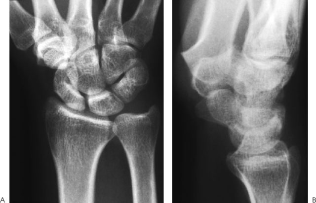
Figure 32–1. Routine anteroposterior (AP) and lateral views of the wrist are normal.
Diagnostic Studies
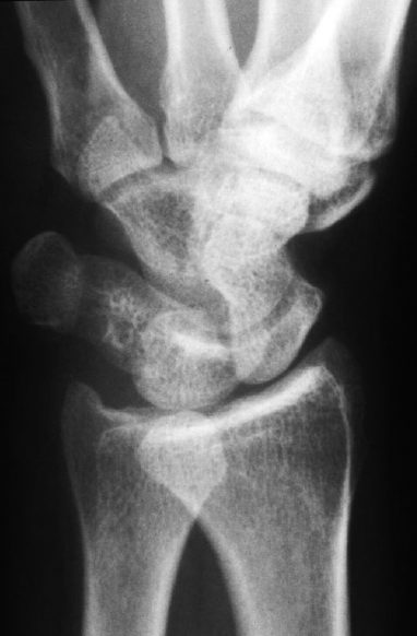
Routine anteroposterior and lateral radiographs of the right wrist were negative (Fig. 32–1). Oblique views demonstrated a small area of calcification just proximal to the pisiform (Fig. 32–2).
PEARLS
- Awareness of the condition is the key to diagnosis.
- Oblique radiographs may be necessary to demonstrate the calcific deposit.
- Treatment is almost always nonoperative.
PITFALLS
- Flexor carpi ulnaris calcific tendinitis can mimic infection.
Differential Diagnosis
Flexor carpiulnaris tendonitis
Septic arthritis
Septic tenosynovitis
Cellulitis
Inflammatory arthritis
Crystalline disease
Diagnosis
FCU Tendonitis
The diagnosis is made by the history, characteristic physical findings, and radiographic findings. The most important factor in making the diagnosis is familiarity with the condition. The history is notable for the acute onset of severe pain, limited wrist motion, and swelling, but no systemic complaints suggesting infection. There is no history of penetrating injury and usually no recent trauma. The physical findings include swelling, loss of motion, and exquisite tenderness of the flexor carpi ulnaris tendon, often just proximal to the pisiform. There may be erythematous streaking extending into the forearm. The physical findings may mimic a septic joint or cellulitis with ascending lymphangitis, but there is no lymphadenopathy or fever. The radiographic findings are often pathognomonic. Often, there is a large fluffy calcific deposit in the region of the flexor carpi ulnaris tendon just proximal to the pisiform. The calcification may be subtle and visualized only on oblique radiographs as in this case. White count, differential, and sedimentation rate are almost always normal.