Fig. 43.1
Posterior view of shoulder. The anterior and posterior branches of axillary nerve are seen
Posterior Approach
This approach provides good access to the lower three-fourths of the humerus. The main issue with this approach is that the radial nerve can be damaged easily.
Open reduction and internal fixation of fractures of the humerus.
Treatment of nonunion of fractures.
Biopsy and excision of tumors.
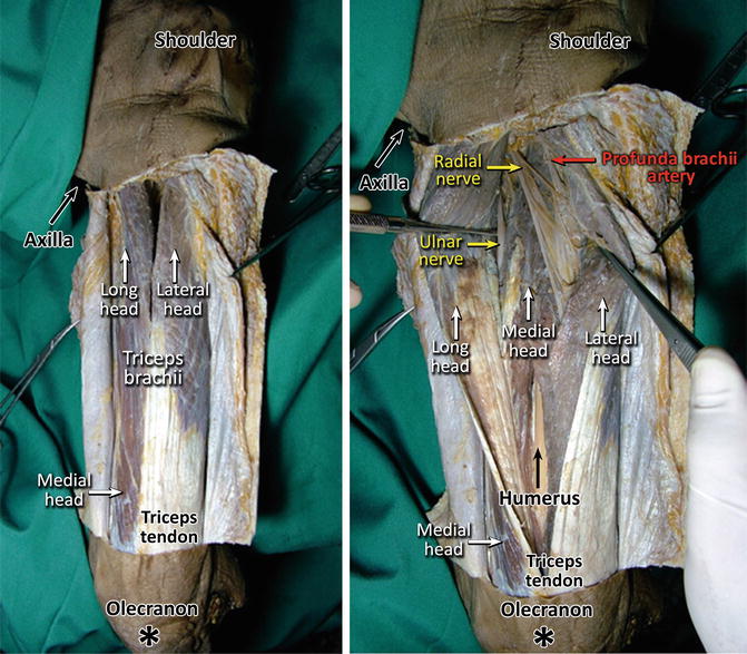
A longitudinal incision starts from 8 cm below the acromion to the olecranon fossa. The lateral and long heads of the triceps brachii must be separated to explore and protect the radial nerve, because it enters the posterior compartment of arm from close to the muscle heads’ origins and the nerve braches pass between this two head (Fig. 43.3) [13, 14].
A deep dissection should be avoided due to the danger of damaging the radial nerve and profunda brachii artery (Fig. 43.3).

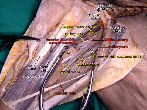
Fig. 43.2
Anterior view of upper arm. The branches of the brachial plexus are seen. CB coracobrachialis, LD latissimus dorsi
Lateral Approach
This approach uncovers the lateral epicondyle and the muscles that originated from that area.
Open reduction and internal fixation of fractures of the lateral condyle.
Surgical treatment of tennis elbow.
A curved or straight incision is done on the lateral aspect of the elbow over the lateral supracondylar ridge.
Here, the radial nerve pierces through the lateral intermuscular septum of the arm. This has to be cautiously handled. While approaching the arm, neurovascular structures do not run down a definite path; therefore, they are considered separately. Thus, muscles have to be considered into two important compartments as flexor and extensor muscles.
Anterior compartment consists of the coracobrachialis, biceps brachii, and brachialis muscles, and innervation is done by the musculocutaneous nerve. Posterior compartment consists of triceps brachii. These two compartments are separated by lateral and medial intermuscular septums.
Innervation of brachial muscle varies. The lateral part is innervated by radial nerve, whereas the medial part is innervated by musculocutaneous. Therefore, while separating the muscle, some parts may be denervated.
Similarly, the long head of triceps brachii is innervated by the superior branches of the radial nerve, whereas the lateral head is innervated by the inferior branches. The medial head is innervated both by the radial nerve and the ulnar nerve.
Coracobrachialis may sometimes have an extra head that may be attached to ligaments and other structures. These ligaments may attach themselves to the medial supracondylar ridge and medial epicondyle. As a result, entrapment neuropathy can be observed in the median nerve, which is similar to the symptoms seen in carpal tunnel syndrome.
Forearm Approaches
Anterior Approach
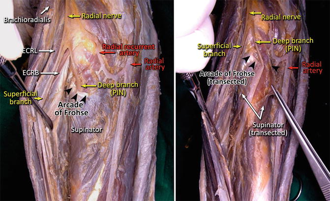
Radius is clearly visible in this approach. The most important aspect is the posterior interosseous nerve (PIN). Damage is possible to this nerve as it is located under the supinator (Fig. 43.4) [15–18].
Open reduction and internal fixation.
Bone graft and nonunion fracture treatment.
Radial osteotomy.
Bone tumor treatment and biopsy.
Osteomyelitis excision.
This approach is used for exposing radial tuberosity. Keeping biceps brachii on the lateral side, the incision can be extended until the radial styloid process. During this incision, the superficial branch of radial nerve and PIN is important (Fig. 43.4). Additionally, radial artery is located below the brachioradialis.

Fig. 43.3
Posterior view of arm. The radial nerve is visualized entering between the long and the lateral heads of triceps

Fig. 43.4
Anterolateral view of forearm. Brachioradialis, extensor carpi radialis longus (ECRL), and extensor carpi radialis brevis (ECRB) were retracted laterally. The branches of the radial nerve are seen. PIN posterior interosseous nerve
Posterior Approach
The main reason for preferring this approach is the ease by which PIN can be retracted.< div class='tao-gold-member'>Only gold members can continue reading. Log In or Register to continue
Stay updated, free articles. Join our Telegram channel

Full access? Get Clinical Tree


