Fig. 6.1
Muscle architecture
PCSA of a muscle is calculated using the formula
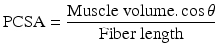 Muscles with small pennation angle and long muscle fiber size (e.g. hamstrings and tibialis anterior) provide large excursion and higher contractile velocity. However muscles with greater pennation angle, small muscle fiber length and large muscle physiological cross-sectional area (e.g. soleus) produce higher force at the expense of excursion and contractile velocity [13].
An alpha motor neuron in the anterior horn of the spinal cord and the muscle fibers innervated by the axon of this neuron form a motor unit. The motor unit is the smallest subunit that is under neural control in a muscle. The number of the muscle fibers in a motor unit is related with the function of that muscle. Muscles contributing fine motor movement have less muscle fibers in a motor unit than the muscles exerting crude movement. The alpha motor neuron axon gives branches to synapses with the muscle fibers of a motor unit. When an alpha motor neuron is evoked, the electrical signals pass to the muscle fibers via neuromuscular junctions. These electrical signals may generate an action potential in the membrane of the muscle fibers. The sum of the action potentials of the muscle fibers forms a motor unit action potential (MUAP). The result is a mechanical twitch of tension. Tension increase in a muscle occurs via consecutive mechanisms. First, stimulation rate increases in the related motor unite, and then the other motor units are activated (recruitment). Reverse sequences cause decreases of tension in the muscle. While performing movements the human body tries to spend little energy as possible to conserve energy expended. Entire motor units in a muscle are not necessarily activated during performing a movement. Needed tension force for a movement determines the amount of motor unit recruitment via neuromotor control mechanism. When tension force requirement increases, a greater number of motor units will be activated. First, the smallest motor units are recruited, and then large motor unites are recruited. This is called as “size principle” [2].
Motor units are divided into two main types. The alpha motor neuron innervating the muscle fibers determines the type of motor unit. Smaller slow-twitch motor units (type 1) are called tonic units. They produce twitches with lower peak tension in a long time. The large fast-twitch motor units (type 2) are called phasic units. They have larger peak stress with a short time to peak. Muscles performing strong and phasic contractions have type 1 and type 2 muscle fibers equally. The gastrocnemius is an example to this type of muscles. During the terminal stance phase of the gait, gastrocnemius muscle contracts strongly in a phasic manner. Type 1 fibers especially exist in the tonic postural muscles. An example of such muscles is soleus. It creates tonic contraction during the second rocker of the gait [13].
As noted above, muscles contain contractile active cells (muscle fibers) and passive elements of connective tissue. The force-length curve of a muscle consists of a combination of the force-length characteristics of both active and passive structures in the muscle (Fig. 6.2). Interactions between myofibrillar structures at sarcomere level determine the shape of the force-length curve. Resting sarcomere length is 2.5 μm. At this situation, maximum number of cross-bridges occurs between the filaments; hence, maximum tension is generated by the active elements. As the muscle undergoes lengthening or shortening, cross-bridges between the filaments are reduced, and the tension produced by muscle fibers decreases. Connective tissue of the muscle whether in parallel or series has significant effects on the tension generated. Connective tissue that surrounds the contractile elements is called as parallel elastic component and behaves like an elastic band. It is in a slack state when the muscles are at rest or in a shorter position. In this situation parallel elastic component does not constitute to total tension. As the muscle length increases, elastic component causes increases in the tension. This increase occurs first slowly then quickly. All connective tissues, including tendon, in series with the contractile elements, are called the series elastic component. During the isometric contraction the series elastic component is under tension and undergoes limited stretching. Total length of the muscle and tendinous structure does not change as the muscle shortens during this kind of contraction. Shortening of the muscle fibers stretches the serial elastic component of the muscle. This is called internal shortening. When the muscle isometrically contracts, stretched series elastic component stores large amounts of energy. As an example of this, the gastrocnemius muscle contracts and stores energy in the terminal stance phase of the gait to generate propulsion power during push off. Series elastic component stores large amounts of energy at the terminal stance phase during contraction of the gastrocnemius muscle. This contraction causes only little plantar flexion movement at the ankle, because the body weight is still supported by the ipsilateral lower extremity and there is considerable amount of dorsiflexor moment generated by ground reaction force. In the preswing phase of the gait, considerable part of the body weight is transmitted to the contralateral limb. Hence, the dorsiflexor moment of ground reaction forces around the ankle is reduced significantly. Then, energy stored in the series elastic component causes rapid plantar flexion at the ankle joint and propulsion is provided. Muscle length is shortened during concentric contraction. Besides, intramuscular tension also decreases. There is an inverse relationship between muscle shortening velocity and tension [13].
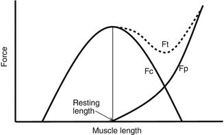
Fig. 6.2
Force-length characteristics of a muscle
Gait Cycle
Lower extremities move in a specific order during gait. A gait cycle (stride) begins with the ground contact of the foot and ends when the same foot touches the ground. According to the foot contact with the ground, a gait cycle is divided into periods and phases. There are two periods (stance and swing) in a gait cycle (Fig. 6.3).
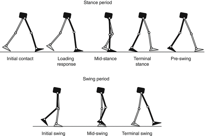
Fig. 6.3
Gait cycle
Stance period constitutes about 60 % of a gait cycle. In this period the foot is in contact with ground. In the first and last 10 % percentage of stance period, the feet are in contact with the ground. These phases are called as double-limb stance. During these phases body weight is transferred between two lower extremities. During the middle of the stance period (40 % percentage of gait cycle), one foot has a contact with the ground. This phase is defined as single-limb support. Meanwhile contralateral foot is in the swing period. Ratio between double-limb stance and single-limb support depends on the walking speed. Ratio decreases proportionally with the increase of walking speed. The last 40 % percentage of a gait cycle is called as swing period. In this phase the foot does not make any contact with the ground and exerts a pendular movement. Contralateral foot is in single-limb support phase. Furthermore the stance and swing period is also divided into several phases (Fig. 6.3). In order the phases of stance period are initial contact, loading response, mid-stance, terminal stance and preswing. The phases of swing period are initial swing, mid-swing and terminal swing. In a normal walking, stance period begins with heel strike and ends with toe off. The swing period begins with the toe off ends with the heel strike. However in neuromuscular pathology initial ground contact of the foot may be with forefoot rather than the heel. For that reason, “heel strike” terminology has not been using to define the first contact of the foot with the ground.
A gait cycle can also be evaluated in three different tasks. These are weight acceptance, single-limb support and limb advancement. Weight acceptance includes the first two phases of stance period – initial contact and loading response. Single-limb support includes the mid-stance and terminal stance phases. Advancement includes the last phase of stance (preswing) and whole phases of swing period [10].
Centre of gravity (COG) of the body moving horizontally during walking also changes its position in vertical (approximately 3.2 ± 0.8 cm) and lateral (3.5 ± 0.9 cm) directions. COG reaches its maximum height in the mid-stance phase. Then it falls through the initial contact phase. Meanwhile, the potential energy of the body endowed by the height from the ground transforms into kinetic energy. Impact at the initial contact must be absorbed to protect musculoskeletal structure from injury. Heel fat pad and tibialis anterior and quadriceps eccentric contractions absorb this impact during initial contact and loading response, respectively. This is defined as weight acceptance. After initial contact COG again begins to rise to its peak height at the mid-stance phase. Meanwhile, kinetic energy is stored as potential energy. Energy required for horizontal and vertical movement of COG is produced by strong contraction of ankle plantarflexor muscles in the terminal stance phase. During single-limb support, hip abductors of the lower limb contract to protect the pelvis from falling to the other side. In limb advancement, the lower extremity acts as a compound pendulum and moves forward due to the repulsive effect of the gastrocnemius muscle contraction. The inertial force has an important role in this progress [5, 11].
Four rockers occurring during the stance period of the gait yield the body’s forward progress. During rockers movements are performed around the points called fulcrum [11]. The first rocker appears during initial contact and loading response. In this rocker the foot moves to the ground, and the tibia goes forward. The fulcrum is the heel. Ground reaction force (GRF) acts through the heel and passes behind the ankle. It generates a plantar flexion moment at the ankle. If the movement was made only under the effect of this force, the forefoot would hit the ground strongly. To prevent this, the ankle dorsiflexor muscles contract to yield smooth and controlled dropping of the foot. While the foot moves to the ground, the contracting tibialis anterior muscle pulls the tibia forward. As mentioned earlier, during terminal swing the foot falls to contact with the ground in the beginning of the stance phase. The body has momentum due to its mass and velocity. Some part of the force generated during the collision is absorbed by the fat pad of the heel. The other part is absorbed by foot dorsiflexors contracting eccentrically. The purpose of the first rocker is shock absorption. The second rocker occurs in mid-stance phase. The fulcrum is the ankle. The foot has a full contact with the ground, and the tibia moves forward around the ankle joint. Motion of the tibia causes dorsiflexion of the ankle. GRF vector passes in front of the ankle joint resulting in external dorsiflexor moment. Meanwhile, the soleus muscle contracts eccentrically to control forward falling of the body. Thus excessive forward movement of the tibia is prevented, and the GRF vector is held in front of the knee. In the second half of the mid-stance, the GRF vector passes behind the hip joint and contributes the stability of the hip. The third rocker occurs in the terminal stance phase. At the end of the mid-stance, plantarflexors contract concentrically and stop the forward movement of the tibia and foot dorsiflexion. Metatarsal heads are the fulcrum in this rocker. The foot goes to plantar flexion, and the heel moves upward from the ground. In the first two rockers, muscles contract eccentrically to cause deceleration, but in the third rocker strong concentric contraction of the plantarflexors is seen to accelerate the lower limb. Power required for forward progression of the body is provided by this strong concentric contraction of the ankle plantarflexors in the terminal stance. The fourth rocker is formed in preswing phase. The fulcrum is the most anterior margin of the medial forefoot and great toe.
The purpose of the swing period is to advance the lower extremities, provide foot clearance, allow the cadence change and ensure energy conservation. The lower extremity moves as a compound pendulum during the swing period. At the first part of pendular movement, muscles contract for acceleration, but at the second part the muscle contracts for deceleration [5].
< div class='tao-gold-member'>
Only gold members can continue reading. Log In or Register to continue
Related posts:

Stay updated, free articles. Join our Telegram channel

Full access? Get Clinical Tree


