Unidirectional Anterior Instability
Russell F. Warren
William D. Prickett
INTRODUCTION AND HISTORICAL REVIEW
The act of throwing places unique demands on the stabilizing structures of the glenohumeral joint, and over 50% of college or professional baseball pitchers report shoulder pain (1). The overhead athlete must maintain precise control of the ball while generating significant stresses upon the bony and soft tissue restraints of the shoulder. These athletes may display a spectrum of instability ranging from asymptomatic translation to symptomatic subluxation. This difference is often subtle, especially considering the relative increased joint laxity present in throwers compared with nonthrowers (2). Instability is defined as a pathologic increase in glenohumeral translation that results in symptoms (3).
Anatomy and Biomechanics
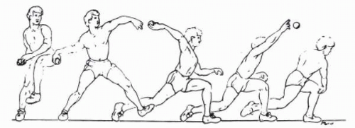
The act of throwing involves a transfer of kinetic energy from the lower extremities and trunk to the upper extremity. The sequence of events in overhand throwing has been well documented and consists of six phases (Fig. 12-1): windup, stride, arm cocking, arm acceleration, arm deceleration, and follow-through (4, 5, 6). Throughout the process, the arm is maintained in approximately 100 degrees of abduction with external rotation reaching 175 degrees in the cocking phase and 105 degrees of internal rotation in the follow-through phase (7,8). Dillman and colleagues (4) estimated the speed of ball acceleration to be 7,000 degrees per second, with a torque estimate of 52 Nm in baseball pitchers. Additional analysis has shown that the humeral head experiences an anterior translation force of 40% body weight during the cocking phase and a distraction force equivalent to 80% body weight during follow-through. Given the supraphysiologic loads placed on the surrounding soft tissues of the shoulder, subtle increases in translation may result in marked symptoms in overhead athletes.
Shoulder stability is a function of static and dynamic restraints. The static restraints are primarily the capsuloligamentous structures, with mild contributions from articular congruity and negative intraarticular pressure. The dynamic restraints consist primarily of the periarticular musculature. The anterior capsuloligamentous complex, as described by Flood in 1829 (9), consists of the anterior shoulder capsule with its relatively constant capsular thickenings that comprise the superior, middle, and inferior glenohumeral ligaments. The coracohumeral ligament (CHL), another thickening of the anterior capsule, originates from the base of the coracoid and inserts into the greater and lesser tuberosities (10). The superior glenohumeral ligament (SGHL) originates anterior to the long head of the biceps at the supraglenoid tubercle and inserts slightly anterior to the biceps tendon into the lesser tuberosity (10). The middle glenohumeral ligament (MGHL) originates from the anterior labrum between the 1-and 3-o’clock positions and inserts into the lesser tuberosity with the subscapularis (11). As described by DePalma and co-workers (12), the MGHL has the greatest variation in size of all the ligaments of the shoulder and has two morphologic variations: (a) sheetlike, which is confluent with the inferior glenohumeral labral (IGHL) complex, or (b) cordlike with a sublabral foramen. The IGHL complex consists of an anterior band, posterior band, and an intervening pouch. The anterior band originates from the anteroinferior glenoid labrum and periosteum and inserts into the anteroinferior humeral anatomic neck. The posterior band originates from the posteroinferior glenoid and inserts into the posteroinferior anatomic neck of the humerus. The intervening pouch connects the anterior and posterior bands of the IGHL while cradling the humeral head inferiorly (13).
The individual ligaments of the shoulder become taut at varying degrees of glenohumeral motion. The anterior band of the IGHL complex dampens anterior and inferior humeral translation with increasing amounts of humeral abduction (14,15). At lower levels of abduction, the
MGHL limits anterior humeral translation and when the arm is at the side, the SGHL and CHL resist inferior humeral translation (14,15). The function of the biceps tendon with respect to shoulder stability is controversial. It has been reported to depress the humeral head and decrease the load sustained in the IGHL complex, (16,17) while others have noted essentially no functional role in dynamic stability and only a questionable role as a static restraint to excessive glenohumeral motion (18).
MGHL limits anterior humeral translation and when the arm is at the side, the SGHL and CHL resist inferior humeral translation (14,15). The function of the biceps tendon with respect to shoulder stability is controversial. It has been reported to depress the humeral head and decrease the load sustained in the IGHL complex, (16,17) while others have noted essentially no functional role in dynamic stability and only a questionable role as a static restraint to excessive glenohumeral motion (18).
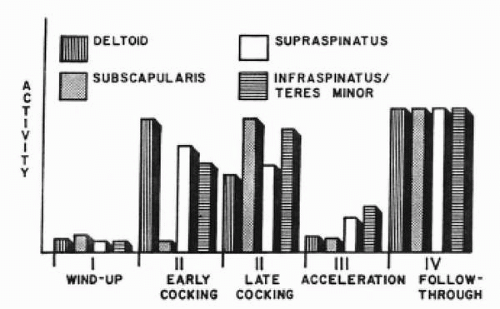
EMG (electromyography) analysis has helped provide insight into the dynamic constraints and firing patterns of the shoulder musculature during the overhead throw (19) (Fig. 12-2). The pitching motion is a complex interplay of several muscles, which combine in precise firing patterns to provide a fluid and forceful overhand throw. The rotator cuff functions to maintain stability and initiate motion, while the other muscles generate force during acceleration and dampen distraction during follow-through (5,20, 21, 22, 23, 24).
ETIOLOGY OF INJURY
In the overhead athlete, mild incompetence of the anterior restraints (secondary to repetitive microtrauma or fatigue and dyssynchrony of the dynamic stabilizers) may allow subtle amounts of increased humeral head translation with respect to the glenoid. Normally, with the arm abducted 90 degrees as it is externally rotated, the humeral head translates posteriorly. This coupled posterior translation occurring with external rotation at 90 degrees of abduction may be decreased or anterior translation may occur in the setting of anterior laxity. These altered mechanics may strain the posterosuperior rotator cuff to an increasing degree with resultant rotator cuff tears. This concept has been described by Walch and colleagues (25) and by Jobe and co-workers (26) and has been coined as “internal impingement.” The increased contact of the posterior supraspinatus and infraspinatus on the posterosuperior glenoid in the late cocking/early acceleration phase may cause pain and weakness that further accentuate the athlete’s underlying anterior microinstability (27).
Shoulder instability in the thrower rarely reaches proportions of frank dislocation unless the athlete experiences an isolated high external load such as a quarterback sack or sliding headfirst into a base. When macroscopic instability occurs, detachment of the IGHL complex from the anterior glenoid is the most common defect noted (28). This injury has been referred to as the Bankart lesion (29), although it has been proven to be more than one defect and it exists as a spectrum of lesions that render the IGHL complex incompetent. These defects may occur along the entire course of the anterior IGHL complex, ranging from anterior labroligaruentous periosteal sleeve avulsions (ALPSA) to humeral avulsion of the glenohumeral ligament (HAGL). Bigliani and co-workers (30) loaded the IGHL complex to failure and noted defects at the glenoid insertion (40%), midsubstance (35%), and humeral origin (25%). Importantly, when the complex failed at one location, it underwent significant plastic deformation in other regions. This was clinically evident in Rowe’s series (31,32), which showed greater than 50% of patients with instability had abnormal capsular redundancy. Additional studies by Turkel and colleagues (33) and Warren (34) have emphasized the importance of a circumferential injury pattern in the shoulder with macroscopic instability; thus, not only does an anterior dislocation occur with an anterior injury (Bankart lesion) but also some form of posterior injury often exists. The posterior injury may take the form of a capsuloligamentous disruption or a bony injury (posterosuperior humeral impaction fracture).
In addition to the attenuation of the anterior capsulolabral tissue and the articular sided partial thickness rotator cuff tears of the infraspinatus, other structures may also be affected by the increased humeral translation with respect to the glenoid. Abnormal superior elevation of the humeral head may allow for subacromial impingement, and excess distraction during follow-through may place increased tensile stress on the rotator cuff or the long head of the biceps and its origin from the superior glenolabral complex.
PRESENTATION AND PHYSICAL EXAMINATION
The symptoms of shoulder instability in the throwing athlete are usually more subtle than those of the nonthrower. Throwers often present with symptoms of pain, decreased ball velocity, decreased control, occasionally “dead arm” syndrome, and, rarely, actual episodes of subluxation or dislocation. It is unlikely that an elite thrower would present with recurrent gross instability because the athlete would be unable to function at or even near their maximal throwing effort and would thus gravitate to nonthrowing positions.
The location of the athlete’s pain and the phase of throwing are important clues as to the etiology of the thrower’s symptoms. Internal impingement with anterior capsulolabral stretch typically results in pain at the posterior joint line during the cocking phase and early acceleration phase when the humerus experiences maximal external rotation (25,35). Throwers who note pain during follow-through often have posterior capsulolabral injury because the internal rotation of follow-through stresses the posteroinferior capsulolabral complex (3).
The bench examination begins with observation for soft tissue symmetry and for any evidence of muscular atrophy or previous incisions. Neck range of motion, strength testing, and provocative maneuvers such as Spurling’s test (36) may elicit symptoms of a nerve root compression that may cause pain and affect the dynamic restraints of the glenohumeral joint. Additional pain generators such as the long head of the biceps and superior labrum are tested with provocative maneuvers such as Speed’s, Yergason’s, and “clunk” (37) tests, the anterior slide test of Kibler (38), and the active compression test of O’Brien (39). The acromioclavicular joint is evaluated by direct palpation and cross body adduction. Subacromial pathology may be evaluated with Hawkins (40) and Neer’s (41) tests, and periscapular muscular strength is evaluated by manual muscle force testing.
When evaluating shoulder stability, examination and comparison of the asymptomatic shoulder to the symptomatic shoulder is important. Athletes with anterior microinstability may show increased external rotation and limited internal rotation with the arm abducted 90 degrees secondary to contracture of the posterior rotator cuff or posterior capsule, or due to developmental changes in glenohumeral geometry (42). Anecdotally, it has been suggested that a substantial decrease in internal rotation of the dominant shoulder may predispose the athlete to shoulder pain (3).
Active and passive motion are helpful in assessing the overall function of the shoulder; however, they may be limited
by patient apprehension as a result of instability. Strength testing of the periarticular musculature is then carried out with an emphasis on the rotator cuff.
by patient apprehension as a result of instability. Strength testing of the periarticular musculature is then carried out with an emphasis on the rotator cuff.
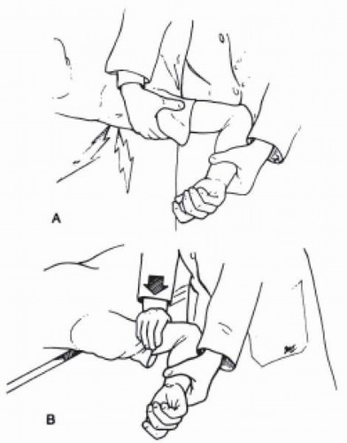
Provocative testing may help localize the athlete’s pain. With the arm adducted in the neutral position, inferior stress is applied to the humerus and the acromiohumeral “sulcus” is evaluated. Some authors recommend performing this test in neutral (evaluate inferior capsular and rotator interval laxity) and in external rotation (tensions the rotator interval). Thus (a decrease in the sulcu as) one goes from neutral to external rotation implies inferior capsular laxity. By contrast, if the sulcus stays constant, this suggests excessive laxity in the rotator interval. The sulcus may be graded as 0 to 3+ (grade 0 = no increased translation, grade 1+ = mild increased translation of less than 1 cm, grade 2+ = 1 to 2 cm of inferior translation, and grade 3+ = greater than 2 cm of inferior translation (8). The load and shift test centers the humeral head on the glenoid, then anterior and posterior translational stresses are applied to the humerus. The amount of humeral translation during the load and shift is also graded as 0 to 3+ (grade 0 = no increased translation, grade 1+ = mild increased translation to the glenoid rim, grade 2+ = distinct subluxation that reduces passively, and grade 3+ = complete dislocation that requires manual reduction) (8). This test may also elicit mechanical symptoms related to labral or articular defects or apprehension as the athlete anticipates glenohumeral subluxation. The examiner should expect a normal thrower’s shoulder to display some increased laxity with true instability existing when a 2+ load and shift test is present (3). The quality of the endpoint in the direction tested is also valuable, in that a solid endpoint essentially excludes macroscopic instability. The apprehension and relocation maneuvers are also valuable in evaluating anterior shoulder instability (Fig. 12-3). The apprehension test is performed by placing the humerus in a position of maximal external rotation at 90 degrees of humeral abduction and assessing the athlete’s response to feelings of subluxation or dislocation (43). In this apprehensive position, a posteriorly directed load is applied to the shoulder and an assessment of the symptomatic relief (sense of dislocation) is recorded (43). Positive apprehension and relocation tests are markers for anterior instability. However, if in the position of apprehension the athlete notes posterior shoulder pain that is relieved with the relocation maneuver, then the athlete’s symptoms may be due to internal impingement with rotator cuff injury (25). Before the completion of the physical examination, signs of generalized ligamentous laxity are evaluated that may provide clues as to a multidirectional component to the thrower’s instability. Following the physical examination, in the setting of a complicated clinical scenario, the physician may augment the clinical evaluation with the use of differential injection techniques. Lastly, upon completion of the bench examination, the physician observes and interacts with the athlete while the athlete is throwing. This allows evaluation of the mechanics of the throw and confirms the location of pain during the throwing cycle.
RADIOGRAPHIC AND DIAGNOSTIC EVALUATION
Plain radiographs are an essential supplement to the history and physical examination of the patient with suspected anterior instability. They demonstrate bony integrity, glenohumeral alignment, arthritic changes, loose bodies, and periarticular calcification, and the potential exists to visualize signs of prior subluxations or dislocations. Our standard series includes a true anteroposterior (AP) view in neutral rotation, West Point, outlet, and Stryker notch views. The AP in neutral rotation allows the evaluation of the glenohumeral joint space and large Hill-Sachs lesions may also be evident. The West Point view is especially useful to evaluate
anterior or posterior subluxation or the presence of a bony Bankart lesion. The outlet view confirms humeral location with respect to the glenoid and defines the supraspinatus outlet. The Stryker notch view is used to evaluate the contour of the posterosuperior aspect of the humeral head and evaluate for the presence of a Bennett’s lesion due to injury adjacent to the posteroinferior glenoid rim.
anterior or posterior subluxation or the presence of a bony Bankart lesion. The outlet view confirms humeral location with respect to the glenoid and defines the supraspinatus outlet. The Stryker notch view is used to evaluate the contour of the posterosuperior aspect of the humeral head and evaluate for the presence of a Bennett’s lesion due to injury adjacent to the posteroinferior glenoid rim.
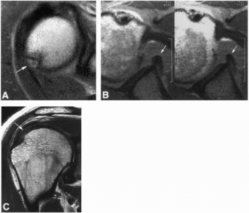
 FIGURE 12-4. Axial fast spin-echo sequence in a patient following an acute dislocation demonstrates a defect at the posterolateral aspect of the humeral head (arrow) (A) and a displaced anterior labral fragment (arrows) (B) on two consecutive images. C: Oblique coronal fast spin-echo sequence demonstrates abnormal signal on the articular side of the rotator cuff (arrow). (See corresponding arthroscopic image Fig. 12-8.) |
Magnetic resonance imaging (MRI) is an invaluable tool in the assessment of the throwing athlete. Using high-quality imaging equipment and software, bony and soft tissue injuries about the throwing shoulder can be evaluated with greater than 90% accuracy (44, 45, 46). Precise depiction of the injury pattern using MRI helps with patient education and preoperative planning. Patients may benefit from a more accurate understanding of their injury, prognosis, and, when appropriate, their postoperative expectations. In the throwing athlete with anterior instability, particular attention is directed to the anterior capsulolabral structures, the posterior rotator cuff, and the posterosuperior humeral head (Fig. 12-4).
Stay updated, free articles. Join our Telegram channel

Full access? Get Clinical Tree


