22
Ulnar Nerve-Tendon Transfer
Mark S. Cohen
History and Clinical Presentation
A 57-year-old man presents for evaluation of his dominant hand. He complains of a deformity involving his small and ring fingers with loss of function, especially fine motor skills. He relates his problem to a laceration he suffered at the wrist level several years prior. He also reports numbness in his small and ring fingers. He denies any radicular symptoms or problems with his contralateral hand.
PEARLS
- Ulnar nerve dysfunction leads to a claw deformity with asynchronous motion, weakened grip, and loss of lateral finger mobility and sensation.
- Various tendon transfers can restore clawing and asynchronous digital flexion. To improve weakness of grasp, the transfer must utilize a wrist muscletendon unit. Using a digital motor simply redistributes balance within the hand.
- Transfer selection is based on patient age, requirements, tendon availability, and compliance.
PITFALLS
- Claw deformity can result from a variety of conditions including lesions of the ulnar nerve, lower cervical roots, inferior brachial plexus, and more generalized neurologic conditions (e.g., motor neuron disease).
- The diagnosis requires a careful history, physical examination, and often electrodiagnostic studies.
- Contraindications to tendon transfers include an uncooperative patient, a lack of expendable donor tendons, and stiff digits without full passive mobility.
Physical Examination
Obvious atrophy is visible in the hand involving the intrinsic musculature. The small and ring fingers are positioned in a claw deformity, although passive interphalangeal joint extension is present and the joints are supple. The patient has difficulty abducting and adducting his digits. His flexor digitorum profundus function to the small and ring fingers is intact (he can actively flex his small and ring distal interphalangeal joints). Sensory examination reveals diminished two-point discrimination in the small and ulnar half of the ring fingers.
Diagnostic Studies
Roentgenograms were obtained of the hand including the wrist. They were within normal limits. Electrical studies were obtained and revealed a complete ulnar nerve lesion at the wrist level with denervation present in all ulnar nerve innervated intrinsic muscles tested (interossei and hypothenar muscles). No reinnervation potentials were present. The thenar muscles were electrically normal and there was no evidence of a peripheral neuropathy or radiculopathy.
Differential Diagnosis
Brachial plexus injury
Upper plexus
Lower plexus
Cervical root compression
Peripheral nerve dysfunction
Charcot-Marie-Tooth disease
Ulna nerve tunnel compression
Cubital tunnel (source)
Pancoast tumor
Tendon lacerations
Laceration of the ulna nerve at the wrist
The differential diagnosis of a claw deformity of the hand with loss of sensation in the ulnar nerve distribution is somewhat limited. One assumes a lesion of the ulnar nerve or possibly the lower cervical nerve roots (C8–T1) or inferior brachial plexus. Cervical nerve root compression typically results in neck pain with radicular symptoms down the arm. Weakness and atrophy would be expected in the thenar and hypothenar musculature, which are both innervated by the lower cervical and first thoracic nerve roots. A lesion of the lower brachial plexus (e.g., a Pancoast tumor of the lung apex) would result in similar findings. The ulnar nerve is most commonly compressed at the elbow (cubital tunnel syndrome) or at the wrist (ulnar tunnel syndrome). Intact function of the two ulnar flexor digitorum profundus tendons would point to a more distal lesion in the lower forearm or wrist.
Diagnosis
The diagnosis is a low complete ulnar nerve palsy. The ulnar nerve innervates the four dorsal interossei, the three volar interossei, the two ulnar lumbricals, the hypothenar muscles and typically the deep head of the flexor pollicis brevis. It supplies sensation to the small finger and ulnar half of the ring finger.
A claw deformity develops due to imbalance between absent intrinsic (interossei and lumbricals) and intact extrinsic muscle function. The claw deformity is more pronounced in the ring and small fingers due to the intact index and middle finger lumbricals, which are innervated by the median nerve. Clawing is more significant in a low ulnar nerve palsy than a lesion at the elbow due to the intact flexor digitorum profundus tendons to the ring and small fingers. These tend to accentuate the flexion posture of the interphalangeal joints.
Loss of ulnar nerve function can be quite disabling. The intrinsic muscles are responsible for simultaneous flexion of the metacarpophalangeal (MP) joints and extension of the interphalangeal (IP) joints (the intrinsic-plus position). Although full finger flexion and extension is present, with intrinsic loss the fingers tend to roll up during flexion due to asynchronous motion of the MP and IP joints (MP flexion does not begin until IP flexion has been completed). IP joint extension requires contraction of the extrinsic extensor tendons (extensor digitorum communis). This leads to MP joint hyperextension and contributes to the claw posture.
The ability to place the hand around objects such as a glass or doorknob is lost. These activities requires the intrinsic-plus posture. All fine motor skills that require simultaneous MP flexion and IP extension, such as writing or threading a needle, are similarly impaired. Lastly, pinch and grip strength are markedly diminished due to loss of the interossei and hypothenar muscles.
Nonsurgical Management
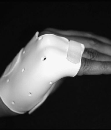
Conservative treatment of ulnar nerve dysfunction relies on the use of external splints to block clawing of the digits. A lumbrical bar splint is commonly utilized for this purpose (Fig. 22–1). It is a static splint, usually made of Orthoplast by a hand therapist, that fits over the dorsum of the metacarpal head and proximal phalanges. It does not improve grip strength or integrated MP and IP motion. It does, however, block MP hyperextension of the ring and small fingers, thereby correcting the claw deformity. This enables the extrinsic extensor tendons to fully extend the IP joints. It also prevents fixed proximal IP joint contractures from occurring.