Traumatic Brain Injury


Traumatic Brain Injury


John Whyte

Jennie Ponsford

Thomas Watanabe

Tessa Hart


NATURE OF TRAUMATIC BRAIN INJURY

Traumatic brain injury (TBI) is one of the major causes of disability worldwide. The systematic study of the residual effects of TBI can be traced to World War II and the work of Alexander Luria, Kurt Goldstein, and others (1, 2). In this early work, much was learned about the deficits following penetrating injuries to the brain in soldiers with gunshot wounds. The pattern of residual dysfunction often corresponded to a focal lesion caused by the bullet passing through the brain. These focal deficits were similar to those observed in strokes.

Most TBIs are classified as closed head injuries, wherein the skull is not actually penetrated. The nature of injury sustained in vehicular accidents and falls (e.g., blunt impact, acceleration-deceleration) often results in multifocal lesions and diffuse brain damage with a variety of physical, cognitive, and neurobehavioral impairments that are unique to each person and pose formidable obstacles to community integration. Modern international conflicts involving the use of improvised explosive devices have led to a new injury mechanism—blast injury—the biophysics and clinical outcomes of which are not yet fully understood (3).

TBI affects all age groups. However, complex issues are posed by children with TBI because the injury interacts with the processes of biological, psychological, and social development. The special problems of children with TBI are not treated in detail here, but there are excellent sources for additional information on this topic (4, 5). At the other end of the age spectrum, one finds the older adult with brain injury, whose recovery tends to be slower than the younger population and may be complicated by preexisting comorbidities and the reduced plasticity of the aged brain (see also Englander and Cifu, 1999 for a review of this topic (6)). Given the increasing percentage of older persons within the general population, it is likely that a growing number of survivors of brain injury will be in this subgroup.


The Range of Outcomes: Death to Complete Recovery

Injury type and severity vary widely. In some injuries, commonly referred to as concussions or mild traumatic brain injuries, the person may suffer only a period of confusion or a brief episode of unconsciousness. Most of those with mild TBIs return to normal activity within days. In a minority of cases, somatic, cognitive, and affective symptoms may persist for weeks, months, or longer. Mild brain injuries constitute the vast majority of TBIs. At the other extreme, death from severe brain injury is estimated at 20.7/100,000 population, which represents a 22% decline from 1979 (7). Most rehabilitation efforts are focused on survivors of moderate and severe TBI, among whom 80 to 90,000 survive each year with disability. Nearly 2% of the U.S. population requires ongoing assistance with activities of daily living (ADL) as a result of a TBI (8, 9).


Measures of Injury Severity

Both depth and duration of coma have been considered to be indices of the severity of TBI. Clinical assessment of coma was made more precise and objective with the advent of the Glasgow Coma Scale (GCS) in the 1970s (10), a quantitative measure of the depth of unconsciousness (Table 24-1). Coma is defined as not opening the eyes, not obeying commands, and not uttering understandable words. A GCS score of 8 or less in the acute period is operationally defined as a comatose state (11). Ironically, however, the use of the GCS as a marker of injury severity has become more problematic with changes in emergency management, in that increasing numbers of patients are intubated and subjected to chemical paralysis in the field, making it impossible to record an accurate GCS score. The Full Outline of Unresponsiveness (FOUR) is a recently developed measure of depth of unconsciousness that adds assessment of brainstem reflexes, and does not require verbal responding, thus avoiding the confound of intubation. The FOUR is of comparable reliability to the GCS (12), but it does not avoid the problem of chemical paralysis and is too new to have accumulated a large volume of prognostic data.

Duration of coma is often defined as the time until the patient resumes following commands. Both duration of coma and initial GCS score have been reported to predict neurobehavioral outcome from TBI (13, 14), varying with the outcome measures selected and the time since injury. Stein and Spettell (15) showed that when the GCS was modified slightly to include complications such as intracranial lesions, its predictive power was improved.

The duration of posttraumatic amnesia (PTA) has been used as a measure of injury severity as long ago as the 1930s (16). During PTA, patients are out of coma but remain disoriented and amnesic for day-to-day events. Duration of PTA is measured from the onset of TBI to the resumption of ongoing memory; the duration of coma, if any, is thus included. Researchers have used PTA as an index of injury severity and an important predictor of outcome (17, 18). Retrospective
measurement of PTA duration from examination of medical records, or by asking the person to estimate the amnesic interval, can be unreliable. The Galveston Orientation and Amnesia Test (GOAT) provides an objective, widely used, and reliable way of measuring PTA prospectively (19). However, it has been criticized on the grounds that although it assesses orientation, it fails to capture the amnesia characteristic of PTA. An alternative, the Westmead PTA Scale, includes a measure of day-to-day memory as well as standard orientation questions (20). Duration of PTA, assessed prospectively with the Westmead, is a strong predictor of long-term outcome variables such as employment status (21, 22, 23, 24).


TABLE 24.1 Glasgow Coma Scale


Examiner’s Test


Patient’s Response


Assigned Score


Eye opening


Spontaneous


Opens eyes on own


4


Speech


Opens eyes when asked in a loud voice


3


Pain


Opens eyes when pinched


2


Pain


Does not open eyes


1


Best motor response


Commands


Follows simple commands


6


Pain


Pulls examiner’s hands away when pinched


5


Pain


Pulls a part of the body away when pinched


4


Pain


Flexes body inappropriately when pinched (decorticate posturing)


3


Pain


Body becomes rigid in an extended position when pinched (decerebrate posturing)


2


Pain


Has no motor response to pinch


1


Verbal response (talking)


Speech


Carries on a conversation correctly and tells examiner where he or she is, month and year


5


Speech


Seems confused or disoriented


4


Speech


Talks so examiner can understand victim but makes no sense


3


Speech


Makes sounds that examiner cannot understand


2


Speech


Makes no noise


1


From Teasdale G, Jennett B. Assessment of coma and impaired consciousness. Lancet. 1974;2:81.


The predictive power of PTA duration is better for patients whose brain damage is caused primarily by diffuse axonal injury (DAI) than for those with primarily contusions or other focal brain injury (25). Another alternative, known as the Orientation Log (O-Log), has been used increasingly and found to be easier to administer than the GOAT. Unlike the GOAT, the O-Log allows for cueing to assist individuals with language or severe memory impairments, and it does not require the patient to verify information about events relating to the injury (26). The initial O-Log score, combined with measures such as time since injury and number of O-Log administrations, accurately predicted resolution of disorientation in 76% of a sample of 389 individuals aged 10 to 93 years (27).


EPIDEMIOLOGY OF TBI

The epidemiology of TBI varies with injury severity and mechanism. Based on 2003 estimates from the United States Centers for Disease Control and Prevention (CDC), the overall incidence of TBI resulting at least in an emergency department visit was 538.2/100,000, meaning that approximately 1 in every 200 individuals sustains a TBI annually. The incidence of mild TBIs that do not lead to emergency department visits (i.e., are unevaluated or evaluated in a private physician’s office) is unknown. The vast majority of individuals—nearly 80%—sustain a “mild” TBI (i.e., a concussion) and are discharged directly from the emergency department. The rate of hospitalization for mild TBI has declined substantially from 130/100,000 population in the 1970s to 51/100,000 in the 1990s, perhaps due to changes in admission criteria (8). Approximately 290,000 individuals are hospitalized and discharged alive annually (about 18.6% of the total), most of whom have moderate or severe injuries. More than 50,000 individuals (about 3.25% of the total or 15% of those with serious injuries), die prior to hospitalization or before hospital discharge (28) (Table 24-2).

The current rate of hospitalization represents an approximately 50% decrease from studies conducted 20 to 25 years ago. It is considered to be a reflection of substantially fewer “mild” cases being hospitalized, rather than a drop in incidence of TBI. Precise measures of prevalence within the population are unknown; however, the CDC estimates that approximately 5.3 million Americans are living with a TBI-related disability (29), with about 125,000 new individuals annually requiring assistance with ADL at least a year after their injuries (30).

These overall figures obscure significant differences in injury severity by age, gender, ethnicity, and mechanism of injury. For example, adults and men, on average, sustain more
severe injuries than children and women (28). In addition, the rate of TBIs is elevated among African-Americans and Native Americans compared to other ethnic groups (30, 31).


TABLE 24.2 Numbers and Rates of TBI-Related Emergency Department Visits, Hospitalizations, and Deaths by Age Group, United States 2003


Emergency Department Visits


Hospitalizations


Deaths


Totala


Age (years)


Number


Rateb


Column %


Number


Rateb


Column %


Numberc


Rateb


Column %


Number


Rateb


Column %


0-4


216,000


91.2


17.6


18,000


92.1


6.3


1,035


5.2


2.0


235,000


1188.5


15.0


5-14


188,000


458.2


15.3


24,000


59.3


8.4


1,250


3.1


2.5


213,000


520.5


13.6


15-24


313,000


760.1


25.6


56,000


135.4


19.2


9,053


22.0


17.8


378,000


917.5


24.2


25-44


254,000


301.0


20.7


58,000


69.1


20.1


13,904


16.5


27.4


326,000


386.7


20.8


45-64


163,000d


237.9d


13.4


50,000


72.4


17.1


11,698


17.0


23.0


225,000


327.3


14.4


≥65


90,000d


251.8d


7.4


84,000


234.1


29.0


13,796


38.4


27.2


188,000


524.3


12.0


Totala


1,244,000


420.9


100.0


290,000


99.9


100.0


50,757


17.5


100.0


1,565,000


538.2


100.0


a Numbers and rates may not sum to totals due to rounding.

b Rate per 100,000 population.

c Total includes 21 deaths with unknown age.

d Sample size is 30 to 59; the value of the estimate was reported but may not be stable.


Note: Emergency department visits does not include 247,000 hospitalizations, deaths, transfers, or persons with an unknown disposition. Hospitalizations do not include 21,000 persons who died in-hospital.


The most common etiologies of TBI include falls, motor vehicle accidents, and assaults, with falls now outnumbering motor vehicle injuries in many regions, as a result of the aging of the population. Motor vehicle accidents continue to have the highest fatality rates, however, and 40% to 50% of those injured were not wearing protective equipment (seat belts or helmets) at the time of injury (28). The mechanism of injury, however, also differs for different social groups, with injuries from firearms seen at the highest rate among middle-aged men (with rates on the decline), motor vehicle injuries seen most among adolescents and young adults (with rates also declining), and falls seen most among the elderly (with rates on the rise) (32). Elderly patients have higher death rates and a slower and less certain recovery process, compared with the young adult population (33).

A variety of risk factors have been identified as influential in determining who is likely to sustain a TBI. The most common factor cited is alcohol intake before the TBI (34). In the TBI Model System database as of 2001, approximately 50% of those screened for blood alcohol level (which includes about 80% of cases) were legally intoxicated at the time of injury (35). Other factors have been noted, such as preinjury personality disturbance, history of attention deficit hyperactivity disorder (ADHD) (36), family discord, or antisocial behavior, but little systematic research has been done to relate these factors to risk of injury (37). Helmet use by both motorcyclists and bicyclists reduces the severity of injuries that occur (38, 39).

The economic and social impact of TBI is considered enormous but has not been extensively researched to date. In a study based on more than 300,000 individuals with TBI, Max et al. estimated that the total lifetime cost for all people who sustained TBI in 1985 in the United States was $37.8 billion (40). Charges for acute care and rehabilitation alone in the Model System database averaged about $120,000 per patient, excluding physician charges (41). Estimates of return to work vary greatly, from 15% to 100%, depending on the admission criteria of the vocational program under study (42, 43). In a population-based study, conducted without regard to whether or not individuals received vocational rehabilitation services, 47% of those with severe injuries who were working prior to injury, were working again 1 year later. Comparable figures for those with moderate and mild injuries were 78% and 81%, respectively (44). TBI creates strain in intimate relationships, affects role functioning, fosters economic hardship, and creates a great burden on the family (45). These issues are discussed further later in this chapter.


PATHOPHYSIOLOGY OF TBI


Primary Injury

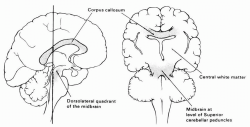
Primary injury is defined as damage that occurs directly and immediately as a result of trauma to the brain. Cortical contusion and DAI are the two subtypes of primary injury. DAI is the distinguishing feature of TBI. Acceleration-deceleration and rotational forces that commonly result from motor vehicle accidents produce diffuse axonal disruption. The direction in which the force is applied affects the severity of injury, with lateral impact leading to poorer outcome than head-on or rear-end collisions (46). Depending on the severity of injury, such lesions may be microscopic, or they may coalesce into focal macroscopic lesions, with a preponderance in the midbrain and pons, corpus callosum, and white matter of the cerebral hemispheres
(47, 48) (Fig. 24-1). DAI is also seen in areas where there is a change in the underlying tissue density, such as the gray-white interface, or where axons bend or change direction (49).


FIGURE 24-1. Brain regions particularly involved by diffuse axonal injury include the corpus callosum and parasagittal white matter as well as the dorsolateral quadrants of the midbrain. (From Auerbach SH. Neuroanatomical correlates of attention and memory disorders in traumatic brain injury: an application of behavioral subtypes. J Head Trauma Rehabil. 1986;1:1-12. Reprinted with permission of Aspen Publishers, Inc. © 1986.)

DAI is primarily responsible for the initial loss of consciousness (50). The precise mechanisms of axonal damage remain controversial but include direct axonal shearing and disruption of the intra-axonal cytoskeleton that may lead to axonal swelling and disconnection (51). Such injury may be a risk factor for development of Alzheimer’s dementia (52). Animal data suggest that some of the loss of axonal integrity may happen after a delay (51), allowing the possibility that preventive treatments may be developed.

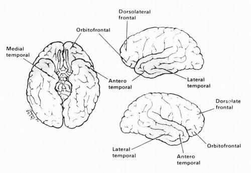
Cerebral contusion is the other main type of primary injury. These cortical bruises commonly occur at the crests of the gyri and extend to variable depths, depending on severity. Contusions often occur on the undersurface of the frontal lobes, as well as the frontal and temporal tips, regardless of the site of impact, due to the internal architecture of the skull (Fig. 24-2). The lesions usually are bilateral but may be asymmetric. Coup-contrecoup injuries are more likely seen when the moving head hits a stationary object, such as with a fall. In this situation, one may see a contusion at the site of impact as well as another, often larger, contusion on the opposite cortex. Cerebral contusions may produce focal cognitive and sensory motor deficits and are risk factors for seizure disorders but are not directly responsible for loss of consciousness. In contrast to DAI, contusions may result from relatively low-velocity impact such as blows and falls. A given patient’s pattern of functional
deficits may be more focal (e.g., from contusions) or diffuse (e.g., from DAI) or may include features of both. The balance of these two pathologic features influences the nature of neurobehavioral deficits. Deficits related to DAI tend to recover gradually, with the pace of recovery inversely related to the duration of coma, whereas recovery from deficits related to cortical contusions depends more on the size and location of the focal injury (25).


FIGURE 24-2. Areas predominantly affected by cortical contusions. Shading represents more frequently involved areas. Anterotemporal and orbito frontal regions are particularly involved. Note relative sparing of dorsolateral frontal lobe and medial temporal lobe. (Adapted from Courville CB. Mythology of the Central Nervous System. Mountain View, CA: Pacific Press; 1937; with permission.)


Secondary Injury and Initial Neurosurgical Management

Secondary injury can be defined as any damage to brain tissue that takes place after the initial (primary) injury. Whereas the only intervention for primary injury is prevention (with the possible exception of regenerative treatments), secondary injuries are at least to some degree treatable and theoretically preventable. The primary injury may set in motion a variety of pathologic processes that result in more severe and widespread brain damage. These processes range from subcellular events to those involving multiple organ systems and often work in concert. Apoptosis and excitotoxicity are examples of intracellular processes that lead to secondary injury, but can be driven by factors taking place at the tissue or systemic level. Local ischemia may lead to direct neuronal necrosis or initiate excitotoxicity by increasing levels of extracellular glutamate (53). This, in turn, leads to influx of calcium into neurons, which activates a number of proteases and increases the production of free oxygen radicals, resulting in intracellular injury and cell death (54). The local ischemia may be a result of several factors including direct trauma to blood vessels, systemic hypoxia, and cerebral hypoperfusion.

Cerebral hypoperfusion is assessed by evaluating the cerebral perfusion pressure (CPP), which is defined as the difference between mean arterial pressure (MAP) and intracranial pressure (ICP). Any factor that increases ICP or decreases MAP can decrease CPP, leading to increased ischemic injury. Management of CPP requires consideration of the pressure dynamics within the cranium, which is essentially a closed structure that contains brain tissue (cells and extracellular fluid), blood, and cerebrospinal fluid (CSF). Increases in the volume of any of these components will increase ICP. Expanding extra-axial or intraparenchymal hematomas, acute hydrocephalus, and brain edema are well-known causes of increased ICP, whereas systemic hypotension reduces CPP by decreasing MAP.

There is class II evidence to support a recommendation for maintaining CPP between 50 and 70 mm Hg (normal value for adults: 70 to 100), whereas data suggest that aggressive efforts to keep CPP above 70 mm Hg increase the risk of developing adult respiratory distress syndrome (55). Current guidelines recommend ICP monitoring in patients with severe head injury (GCS of 3 to 8) and an abnormal computed tomography (CT) scan (hematomas, contusions, edema, or compressed basal cisterns); or a normal CT scan and two out of three adverse features (age >40, unilateral or bilateral motor posturing, or systolic blood pressure <90 mm Hg) (56).

Increased ICP is not treated in isolation. However, current guidelines recommend initiation of intervention when ICP is greater than 20 mm Hg. A number of options exist to decrease ICP. Bolus doses of the osmotic diuretic mannitol may be employed for increased ICP secondary to edema, providing MAP is maintained through adequate fluid replacement (57). There is increasing interest in the role of hypertonic saline as hyperosmolar therapy as well (58). Use of high-dose sedatives such as propofol and pentobarbital is also supported (55). Hyperventilation, which had been a mainstay for the management of high ICP, is now only recommended in very specific cases, while corticosteroids are not recommended at all, and are in fact contraindicated for persons with moderate and severe TBI (55). A craniectomy is another strategy used to decrease ICP, by increasing cranial volume. A recent review did not identify randomized, controlled studies to support this intervention in TBI, although some less rigorously designed trials have suggested utility. Randomized trials are ongoing (59).

Systemic factors such as anemia, hypotension, pulmonary injury, and cardiac or respiratory arrest also may contribute to secondary injury by diminishing the delivery of oxygen to the injured brain. Brain infection may occur from open skull fractures, CSF rhinorrhea, or iatrogenically from ICP monitoring. Protracted seizures also can lead to secondary injury through increased metabolic requirements, disruption of spontaneous respiration, direct injury, and aspiration.


Neurodiagnostic Tools

Many imaging and neurophysiological technologies are available to assist in TBI management. In the acute postinjury period, CT scanning can detect intracranial hemorrhage, brain swelling, hydrocephalus, and infarction. The raw data can be adjusted to better evaluate structures of different radiodensities, for example, bone windows to identify skull fractures. However, CT is not sensitive in identifying small contusions, white matter injury, or in the evaluation of the posterior fossa (60). In the postacute phase, CT scanning can be useful, especially when a patient’s neurological status is deteriorating or failing to progress as anticipated. In such situations, CT may identify progression of hydrocephalus or hygromas, evidence of increased ICP, or new bleeds.

MRI has some advantages over CT, including lack of x-ray exposure, greater resolution in the brainstem, better identification of isodense collections of blood, and detection of small white matter lesions (61). However, MRI takes longer to perform, and requires that the patient not have any MRIincompatible implants or equipment. In the broadest sense, CT is of greatest use in the acute setting, while MRI may be more appropriate in the subacute and chronic setting.

It is important to note that the absence of findings on CT or MRI does not rule out a TBI (62). Newer neuroimaging techniques may be more sensitive to structural and functional changes in the brain after trauma. Diffusion-weighted MRI (DWI) has been utilized to identify cytotoxic edema and to quantify the size of lesions after TBI. Studies have been carried out to correlate this with outcome (63). DWI has also been
used to increase the sensitivity of MRI in detecting DAI (64). More recently, diffusion tensor imaging, a technique derived from DWI, has also been utilized to determine the presence of DAI as well as evaluate changes in white matter tracts over time (65, 66).

It may be that functional imaging can play a role in evaluating the brain after TBI due to its ability to provide information about the metabolic activity of specific regions at rest as well as during mental activities. Single photon emission CT (SPECT), positron emission tomography (PET), and functional MRI (fMRI) can all measure regional cerebral perfusion, although only PET and perfusion fMRI can quantify blood flow in absolute terms. Since perfusion may be compromised in structurally intact brain tissue, either as a result of reduced vascular delivery, or reduced perfusion demand by inactive neural tissue, reductions in flow may identify areas of functional compromise (67). Some fMRI studies conducted later postinjury have found that individuals with TBI demonstrate more widespread cortical activation during mental tasks than uninjured controls, suggesting that increased mental effort is required to perform tasks after injury. This finding has been seen in mild TBI as well as with more severely injured persons (68, 69).

Another technique that is based on MRI technology is magnetic resonance spectroscopy (MRS). Although MRS studies utilize conventional MRI systems, they require different software that detects and quantifies certain brain metabolites that may be markers of particular pathophysiologic processes. For instance, choline can be measured, and levels may be related to the degree of membrane injury from DAI and other pathological processes (70). Glutamate may be a marker for metabolic abnormalities associated with brain injury (71). Shutter et al. have reported that MRS data obtained early in the acute hospital stay can be highly predictive of 6- and 12-month Glasgow Outcome Scale (GOS) scores, especially when combined with clinical data such as GCS motor subscore (72).


Neuroprotection, Neuronal Recovery, and Functional Recovery

Recovery from TBI often is incomplete. Reports have identified subtle but persistent deficits after even mild TBI (73, 74). Yet, many people who sustain TBI make tremendous gains from coma to the reemergence of a variety of complex skills. This recovery is believed to occur at multiple levels, from alterations in biochemical processes to changes in family structure (75). A better understanding of the pathophysiological mechanisms involved in secondary injury, as well an improved understanding of the processes that underlie functional recovery may lead to improved treatments to restore function in injured individuals.

As mentioned previously, there is an increased understanding regarding the mechanisms that contribute to secondary injury. Thus, in theory, interventions could be designed that that block various ion channels, scavenge free radicals, inhibit excitatory neurotransmitters, or block the internal signals for programmed cell death, thereby salvaging a greater proportion of neural tissue that is not irreversibly damaged by primary injury. Despite the theoretical promise, however, the clinical trials that have studied such interventions have been generally disappointing (76). There are many possible reasons for the failure to identify useful neuroprotective treatments, including the heterogeneity of pathophysiologic mechanisms at work, difficulties in extrapolating from animal models, outcome measures chosen, and the possible need to combine different therapeutic mechanisms to maximize impact (77, 78). An active search for additional neuroprotective agents continues, with some preliminary promising results (79, 80, 81, 82).

Once a given degree of brain damage has occurred, a number of processes, overlapping in time and pathobiology, contribute to functional recovery. Restoration of effective perfusion and oxygenation, resolution of edema, and weaning of sedating drugs can lead to improvements in function in the first days and weeks after injury. Latent neuronal circuitry may be unmasked by injury, supporting functions that were previously carried out by neurons that are no longer functioning (83). Experience, often in the form of intense practice, has been shown to alter sensory-motor cortical representations (“maps”) in healthy humans as well as lesioned animals, suggesting that appropriately structured therapy might directly contribute to improved function after brain damage (84, 85, 86). Other interventions such as transcranial magnetic stimulation may also positively influence plasticity and cortical reorganization after TBI but await rigorous assessment (84). Resolution of diaschisis, either spontaneously, or through the actions of neurotransmitter agonists, may also play a role, and receptor antagonists may slow down recovery (85, 86, 87). Animal studies of neural transplantation are in progress and human trials in more focal neurological conditions have already appeared (88). At a more behavioral level, a person with a particular deficit may discover (or be taught) another strategy to accomplish the task that relies on remaining intact neural systems (89). Finally, as long as an individual is conscious and retains some learning capacity, it may be possible for them to learn specific tasks and activities, even though their overall capacity may remain fixed.

Although there is considerable evidence for several recovery mechanisms, particularly in the animal literature, there is as yet limited systematic application of these principles to the design of human rehabilitation efforts. Nevertheless, the growth of basic science research related to neural recovery is increasingly being translated into clinical research, with the anticipation of more theoretically based rehabilitation efforts in the future (90).


OUTCOME AFTER TBI


Types of Deficits

The majority of survivors of severe TBI emerge from coma and achieve remarkable progress toward regaining their preinjury functional abilities. Most, however, are left with a combination of physical and neurobehavioral impairments, which
collectively result in activity limitations and reduced societal participation. Each individual’s specific pattern of deficits is a consequence of the severity of the injury, nature of brain damage, and medical complications, varying greatly from one person to the next. However, deficits in cognition, particularly affecting attention, memory and/or executive function, as well as fatigue are nearly ubiquitous after moderate or severe TBI.

Changes in behavior, mood, and personality after TBI may be the most difficult impairments to manage effectively. Behavior problems range from minor irritability or passivity to labile emotional displays or disinhibited, bizarre, or aggressive behavior. The severely brain-injured person may appear egocentric and childlike, and may display a loss of empathy and concern for others.

Limitations in activities related to self-care, toileting, mobility, basic communication, and feeding commonly occur in severe TBI. In those with less severe injuries, difficulties may not be revealed until more complex community-oriented skills are attempted, such as time and money management, community mobility, and school or vocational tasks. Restrictions in vocational, educational, and interpersonal domains are widespread. There is frequently loss or diminution of friendships and intimate relationships, growing psychological distress, and a devastating impact on the family system. The psychosocial consequences of TBI, and the cognitive and behavioral impairments that underlie them, are reviewed in greater detail later in this chapter.


Outcome Measurement

The measurement and prediction of outcomes have taken on increased importance in health care generally and rehabilitation specifically. There are several distinct uses for outcome measurement, with different implications for the types of outcomes measured and their interpretation. Outcome data for clinical program evaluation systems in brain injury rehabilitation are required by the Commission for the Accreditation of Rehabilitation Facilities (CARF), with the intent of ensuring that the types of functional outcomes achieved by each program are reasonable with respect to programmatic goals. However, the quality of functional outcomes achieved by clinical programs is a function of demographic and clinical characteristics of their client populations as well as the effectiveness of their treatment services. Consequently, comparison of the quality of different programs can only be made after appropriate “case mix adjustment”—a technique that is still in a rather primitive state.

Outcome measurement can also identify treatments that appear promising in producing outcomes superior to the prior standard of care, which can then be confirmed in controlled research. Highly accurate outcome prediction could also, in principle, guide individual treatment decisions. For example, if it could be predicted in the first few days postinjury that a patient would remain permanently vegetative, the physician and the family might choose not to pursue aggressive treatment; if independent ambulation were predicted not to be possible, the energies of physical therapy could be redirected to other goals, and a definitive wheelchair could be prescribed earlier. For outcome prediction to be used in this way, however, it would need to be highly accurate in projecting the specific outcomes of individual patients; unfortunately, this goal has not been achieved.

The outcome scale used in many studies of acute TBI is the GOS. It has been demonstrated to have a high degree of interrater reliability (91). It has been used to correlate early injury severity measures (e.g., GCS, length of PTA) and outcome at 6 months postinjury. Several major drawbacks in the utility of the scale for rehabilitation purposes have been identified:


  • The categories are so broad that it is not a sensitive measure of progress during rehabilitation.


  • The global categories do not provide a real indication of functional abilities.


  • Cognitive and behavioral dysfunctions are poorly addressed in the outcome categories.

Despite these limitations, it continues to have widespread use for its intended purpose—to provide a quantitative, general way of describing outcome. An extension of the GOS, the GOS-extended (GOS-E) seeks to remedy some of the shortcomings of the GOS by providing a broader range of outcome scores to improve sensitivity (92), and by incorporating a comparison with preinjury status in both medical and psychosocial domains (93) (Table 24-3).

Several other outcome measurement scales have been developed to be more sensitive to functional changes that are of interest in rehabilitation. Some of these are braininjury-specific, and others are general rehabilitation scales.
The Disability Rating Scale (DRS) was developed specifically for TBI and is intended to assess changes “from coma to community” (94). This scale produces a quantitative index of disability across ten levels of severity, with high interrater reliability, and is considerably more sensitive to clinical change than is the GOS, particularly at the severe end of the spectrum, with less sensitivity to improvement within higher levels of functioning (95, 96).


TABLE 24.3 Glasgow Outcome Scale-Extended


1


Dead


2


Vegetative state (VS)
No cerebral cortical function that can be judged by behavior (not able to follow simple commands or communicate)


3


Lower severe disability (lower SD)
Needs full assistance in ADL throughout the day


4


Upper severe disability (upper SD)
Needs some supervision/assistance in ADL, but can be alone for >8 h/d


5


Lower moderate disability (lower MD)
Independent in ADL, and can shop and travel independently on public transportation, but has not returned to previous position or lifestyle


6


Upper moderate disability (upper MD)
Able to resume previous position or lifestyle with alternative/modified duties or part-time due to injury


7


Lower good recovery (lower GR)
Able to resume previous position or lifestyle (may be modified), but reporting some problems


8


Upper good recovery (upper GR)
Complete return to previous lifestyle with no reported problems


The Rancho Los Amigos Levels of Cognitive Functioning Scale focuses on cognitive recovery after TBI and the capacity to interact effectively with the environment. Each level is accompanied by a lengthy description of behaviors that meet the criteria for placement at that level (97). A comparison of this scale with the DRS revealed that it has slightly lower validity and reliability (98).

The Functional Independence Measure (FIM), an 18-item rating scale, is the most widely used outcome measurement scale in medical rehabilitation, and is now incorporated into the federal prospective payment system for inpatient rehabilitation. Although the FIM contains both motor and cognitive subscales, its scoring is weighted toward the physical items, and it fails to strongly capture the individual’s capacity for self-direction. Because of the relative insensitivity of the FIM to cognitive and behavioral deficits, the Functional Assessment Measure (FAM) was developed to supplement it with more cognitively oriented items. Interrater agreement for the FAM is lower (67%) than for the FIM (88%), and although it extends the range of difficulty somewhat compared to the FIM alone, its items are highly redundant with existing FIM items in terms of their scoring (96).

All the outcome scales discussed until now are well suited to measuring the changes that occur during acute rehabilitation, but they focus primarily on the activity level of the World Health Organization’s International Classification of Functioning, Disability, and Health (ICF) (99) and none of them adequately addresses higher-level functions for those with mild injuries or for those with severe injuries who have been discharged into the community (100).

Measures of behavioral and social functioning in the community that have been developed specifically for TBI outcome measurement include the Neurobehavioral Functioning Inventory (NFI) (101) and the Mayo-Portland Adaptability Inventory (MPAI) (102). The NFI is a self-report measure of the frequency of neurobehavioral symptoms, completed by either the injured person or a family member, that yields subscale scores in six domains of emotional/behavioral function: depression, somatic, memory/attention, communication, aggression, and motor. Both construct validity and criterion-related validity were demonstrated to be acceptable (103). However, this measure lacks sensitivity to the more subtle long-term problems in the domains of executive function and social behavior (104). The MPAI was developed to provide an evaluation of progress during postacute brain injury rehabilitation. It is a 30-item rating scale completed by staff or survivor on two fundamental dimensions derived from a principal components analysis: Physical/Cognitive Impairment Scale and Social Participation Scale. Both person reliability (0.82) and item reliability (0.96) were found to be good to excellent (102).

The first outcome measure developed specifically to measure community functioning of persons with brain injury was the Community Integration Questionnaire (CIQ) (105). This 15-item questionnaire has been demonstrated to consist of three subscales: home integration, social integration, and productivity. It can be completed by the person with brain injury or a significant other. The CIQ has been found useful as a measure of “objective” quality of life and level of productivity, but has also been subject to criticism for its psychometric properties (106). Another measure of community functioning, originally developed for persons with spinal cord injury, is the Craig Handicap Assessment and Reporting Technique (CHART) (107). This measure focuses on measurement of handicap or participation restriction. Original subscales included physical independence, mobility, occupation, social integration, and economic self-sufficiency. A revised version of the scale, which included a dimension identified as Cognitive Independence, was developed more recently (108). This additional scale also showed high test-retest reliability (0.87) in a mixed sample of individuals with neurologic illness including TBI and was also found to be a useful tool in evaluating postacute functioning in persons with TBI (109).

Neither the CHART nor the CIQ measures change relative to preinjury. One measure that does so and has both patient-rated and relative versions is the Sydney Psychosocial Reintegration Scale (SPRS) (Form A) (110, 111). It comprises 12 items covering three domains: occupational activity (OA); interpersonal relationships (IR); and independent living skills (LS), rated on a seven-point scale ranging from 0 (extreme change) to 6 (no change), giving total scores from 0 to 24 for each domain and an overall total score from 0 to 72. Other versions of the scale measure current level of functioning (Form B) and as categorized on three levels: good, limited and poor (Form C) (110, 111). The SPRS has been shown to have high levels of internal consistency, with Cronbach’s alpha ranging from 0.69 to 0.89 for the three domains and reaching 0.90 for the total score (110). High levels of interrater reliability have also been reported for the total score (0.95) and for the three domains (0.86 to 0.94) (110).

Many of the scales mentioned earlier, as well as other instruments, are described in more detail on the Center for Outcome Measurement in Brain Injury (COMBI) website, located at www.tbims.org/combi.


Prediction of Outcome

Prediction of outcome raises questions about both precision and values. While relatively imprecise predictions may still be useful for service planning, modeling reimbursement, or understanding factors associated with recovery, greater precision is required to use outcome prediction for individual clinical decision making and family counseling. Moreover, although many of the outcome measurement scales have levels labeled “good” or “poor,” this should not be assumed to
represent what injured individuals or their families believe are lives worth living.

The GCS is the most widely used measure of injury severity and is a primary basis for most early predictions of outcome. The total coma score when taken at 2 to 3 or 4 to 7 days postinjury is highly predictive of outcome at 6 months, as measured by the GOS (91). Scores less than 8 are usually predictive of poor outcome. Duration of PTA also is highly correlated with ultimate outcome, with PTA greater than 14 days associated with greater likelihood of moderate or severe disability (91). More recent outcome studies have suggested that PTA duration is a stronger predictor of long-term outcome than GCS (22, 112, 113). The rate of early recovery, as reflected in serial DRS scores, is also predictive of final outcome (114).

Multimodality evoked potentials (MEP), a combination of brainstem auditory evoked responses (BAERs), visual evoked responses (VERs), and somatosensory evoked responses (SEPs), also have been used as an early means of assessing neurologic status and predicting outcome. Greenberg et al. found that maximal recovery occurs in approximately 3 months for patients with minimal EP abnormalities, whereas severe EP abnormalities suggest that maximal recovery may extend to 12 months (115). Somatosensory evoked potentials (SEP) can predict the gross outcome of acutely injured patients (116). The bilateral absence of the median nerve SEP suggests a very poor outcome in patients that have sustained severe TBI, whereas preserved SEPs do not guarantee a favorable one (117). While there are those who may advocate the use of evoked potentials to allocate rehabilitation resources to patients with a potential to benefit, studies have shown that evoked potentials should not be the sole basis for prediction of outcome in cerebral injury, because of inadequate sensitivity and specificity (118).

Reactive pupils are associated with better outcomes than nonreactive pupils; 50% of those with reactive pupils achieve the moderate disability or good recovery range, as opposed to 4% with nonreactive pupils (119). An absent oculovestibular response, elicited by injecting ice water into the ear of a comatose patient, is an indication of severe brainstem dysfunction and poorer outcome (11). The presence of an intracranial hemorrhage (120), and high levels of creatine kinase (BB fraction, reflecting destruction of brain tissue) measured early after TBI (121) also suggest poorer outcomes. Hyperglycemia and low levels of thyroid hormones have negative prognostic significance, presumably reflecting the severity of the stress response (122, 123).

The research from Glasgow and other major TBI centers strongly suggests that the bulk of neurologic recovery from acute brain injury occurs within the first 6 months. The maximal duration of the recovery period is more controversial with some researchers affirming that recovery is virtually complete by 1 year, whereas others assert that recovery can extend 2 years or more postinjury (124). It is clear, however, that certain areas of dysfunction recover more quickly than others. For example, recovery of physical abilities and functional skills such as mobility occurs rapidly, often within 3 months after injury (125). Recovery of more complex mental abilities, as assessed by neuropsychological measures, appears more variable. This aspect of recovery has been studied in large samples in the Traumatic Coma Data Bank study (126) and in the TBI Model Systems Project (127). Impairments of attention, information processing speed, memory, and executive function have been shown to persist up to 10 years postinjury (104). Preinjury medical and psychological factors also may affect the prognosis. For example, the presence of a prior TBI or neurologic deficit is likely to slow the recovery process. Also, if cognitive or behavioral abnormalities existed before the injury, there is a greater likelihood of a slower and less complete recovery. Acquired brain damage is thought to exacerbate preexisting behavior disorders (128). Demographic factors, including education, age, and preinjury employment status have also been shown to influence outcome (22, 129, 130).

Multivariate models combining PTA with age, preinjury occupational status, and early physical and cognitive disability have accounted for 60% or more of the variance in employment outcome at 1 and 6 years postinjury (24, 131).

There probably is no final endpoint to the recovery process; rather, the pace of recovery slows, and its scope narrows, though occasional cases of impressive late improvement occur. Even those with permanent cognitive and physical impairments can continue to learn new skills for solving particular functional problems, albeit slowly. Thus, neurologic and cognitive recovery merge imperceptibly into ongoing learning and adaptation.


THE ASSESSMENT AND TREATMENT-PLANNING PROCESS

The rehabilitation needs of the survivor of severe TBI often begin at the emergency department but are not likely to end for many years. Although many medical conditions and physical deficits stabilize within 1 year after injury, the presence of long-term psychosocial disorders often necessitates ongoing intervention (132). One year after injury, a quarter to a third of those with severe injuries report unmet service needs, including need for assistance coping with cognitive impairments, emotional distress, and managing financial matters (133). Survivors of TBI have a continuing need for treatment and support services across the lifespan, particularly to address psychosocial difficulties (134).


Treatment and Support Options

Many hospital and community-based services may be of help to survivors of TBI. Changes in private and governmental funding streams stimulated much growth of specialized TBI rehabilitation services in the 1980s and early 1990s, but subsequent funding cutbacks and managed care practices have reduced their availability.

Initial management takes the form of aggressive neurosurgical intervention to minimize secondary injury. The physiatrist may be called in as a consultant even when the patient is still in the ICU, to assist the acute care team in
preventing complications such as contractures, pressure ulcers, heterotopic ossification (HO), and bowel and bladder problems that may impede later rehabilitation of surviving patients, and to assist in choosing medications that minimize sedation.

A tremendous variety of treatment programs may be appropriate for a given individual at various points in the recovery process. Acute inpatient rehabilitation is typically provided to individuals who are able to participate actively in treatment, and whose array of medical, physical, and cognitive deficits precludes a safe community placement. Less intensive subacute rehabilitation may be most appropriate for those who are vegetative, slow to recover, or cannot tolerate intensive therapy. However, many such patients, even though their rehabilitation participation is limited, have intensive medical needs that are not adequately addressed in subacute facilities. A day treatment program may be provided to individuals who can be managed at home but continue to display a variety of physical or neurobehavioral problems. This type of treatment often combines extensive cognitive rehabilitation, behavior management, daily life skills training, community activities, and prevocational activities (135, 136). Residential programs may be chosen for individuals who display disinhibited, aggressive, or self-abusive behavior that cannot be managed at home. Such programs aim to reduce inappropriate behaviors and teach more effective means of communication and social interaction, through contingency management and/or pharmacological intervention (137). Transitional living programs assist individuals who have mastered basic ADLs and social interaction skills to live increasingly independently in the community. Typically, the brain-injured person lives in a supervised group home or apartment setting and is given instruction and increasing responsibility in skills needed to live independently (e.g., cooking, cleaning, money management, community mobility, job seeking), with fading of supervision over time. The final goal for many individuals is return to work, most typically through job coaching and supported work, funded by state vocational rehabilitation services.

Many clients with TBI have difficulty applying knowledge gained in one setting to related problems or different environments. In such instances, it is particularly important that the independent living and vocational training be given in the actual community or on the actual job that the individual will be attempting. Some postacute programs deliver community-based services, actively involving the client and family in goal setting and delivering most therapy in the relevant community setting in order to maximize the client’s motivation and engagement in the rehabilitation process and the durability of outcomes (138).

Telerehabilitation is an emerging facet of service provision that may allow for more accurate assessment and treatment planning in the homecare context, and may allow rural patients to receive services from specialized TBI rehabilitation centers (139, 140). As one example, a randomized trial comparing the effects of scheduled telephone intervention after discharge from acute inpatient rehabilitation to usual postdischarge care found a positive impact on 1-year outcomes of this relatively low-cost intervention (141).

During the long rehabilitation process, a case manager may follow patients across multiple treatment services, providing crucial coordination and social support. This type of case manager serves as a liaison among the patient, family, and service providers and gathers medical records, arranges for medical visits, screens programs and facilities, and helps coordinate admission and discharge. With the advent of managed care, intensive case management may be even more crucial although, as noted earlier, it is less often provided (142).

Unfortunately, in many regions, these and other needed services are either unavailable or difficult to access due, in part, to the rapidly evolving changes in health care financing noted previously (143). This calls for creative financing strategies, assisted by case managers, along with advocacy efforts to expand the range of available services. Some states have developed Medicaid waiver programs that extend the array of available services, through the use of public funds (144, 145).


Evidence for the Effectiveness of TBI-Specific Services

As in most areas of rehabilitation, there is currently insufficient evidence for the efficacy of the services discussed earlier (146, 147). However, observational data and randomized pilot studies do provide some evidence to support the impact of comprehensive systems of care for TBI, particularly in the postacute period (136, 148, 149). One of the few randomized trials comparing an organized program of cognitive rehabilitation to a limited home program failed to find an overall treatment effect, but did find superior outcomes for the cognitive rehabilitation program, for the more severely impaired subgroup (150).


Patient Assessment and Treatment Planning

Standards of care and practice for the management of acute and postacute care of persons with TBI have been promulgated by organizations such as the American Association of Neurological Surgeons (56), the Commission on the Accreditation of Rehabilitation Facilities (CARF) (151), and the American Congress of Rehabilitation Medicine (152). Rehabilitation standards emphasize a team model to promote coordination and information sharing across therapeutic disciplines, since a specific cognitive impairment may interfere with the performance and retention of a mobility, communication, or ADL skill. Similarly, disruptive behaviors and lack of initiation cut across all therapy domains. Each of these problems requires the development of a unified team view of the patient’s deficits and needs and the creation of a unified treatment plan.

When a patient enters a rehabilitation service, an initial assessment is needed to guide treatment planning. The goals of this assessment vary with time since injury. Early clinical assessment usually is aimed at defining broad functional areas in need of treatment (e.g., impaired mobility, memory deficits). Ideally, these areas are guided by an assessment of
the injured person’s lifestyle, needs, and personal and family goals. As the pace of recovery slows, and discharge to home or another facility approaches, therapy focuses more intensely on the specific skills and behaviors that will be prerequisites in the new environment (e.g., toilet transfers, learning a daily schedule). Assessment also depends on injury severity. It is focused more on physical function and basic sensory processing in the severely impaired and on cognitive, social, and vocational function in the mildly impaired.

Discipline-specific assessments must be melded into a patient-oriented assessment for treatment-planning purposes. Typically, severely injured patients have a large number of medical, physical, cognitive, and behavioral impairments. Therefore, the team must develop priorities for treatment, framed in terms of long- and short-term goals, based on the needs and priorities of the patient and family, as well as such factors as estimated length of stay, functional importance of each impairment, age and developmental stage, and prognosis for improvement. As time passes, the goals become more specific and functionally oriented, such that “the patient will have increased ROM in all joints” becomes “the patient will have adequate hip flexion for erect sitting throughout the day.” Identification of the patient’s physical and cognitive strengths can also be very helpful in determining the best way to circumvent specific deficits. Goals being addressed by a single discipline must ultimately be shared by the team. For example, a communication strategy designed by a speech pathologist should be carried out by nurses, other therapists, and family to promote generalization of the skill.

Treatment planning also must consider the ICF (99), and its hierarchy of body structure and function, activity, and participation, and must choose at which of these levels to intervene. For example, it may be concluded that a patient’s ambulation activity limitation is related to limitations in body structure and function in the form of limited range of motion, abnormal tone, weakness, impaired balance, reduced proprioception, and disordered attention. Clinicians then must choose whether to try to address each of these impairments to improve ambulation, or whether to assess the patient’s mobility skills in a motorized wheelchair instead, which would be an activitylevel intervention.

A neuropsychological assessment may be important in identifying intact cognitive skills and clarifying cognitive mechanisms responsible for various behavior and skill deficits. In the severely impaired patient, formal testing may be impossible; however, the neuropsychologist’s observation of the patient may still be helpful. Higher-level patients should receive formal testing of such core cognitive areas as attention, learning and remembering, language comprehension and production, visual perception, planning, reasoning, and organization. It should be kept in mind that the results of formal neuropsychological testing have limited predictive ability for real-world function (153). Thus, patients’ skills also should be assessed in naturalistic settings. Moreover, it must be understood that results obtained soon after injury only give an indication of current strengths and weaknesses. Reassessment after longer periods is important to provide a clearer indication of cognitive difficulties that may be more lasting.


SPECIAL SUBPOPULATIONS


The Vegetative State

About 20% of survivors of severe TBI remain unresponsive 1 month after injury. After 2 to 4 weeks of unconsciousness, coma evolves into the vegetative state, a condition of wakeful unresponsiveness that is characterized by the presence of spontaneous sleep-wake cycles but absence of cortical activity as judged behaviorally (154).

Patients who are vegetative 1 month postinjury still may experience substantial recovery, but their chances of doing so diminish over time. Of patients who are vegetative at 1 month, there is approximately a 50% chance of regaining some degree of consciousness within a year and approximately a 28% chance of improving to a level of independence (155).

Patients continue to emerge from the vegetative state following trauma for at least a year, with rare individuals showing recovery of consciousness even later (little research is available for follow-up periods greater than a year), but nearly all are severely disabled if their emergence is this delayed (156). The term persistent vegetative state has been used extensively in the literature, but without consensus on its definition. It has been suggested that this term be abandoned because it confuses diagnosis (vegetative) with prognosis (persistent). Emergence from the vegetative state following nontraumatic injuries (such as cardiac arrest) is far less likely overall, and very few patients emerge beyond 3 months postinjury (155). This suggests that patients with traumatic injuries complicated by substantial secondary anoxic injury are also likely to have a poorer prognosis than those with uncomplicated trauma.

The following factors have positive prognostic significance for emergence from unresponsiveness: young age, reactive pupils and conjugate eye movements, decorticate posturing rather than decerebrate or flaccid states, early spontaneous eye opening, absence of ventilator dependence or hydrocephalus, shorter time between injury and rehabilitation admission, better scores (within the vegetative range) on the DRS, and more rapid early functional improvement (157, 158, 159). Unfortunately, no set of prognostic variables is precise enough to guide early clinical decision making. The life expectancy of those who remain permanently vegetative is not precisely known, but one study of patients in vegetative states of mixed etiologies revealed that almost 75% had died within 5 years (160). Similarly, TBI survivors with severe mobility impairments (though not necessarily vegetative) have a reduced life expectancy due primarily to cardiovascular disease, respiratory disease, choking, and seizures (161).

The minimally conscious state (MCS) refers to individuals who show some evidence of awareness in the form of visual tracking and/or motor behavior that is nonreflexive and contingent on environmental events (e.g., intermittent following of simple commands, replicable pulling out of tubes) but who
do not consistently follow commands or communicate intelligibly (162). The MCS, like the vegetative state, can be a transitional state on the way to greater recovery or it can be the permanent functional plateau (163). Misdiagnosing patients who are in the MCS as vegetative may occur more than 40% of the time, highlighting the need for rigorous diagnostic assessment (162, 164).

Initial coma in TBI probably reflects disruption of brainstem-alerting mechanisms, often with relative preservation of higher brain structures. Brainstem-alerting mechanisms tend to recover with time, however, resulting in return of the sleep-wake cycle. Thus, a vegetative state of long duration generally includes extensive damage to subcortical white matter in higher brain regions, including the thalamus (165, 166). Akinetic mutism and the locked-in syndrome may be confused with the vegetative state, but akinetic mutism generally involves damage to the medial frontal lobes, and hypertonia and posturing are absent. The locked-in syndrome generally results from a bilateral pontine stroke, and there is evidence of preserved consciousness and communication through eye movements (167, 168).

Establishing emergence from the vegetative state can be done clinically, by observing for volitional responses to the environment, such as following commands, orienting visually to salient objects, attempting to remove tubes and restraints, and the like. Essentially, any behavior that is nonstereotyped and that indicates some evaluation of environmental stimuli is evidence of emerging consciousness. A formal and quantitative assessment strategy is an absolute requirement in working with vegetative and minimally conscious patients; without this, team and family members will disagree about whether or not evidence of consciousness is present and whether improvement is occurring. Several standardized scales are available for objectively grading responsiveness in vegetative and minimally conscious patients (169, 170, 171, 172), although only the Coma Recovery Scale-Revised (169) incorporates items that explicitly mark the boundary between VS and MCS. In addition, the principles of single-subject experimental design can be used to answer important clinical questions in individual patients, such as whether they can see (173), follow commands (174), or reliably use a yes/no signaling system; or whether they respond to therapeutic medications (175). Assessment should take place repeatedly and at various times of day because patients may respond inconsistently, particularly as they are first emerging from the vegetative state. When family members report observing volitional behavior that has not been witnessed by staff, individualized assessments of the relevant behaviors can be conducted. Family members may overinterpret reflexive or coincidental behaviors, but staff members may fail to elicit the patient’s best performance. Definitive assessment of the vegetative state must await withdrawal of potentially sedating drugs and ruling out peripheral sensory and motor deficits (e.g., blindness, deafness, extensive polyneuropathy) (164, 176).

The possibility that patients who appear vegetative may have some degree of consciousness that is not reflected in observable behavior has been explored recently using functional imaging and event-related potential (ERP) methods. These studies suggest that there exist patients who are capable of following commands to engage in specific mental activities that can be detected physiologically, even though they cannot produce observable behavioral responses (177, 178). It is not yet clear how many patients classified as vegetative may be conscious as assessed by more subtle methods, or whether such patients have a better prognosis for the emergence of functionally useful behavior.

Many treatments have been attempted for patients who are in the vegetative state, but none has been subjected to an adequate controlled clinical trial. The pathologic heterogeneity of unresponsive states makes it unlikely that one treatment will help all affected people. Deep brain electrical stimulation of the mesencephalic reticular formation or nonspecific thalamic activating system has been reported to improve the clinical status in some vegetative patients, but small heterogeneous samples and early treatment make it difficult to rule out spontaneous recovery (179, 180, 181, 182). A more recent report of late recovery of a patient in MCS after thalamic electrical stimulation does not appear attributable to spontaneous recovery (183). Dopaminergic pharmacologic treatments, including L-dopa, bromocriptine, and amantadine, also have been reported to be of help (184, 185, 186, 187). Since all these treatments primarily augment ascending arousing influences, they would seem unlikely to benefit patients with extensive cerebral lesions. Coma stimulation (involving the systematic and frequent provision of sensory stimulation to all sensory modalities) has been widely advocated (75), but studies on it, like those on the pharmacologic agents, suffer from serious methodologic flaws, which have been summarized in review articles (188, 189). Large, multicenter clinical trials will be needed to definitively assess the value of early treatments designed to improve recovery, because of the confounding and variable effects of spontaneous recovery. However, such trials are fraught with practical and ethical challenges, particularly when they involve a placebo treatment (190). A number of case reports have been published in which patients who had been vegetative for several years after traumatic or anoxic injuries paradoxically regained consciousness following the administration of a single dose of zolpidem, and whose consciousness could be maintained by repeated dosing of the drug (191). The mechanism of zolpidem response is currently under investigation. The proportion of VS patients capable of responding to zolpidem is unknown but appears to be small (192).

In the absence of definitive treatments to alter the prognosis in the vegetative state, the main goals for rehabilitation are to optimize medical stability, preserve bodily integrity, and objectively define the patient’s current sensory and cognitive capacities with measures that can be monitored for change over time (156, 176). This includes screening for adverse medical events such as undiagnosed seizures, hydrocephalus, and endocrine disorders. Attempts to optimize pulmonary hygiene and maintain skin integrity are also essential. Aggressive treatment of hypertonia and contractures is warranted early because they predispose to skin breakdown, interfere with positioning, and
are costly and time consuming to manage in the chronic stage. Finally, sedating medications should be avoided until the prognosis has declared itself. This includes a variety of antispasticity, anticonvulsant, antihypertensive, anticholinergic, and antihistaminic agents that may have subtle cognitive effects in susceptible patients.

Regular evaluation with a quantitative assessment scale should be carried out. This will reveal subtle improvement that should lead to updated treatment plans, deterioration that should lead to further diagnostic evaluation, or no change, which should lead to family counseling about prognosis and planning toward home or chronic care placement. When a patient remains permanently vegetative, or when a patient remains minimally conscious but has left a specific advance directive, the possibility of forgoing further medical treatments and even life-sustaining fluid and nutrition may be discussed with the family after careful review of local legal guidelines relevant to this area and any institutional policies and ethical guidelines relevant to end-of-life determinations. However, as mentioned, a small number of vegetative or minimally conscious patients may continue to improve over several years, making the determination of permanence more difficult (193).


Mild TBI

At the other end of the spectrum from unresponsive states is the patient with mild, or minor, TBI (MTBI), which may occur with or without impact to the head. This is generally defined as a TBI with the following characteristics:


  • Loss of consciousness, if any, 30 minutes or less


  • PTA 24 hours or less


  • Initial GCS 13 to 15


  • No focal neurologic deficit


  • Negative CT and/or MRI

The initial symptoms of the cerebral injury may be difficult to disentangle from those of the common coincident injuries to the scalp, neck, and peripheral vestibular apparatus (194). Acute complaints after MTBI typically fall into three symptom clusters (195):


  • Cognitive: difficulties with attention, speed of information processing, and memory


  • Affective: irritability, depression, anxiety


  • Somatic: headache, dizziness, insomnia, fatigue, sensory impairments

Headaches are the most common and persistent symptom and the symptom that most strongly differentiates those with mild TBI from trauma controls (196). These symptoms clear within the first few weeks or months postinjury for the majority of patients. Group studies have generally revealed impaired speed of information processing in the early hours or days after injury, but no decrement, or only modest or transient decrement, on neuropsychological measures for individuals with MTBI compared to uninjured controls beyond the first 2 weeks after injury (197). For a proportion of individuals (15% to 25%), however, difficulties persist and are associated with social and vocational failure seemingly out of proportion to the severity of the neurologic insult (197). The etiology of these persistent complaints (often termed postconcussion syndrome, PCS) has been elusive and remains controversial. Premorbid factors such as substance abuse, psychiatric disorder, and age have been implicated but do not explain all cases of persistent disability. The presence of other stressors, preinjury psychological problems, being a student or being in a demanding occupation, being injured in a motor vehicle accident and/or having neck or back injury have also been associated with poorer outcomes (196, 198). The idea that pending litigation or financial gain accounts for PCS has not been confirmed (195). In one SPECT study, MTBI patients with unusually persistent disability showed hypoperfusion in the anterior mesial regions of the temporal lobes (199). There is probably no one cause of PCS; premorbid variables, idiosyncratic neurologic vulnerability, and psychological reactions to acute symptoms may all be found to play roles.

The treatment of MTBI should include patient and family education about the typical symptoms and their time frame, and guidance on how and when to resume preinjury activities. Patients with persistent symptoms may benefit from psychotherapy (200), pain management protocols (201), or holistic community reentry programs offering these components along with education, vocational counseling, and group support. Many of the somatic symptoms are responsive to interventions that can be provided by an experienced physical therapist in conjunction with judicious use of medications. Therapeutic interventions may include vestibular habituation exercises (202), Rocabado exercises (203), myofascial release, trigger point injections, nonsteroidal anti-inflammatory medications, and muscle relaxants. For more extensive information, the reader is referred to more detailed publications on this aspect of brain injury (197, 204, 205, 206).

Prompt diagnosis and treatment of “concussion” in sports related injuries has received increased public awareness. Practice parameters have been established by a subcommittee of the American Academy of Neurology and recommendations were also derived from the Second International Conference on Concussion in Sport (207, 208). A Standardized Assessment of Concussion was developed to provide a sideline evaluation that could be performed by nonphysicians (209). Computerized neuropsychological tests have been developed for more standardized evaluation and to provide baseline data to which subsequent postinjury data can be compared (210, 211).


TBI in Children and Adolescents

Children and adolescents are a high-risk group for TBI, with etiologies varying by age group. Falls and child abuse are overrepresented among infants and toddlers, accidental injuries and motor vehicle accidents in school-age children, and injuries due to violence and risk-taking behavior among adolescents.

The management and sequelae of TBI in children and adolescents are very similar to those of adults, but there are a number of issues that are specific to children. On the
one hand, it may be challenging to disentangle neurological recovery from normal growth and maturation. On the other hand, young children may appear to have minimal deficits after TBI, since the skills expected of them are minimal, only to reveal increasing developmental lags as they mature. Because “independence” is not expected of healthy children, many children with TBI are sent home without rehabilitation services, with their parents providing the assistance needed to function safely (212).

Although it has been estimated that 20,000 children and adolescents reenter school each year with significant disabilities related to TBI, surveys of special education services estimate that only about 12,000 of the more than 5,000,000 U.S. special education students receive services due to a TBI (213). This discrepancy may have several sources. Children already receiving special education services, particularly those with ADHD, are overrepresented among those injured, and they may retain their original etiology’s label; children may “grow into” their educational disability without the educational system recognizing their prior injury; and some school systems keep records by educational needs rather than etiology (212, 213). Policy recommendations for improving special education services for this population have been published (213).

Problems with attention and concentration appear to be among the most frequent sequelae of TBI in children, as they are in adults, occurring in up to 50% of survivors, although as many as 19% of those injured carried a premorbid label of ADHD (5, 36). Learning and memory difficulties are also relatively common in those with severe injuries (5). Language impairments have also been reported in children injured in the preschool years (214). In children with severe TBI, injury to the frontal lobes may result in executive dysfunction (215). In general, many children with mild injuries make a good recovery. Children with severe injuries show the most persistent cognitive impairments, with those injured prior to age 8 showing less recovery than those aged 8 to 12 years (5). This is not consistent with the premise that younger children show increased cerebral plasticity in the face of injury, but rather suggests there are periods of increased vulnerability (216). TBI may also have important effects on emotional adjustment in children and adolescents, with depression, dysthymia, and anxiety disorders among the most common new problems (36). In a study of college students with a history of mild TBI, intellectual impairments were not found, although the students with prior TBI had increased levels of subjective distress in several subscales of the Symptom Checklist-90—Revised (217).

Family function also has a significant influence on outcome following pediatric TBI (4), and there is also reason to be concerned about the impact of the child’s injury on the family. In particular, it has been suggested that siblings of children with TBI-related disability may experience adverse impact themselves, including greater psychopathology, depression, or reduced emotional well-being and more negative relationships (218).

The preponderance of data suggests that pediatric TBI is underreported and underrecognized in its importance. A high proportion of children with a history of TBI are likely to present with educational special needs, cognitive problems, and psychological/behavioral problems. The fact that some of these children had such problems premorbidly, coupled with the delay between injury and appearance of the problems, may contribute to their underrecognition. Thus, it appears crucial that children with significant TBI be identified and tracked so that the relevance of their TBI to future problems and needs does not go unnoticed.


TBI in the Elderly

As discussed previously, the incidence of TBI is high among the elderly, with up to 86% of TBIs in those over 65 due to falls (219). Although mortality at a given level of severity is higher among the elderly than among younger individuals, there is also some evidence to suggest cognitive impairments are more severe and functional outcomes poorer, in those who are older (127, 220). Considerable controversy exists about the prognosis for functional recovery among elderly survivors, due to the fact that many older patients with TBI were already suffering from functional decline at the time of the injury, and to the fact that those elderly patients admitted to rehabilitation facilities are a highly selected group. Research that avoided this selection bias by enrolling elderly patients in acute care, found that GOS scores of moderate disability, or good recovery never occurred in those with GCS scores less than 11 (221). This was true even among the subgroup with mild original injuries and subsequent deterioration to lower GCS scores. Though it appears likely that the functional prognosis for elderly patients is, indeed, worse than for younger ones, there is also a concern that this may contribute to a self-fulfilling prophecy, in which elderly survivors of TBI are not given the same rehabilitation opportunities as their younger counterparts.

Understanding the rehabilitation problems and needs of the elderly presents some of the same challenges as those seen in the pediatric population. In a retired population, what are the appropriate standards for community integration? If the normal developmental sequence is for an increasing prevalence of disability and need for assistance with aging, how does one assess the outcome of the rehabilitation process against this moving functional target? But, unlike children with TBI, whose parents are already in the role of caregiver, elderly survivors of TBI may have few support options. A spouse may already have died or be too frail to provide the necessary care, and adult children, if any, may be unwilling or unable to provide sufficient assistance. In the absence of family and community supports, therefore, a nursing home destination may seem to be a foregone conclusion, further eroding optimism about the cost effectiveness of aggressive rehabilitation interventions.


MEDICAL PROBLEMS AFTER TBI


Fractures

For patients with TBI who also have long bone fractures, open reduction and internal fixation can promote early mobilization,
simplify patient care, and improve predictability of fracture outcome. Early surgical intervention may reduce health care costs and delay in mobility, but there has been concern regarding risks for potential secondary brain injury due to surgery, including the risks of general anesthesia (222). A recent article compared outcomes of patients with TBI who underwent early surgery (<24 hours after injury) or late surgery (>24 hours), including both functional and neuropsychological measures. Early surgery was not associated with poorer outcomes (223).

While most fractures are diagnosed in the acute injury period, some are not detected until transfer to rehabilitation, because of increased movement, awareness, and communicative ability as the patient improves. Details regarding the initial trauma may increase the level of suspicion for a missed fracture. The incidence of missed fractures has been reported as high as 11% in this population (223). Any unexplained swelling, deformity, or pain response should prompt evaluation for occult fracture. Fractures in TBI predispose to HO. For example, despite appropriate operative management of 23 acetabular fractures, 61% had poor outcomes because of HO (224).


Seizures

Posttraumatic seizures and epilepsy are known complications of TBI. The risk of seizures is related to injury severity, presence of skull fractures, cortical contusion, subdural hematoma, and age (225). The risk of new seizure development is greatest in the first 2 years postinjury and gradually declines. In a population of patients who developed seizures in the first 2 years after injury, 33% of the seizures first occurred within the first month and 80% occurred within the first year (226). In another study, 86% of patients that had one late posttraumatic seizure (>7 days after injury) had one or more additional seizures within 2 years after injury, so the risk of recurrence can be high (227).

Most seizures are diagnosed clinically on the basis of focal or generalized motor activity. Patients with muscle spasms or tremors may present diagnostic dilemmas. In such cases, a routine or sleep-deprived EEG may reveal epileptiform activity. More definitive is a 24-hour EEG correlated with observations of the suspicious activity. Seizures in limbic and association areas may lead only to altered behavior or states of consciousness, presenting further diagnostic challenges.

Treatment during the first week postinjury with phenytoin or valproic acid can be effective for reducing the incidence of early-onset seizures (228, 229). Long-term seizure prophylaxis is not recommended because it has not been shown to be beneficial in preventing late-onset posttraumatic seizures (230).

Carbamazepine and valproic acid have been found to be relatively free of adverse cognitive effects; their superiority to phentoin in this regard is debated (231, 232). None of these drugs is free of cognitive and physical adverse effects, however (233). These older antiepileptic drugs (AEDs) are all hepatically metabolized. Some newer AEDs are renally metabolized, which may be an advantage in certain clinical situations. It is hoped that some of the newer AEDs such as levetiracetam may not have as many deleterious side effects, but they have not been as well studied as some of the older agents. Studies are ongoing to examine utility and safety in the TBI population (234).

There are no standard recommendations on duration of AED treatment. In view of the expense and the potential toxicity of anticonvulsants, most clinicians withdraw medications after 1 to 2 seizure-free years. Of patients with previous seizure disorders of mixed etiologies who had been seizure-free for 2 years, 35% relapsed after tapering of their anticonvulsants (235).


Hydrocephalus

Ventricular dilation occurs in up to 40% of patients with severe TBI and usually begins to appear within 2 weeks of injury. In most instances, ventriculomegaly results from diffuse atrophy or focal infarction of brain tissue (i.e., hydrocephalus ex vacuo) and, thus, is a sign of primary and secondary injury but not a syndrome requiring treatment. The less common communicating hydrocephalus is generally associated with abnormal CSF pressure dynamics, causes neural dysfunction, and warrants treatment.

Unfortunately, the classic symptom triad for the diagnosis of hydrocephalus—incontinence, gait disorder, and dementia—is of little help in severely disabled patients. Failure to improve or deterioration of cognitive or behavioral function should prompt assessment with a CT scan. Flattening of the cortical sulci and periventricular lucency may support the diagnosis of clinically important hydrocephalus, but this can be a challenging determination (236). At present, there is no single test that is felt to be the gold standard in determining whether hydrocephalus is clinically significant. A recent review of the literature looked at the value of prognostic tests for idiopathic normal pressure hydrocephalus (NPH). The tap test, where 40 to 50 mL of CSF is drained and the patient is subsequently assessed to evaluate clinical improvement, was considered specific but not sensitive. A CSF infusion test to determine outflow resistance was more sensitive and specific, while prolonged drainage of CSF from a lumbar catheter may have the highest predictive value (237). The more sensitive tests may carry a higher risk of complications, especially in less medically stable or cognitively compliant individuals, which would include many patients with TBI. Even with an accurate diagnosis of clinical hydrocephalus, the prognosis for improvement from shunting is uncertain. This may be partly because the patient has other cognitive and motor deficits unrelated to hydrocephalus. Mazzini et al. suggest that patients that demonstrate a clinical deterioration may be more likely to benefit from shunting (238).

The etiology of posttraumatic hydrocephalus appears to be multifactorial. One proposed cause is the change in hemodynamics, CSF hydrodynamics, and brain metabolism caused by the presence of a craniectomy defect (239). This is of interest because there has been much discussion in the literature regarding the optimal timing of cranioplasty for patients that have undergone craniectomies, and reported improvements in function after cranioplasty have been attributed to this mechanism (240).


Patients with ventricular shunts may experience complications due to shunt failure, infection, or over/under drainage. The latter complication has been lessened through use of programmable shunt valves. Though not problem free, they have improved the ability to make minor changes in CSF flow noninvasively (241). The changes associated with hydrocephalus and shunt failure may be subtle and it is critical that the entire rehabilitation team’s assessments of cognitive and behavioral fluctuations be taken into account in this evaluation.


Cardiac Abnormalities

Cardiac complications may appear acutely, particularly in the setting of multiple trauma, and may involve direct injury to cardiac muscle, vessels, or valves, as well as the generation of arrhythmias, any of which may lead to impaired perfusion and increased secondary brain injury. Because of the multitude of potential injuries, there is no gold standard for cardiac evaluation in the setting of blunt trauma (242).

Hypertension, tachycardia, and increased cardiac output in the acute postinjury period may result from the increased release of epinephrine and norepinephrine (243). Central sympathetic hyperactivity can lead to ongoing myocardial necrosis (244). Although β-blockers may be considered, some medications in this class, such as propranolol, have been shown to cause cognitive impairments in hypertensive patients (245). It is unclear whether highly polar β-blockers such as atenolol or nadolol have fewer side effects in this population due to decreased ability to cross the blood-brain barrier.

Autonomic disturbances after brain injury may lead to dramatic increases in blood pressure and heart rate. These disturbances have been known by a number of names. More recently, the term paroxysmal autonomic instability with dystonia (PAID) has been coined (246). It is suggested that the diagnosis of this syndrome should be made when at least five of seven symptoms are present (fever, hypertension, hyperhidrosis, tachypnea, tachycardia, posturing, and dystonia). The etiology and treatment of this syndrome remains controversial. It has been suggested that environmental factors such as noxious stimuli may play a role in triggering the dysautonomia (247).


Pulmonary Disorders

Multiple trauma often causes pneumothoraces, pulmonary contusions, and lacerations. In addition, adult respiratory distress syndrome, excessive fluid administration during resuscitation, and intense α-adrenergic outflow may lead to noncardiogenic pulmonary edema (248). These and other problems can further compromise cerebral oxygenation.

Many brain-injured patients require tracheostomies for ventilation and suctioning. Humidified air may be delivered through a tracheostomy collar to maintain moist secretions and prevent tracheitis, and frequent suctioning may be required. Patients who initially require supplemental oxygen usually can be weaned from it by assessing pulse oximetry on oxygen and again after it has been stopped. Patients who require long-term tracheostomies but have begun to vocalize may benefit from one of the varieties of tubes that permit vocalization.

Most TBI patients eventually can be decannulated. Indirect laryngoscopy screening can be used to check for adequate vocal cord abduction and to rule out subglottic stenosis (249). In some instances, tracheal or subglottic stenosis will prevent decannulation and will require dilation or surgical management. If there is no sign of anatomic obstruction, a small-caliber tracheostomy tube will allow air to bypass the tube while it is plugged for progressive intervals. The patient is checked at the end of each interval or at any sign of distress with pulse oximetry. Once the patient has tolerated 24 hours of plugging without incident, decannulation can take place, and the tracheostomy stoma can be covered by a gauze pad or occlusive dressing until it heals. Some patients will have difficulty with decannulation because of the quantity of secretions and inability to cough them up into the pharynx. Thus, persistent suctioning is required. The tracheostomy tube itself may be an irritant that evokes secretions, however. To evaluate this possibility, a tracheal button (i.e., a small plug that keeps the stoma open) can replace the tube temporarily to see if the elimination of the tube allows patients to manage their own secretions; if not, the tube can be replaced.

Long-term survivors of severe TBI have been reported to show decreased lung capacity, vital capacity, and forced expiratory volume. The etiology of these abnormalities is not entirely clear but appears to be a combination of muscle weakness and incoordination, decreased chest compliance, and deconditioning (250). Guidelines for cardiopulmonary conditioning specifically for patients with brain injury have not been clearly established.

Pulmonary embolism is a potentially fatal end result of venous thromboembolism. Because persons with TBI who are hospitalized have many risk factors, it is not surprising that the incidence of deep vein thrombosis (DVT) is elevated in this population (251). The problem is compounded by a reluctance to use pharmacological prophylaxis because of the risk of intracerebral bleeding. The literature is too limited to allow development of a treatment standard (252). A recent survey of a group of TBI rehabilitation centers failed to identify a consensus regarding prophylaxis or screening (253).

Only gold members can continue reading. Log In or Register to continue

Stay updated, free articles. Join our Telegram channel

May 25, 2016 | Posted by in PHYSICAL MEDICINE & REHABILITATION | Comments Off on Traumatic Brain Injury

Full access? Get Clinical Tree

Get Clinical Tree app for offline access