Spinal Injection Procedures


Spinal Injection Procedures


Boqing Chen

Todd P. Stitik

Patrick M. Foye

Jong-Hyun Kim

Michael Mehnert

Christopher Castro

Steve Aydin

Glen Cheng

Evish Kamrava


Spinal pain, especially low back pain (5% incidence and 60% to 80% lifetime prevalence in the United States), is very common (1). Low back pain is the leading cause of disability in people younger than 45 years. Although spinal pain often improves and resolves, a significant proportion of patients have ongoing symptoms and pain recurrence. Low back pain is a costly disorder with an annual cost approaching $50 billion (1). Therefore, a comprehensive rehabilitation approach that improves outcomes for patients with spinal pain can have a significant positive medical and economic impact. Adequate pain control can minimize disability, maximize function, improve quality of life, and potentially improve long-term outcomes by preventing the development of chronic pain syndromes. Spinal injection procedures have become an integral part of comprehensive rehabilitative management for individuals with spinal pain. Judicious use of these interventional procedures on carefully selected patients can provide optimal pain control, reduce disability, and improve functional outcome. This chapter is intended to discuss common spinal interventional procedures in an evidence-based manner and provide some instruction on performance of these procedures.


EPIDURAL STEROID INJECTION

Epidural steroid injections (ESIs) for the management of lumbar radicular pain provide the advantage of delivering potent anti-inflammatory agents in a localized fashion to the area of affected nerve roots, thereby decreasing the systemic side effects often seen with orally administered steroids. Lumbar ESIs have been endorsed by the North American Spine Society (2) and the Agency for Health Care Policy and Research (3) as an integral part of nonsurgical management of radicular pain from lumbar spine disorders. Cervical and thoracic ESIs are also being used to treat cervical and thoracic radicular pain respectively, but have not, as of the time of this writing, received similar endorsement.


Pathophysiology of Radicular Spinal Pain

Radicular pain is the result of nerve root irritation or inflammation (4, 5, 6, 7, 8, 9, 10, 11, 12, 13). It is often described as a sharp, lancinating, and cramping discomfort that may feel like it is “shooting” from the spine along a dermatomal distribution. Other clinical manifestations of nerve root inflammation may include dermatomal hypesthesia or hyperalgesia, weakness of muscle groups innervated by the involved nerve roots, diminished deep tendon reflexes, and positive neural tension signs such as the straight-leg-raising test or Spurling’s maneuver.

Animal research in rats has revealed severe local inflammation in the epidural space and nerve root after injection of autologous nuclear material into the epidural space (6). High levels of PLA2, an enzyme that regulates the initial inflammatory cascade, have been demonstrated in herniated disc material from surgical samples in humans. Leukotriene B4, thromboxane B2, and inflammatory products also have been discovered within herniated human discs after surgery (4). Animal models have demonstrated that injection of PLA2 into the epidural space induces local demyelination of nerve roots and subsequent ectopic nerve discharges, which is considered to be the primary pathophysiologic mechanism of radicular pain (5). Interleukin 1 and tumor necrosis factor-α (TNF-α) have been detected in the herniated nucleus pulposus and found to play an important role in the pathogenesis of radicular pain from a herniated nucleus pulposus (11,12,14,14a).

Radicular low back pain caused by spinal stenosis probably occurs through the impedance of normal nerve root vascular flow and subsequent development of nerve root malnutrition, nerve root edema, and nerve root dysfunction (13). Chronic nerve root compression can induce axonal ischemia, impede venous return, promote extravasation of plasma proteins, and cause local inflammation (7). Venous congestion and arterial compromise can induce radiculopathy.

In a rat study, the compression of dorsal root ganglion (DRG) resulted in the reduction in K+, a hyperpolarizing shift in TTX-S Na+ current activation, and an enhanced TTX-R Na+ current. These phenomena may all contribute to the enhanced neuronal excitability and thus to the pain and hyperalgesia associated with the compression of DRG. In a separate study, an inflammatory soup (IS) consisting of bradykinin, serotonin, prostaglandin E2, and histamine (each 10-6 M) was applied topically to the DRG that had undergone chronic compression. IS remarkably increased the discharge rates of somata of a chronically compressed dorsal root ganglion (CCD) in rat neurons and evoked discharges in more silent-CCD than control neurons. Inflammatory mediators, by increasing the excitability of DRG somata, may contribute to chronic compressioninduced neuronal hyperexcitability and to hyperalgesia and tactile allodynia (15). That these findings of lumbar radicular pain may be associated with increased excitability of involved DRG neurons was confirmed by a similar animal study (16).


In summary, clinical practice and animal research suggest that radicular spinal pain is the result of nerve root inflammation in the epidural space that is provoked by leakage of disc material, compression of the nerve root, and/or irritation of DRG from spinal stenosis. In addition, the pain may also relate to the enhanced neuronal excitability of the DRGs associated with chronic compression that occurs in neuroforaminal stenosis due to spondylosis or nucleus pulposus herniation.

Because radicular pain appears to originate from inflammation within the epidural space and nerve root, analgesic effects of epidural corticosteroids most likely are related to their anti-inflammatory effect. Underlying corticosteroid anti-inflammatory mechanisms include inhibition of phospholipase A2 and inflammation (14, 14a), inhibition of neural transmission in nociceptive C fibers (5), reduction of capillary permeability, and nonselective inhibition of TNF-α (12) and IL-1 (17).


Rationale for Epidural Injection of Corticosteroids and Local Anesthetics

Given the fact that the efficacy of corticosteroids apparently depends on their anti-inflammatory mechanism, it stands to reason that epidural injection should have higher efficacy in reducing nociceptive-induced radicular pain if applied earlier in the inflammatory process. In contrast, at least some of the nerve root fibers undergo fibrosis and necrosis in chronic radicular pain states. This perhaps explains in part why corticosteroid injections are less beneficial in the chronic setting. In fact, one study demonstrated little efficacy of cervical ESIs in atraumatic (i.e., stretched or compressed nerve roots) neuropathic radicular pain (68). In addition to timing of administration, the actual route of administration is important in treating radicular pain. For example, one study found that corticosteroid administration through the intravenous approach only offered short-term (<3 days) benefit (18). However, an open trial without control patients showed that intravenous methylprednisolone resulted in improvement in all 11 patients at the median follow-up time of 3.8 months (19). At the time of this writing, there have been only a couple case reports and no peer-reviewed, randomized controlled studies on the benefit of oral steroids in the treatment of radiculopathy. Some case reports provide initial data (20,21).

Local anesthetics are often administered along with corticosteroids during epidural injections. By blocking sodium channels, local anesthetics impair peripheral neurotransmission of pain impulses, normalize the hyperalgesic state of the nervous system, and prevent and/or reduce the neuronal plasticity in the central nervous system by reducing the peripheral nociceptive input. Perhaps this, in part, explains the well-recognized phenomenon in clinical practice of pain relief after injection of local anesthetic often outlasting the physiological action of the anesthetics. In chronic (>6 months in duration) radiculopathy, neuropathic pain likely plays a greater role than pain due to inflammation. Since local anesthetics act directly on axons rather than acting as anti-inflammatory agents like corticosteroids, one would anticipate that they have efficacy in chronic radiculopathy. In fact, one study demonstrated the important role of local anesthetics in reducing chronic cervical radicular pain and discussed their possible mechanism via the neuronal plasticity that has been proposed to play a role in chronic radiculopathy (22).


Indications for ESIs

The primary indication for ESIs is radicular pain associated with a herniated nucleus pulposus or spinal stenosis. A variety of other indications have been reported with variable results (23,25, 26, 27, 28, 29). These include radicular pain associated with lumbar spine compression fracture, facet or nerve root cysts, postlaminectomy back pain, cervical strain syndromes with associated myofascial pain, and postherpetic neuralgia (23,25, 26, 27, 28, 29).


Contraindications for ESIs

Contraindications for epidural corticosteroid injections include systemic infection, local infection at the site of planned injection, bleeding disorder or full anticoagulation, history of significant allergic reactions to the components of the solution for injection, severe central canal stenosis at the level of planned injection, and lumbar ESI in pregnant women (23,25, 26, 27, 28, 29). Caution should be used when performing injections in patients with poorly controlled diabetes and in individuals who have a history of severe or uncontrolled hypertension or congestive heart failure (CHF), because of the potential for steroid-induced fluid retention.


Techniques

Cervical, thoracic, and lumbar epidural injections may be performed through either interlaminar or transforaminal approaches, and lumbosacral injections may also be performed through the caudal route (23,25, 26, 27, 28, 29).


INTERLAMINAR EPIDURAL INJECTION


Technique

The patient is placed in a prone position, ideally with a pillow or abdominal roll under the abdomen to help open up the lumbar interlaminar space by reversing the lumbar lordosis. The skin is then prepped and draped in a sterile manner. The targeted interlaminar space is identified using an anteroposterior (AP) fluoroscopic view, the vertebral body endplates at the targeted level are “squared off ” by adjusting the relative cephalad-caudad orientation of the fluoroscope, and the fluoroscope position is further adjusted so that the proposed needle entry site into the epidural space is centered with respect to the fluoroscopic view in order to reduce parallax error. After the local skin and underlying tissues are anesthetized with 1% lidocaine, a 17- or 20-gauge epidural needle (e.g., Tuohy or Crawford) of appropriate length, depending upon body habitus, is inserted at the injection site. The epidural needle then penetrates the skin, subcutaneous tissue,
paraspinal muscles (paramedian approach) or the interspinous ligament (midline approach), and ligamentum flavum, where increased resistance is usually felt. At this point, the needle stylet is removed and the epidural needle is connected, ideally via extension tubing, to a Luer-Lok low friction glass or plastic syringe filled with about 2 mL of preservative-free saline. (Although the syringe can alternatively be filled with air, this can theoretically lead to an air embolus with inadvertent intrathecal injection and is believed to cause a higher incidence of postepidural headaches.) As the operator’s one hand advances the needle slowly into the ligamentum flavum, the other hand exerts steady gentle pressure on the plunger of the syringe. Depending upon the experience of the injectionist and the patient’s body habitus, the entire procedure can either be done using an AP view, or additional lateral views can also be obtained to help judge the depth of penetration. Once the needle penetrates the ligamentum flavum, loss of resistance should be detected by the hand holding the Luer-Lok syringe because saline will be suddenly injected owing to the negative pressure within the epidural space. Aspiration is then performed to ensure no CSF or blood return. (If blood is present, the needle position should be readjusted until no blood return is found. If CSF return is present, the needle is either withdrawn and the procedure attempted at an adjacent level or a caudal or transforaminal approach considered for the epidural.) A small amount of contrast (usually in the range of up to several milliliters) is then injected to visualize an epidurogram pattern that can be described as a Christmas tree, a bunch of grapes, or a vacuolated pattern (Fig. 68-1). Two other contrast patterns are possible if there has been false loss of resistance (in which the needle has not yet penetrated into the epidural space) or accidental needle penetration through the subarachnoid membrane. In these cases, contrast pattern recognition is essential. For example, in situations of false loss of resistance, the injected contrast typically appears as a local accumulation of contrast, whereas a typical myelogram revealing a relatively tubular (column-shaped) contrast pattern is generated when there has been subarachnoid membrane penetration. In the latter situation, the needle should be withdrawn, and the injection can be reattempted at an adjacent interlaminar space or by switching to a caudal or transforaminal approach. Once the needle is confirmed in the epidural space and no vascular pattern is observed upon contrast injection, a mixture of 4 to 10 mL of solution containing 80 to 125 mg of preservative-free methylprednisolone or 12 mg of preservative-free betamethasone sodium phosphate (Celestone Soluspan) and preservative-free 1% lidocaine with or without saline is injected into the epidural space through the epidural needle.


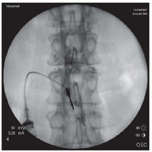
FIGURE 68-1. Lumbar interlaminar epidural injection. AP view showing a typical vacuolated epidurogram.

Several procedural modifications are recommended for cervical or thoracic interlaminar epidural injections due to the presence of the underlying spinal cord. For example, the cervical or thoracic interlaminar epidural injections should not be performed at the level of herniated nucleus pulposus or spinal stenosis, to avoid further potential spinal canal compromise and spinal cord compression. Furthermore, consideration should be given to directing the needle so that it contacts the inferior aspect of the lamina, to provide a clearly felt sense of depth prior to engaging the ligamentum flavum. The needle is then withdrawn slightly and directed into the ligamentum flavum. Further needle advancement should be performed using a lateral view and in addition to using the loss-of-resistance technique, the needle tip should not be advanced further than the laminar line to avoid the potential penetration of the dura mater or spinal cord injury. Epidural dye pattern recognition should be performed after a minimal amount of contrast has been injected since a total volume of less than 4 mL is recommended in these body regions (Figs. 68-2 and 68-3).


FIGURE 68-2. AP view of cervical interlaminar epidural injection demonstrating typical “honeycomb” pattern of epidurogram.


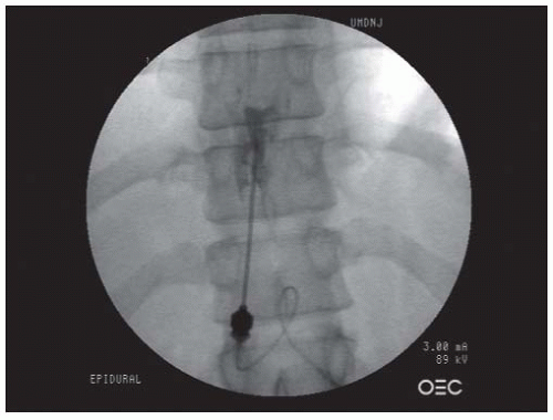
FIGURE 68-3. AP view of thoracic interlaminar epidural injection at the T10-11 level. Note the angle of the needle relative to the axis of the spine.


TRANSFORAMINAL EPIDURAL INJECTION


Comment

In one systematic review comparing transforaminal, interlaminar, and caudal ESIs, the authors concluded that there was moderate evidence for long-term (>6 weeks) relief of lumbar radicular pain using the transforaminal and caudal approaches, but limited evidence using the interlaminar approach (30). The authors also concluded that there was moderate evidence for relief of cervical nerve root pain using both the transforaminal and interlaminar approaches (30). Another study concluded that the transforaminal approach was more effective than the interlaminar or caudal approaches in treating lumbar pain (31). A review showed transforaminal lumbar ESIs under fluoroscopic guidance to be more cost effective than blind interlaminar and caudal ESIs (32).

As of this writing, no peer-reviewed, randomized controlled studies have compared transforaminal and interlaminar ESIs in the thoracic region.


Technique

For lumbar transforaminal epidural injection (TEI), the patient is placed in a prone position with a pillow or abdominal roll under the abdomen to at least reduce and ideally reverse the lumbar lordosis in order to open up the foramen. Using an ipsilateral oblique fluoroscopic view, the x-ray tube (source) of the C-arm fluoroscope is generally angulated in either a caudal direction (for L5-S1 and L4-5 TEI) or cephalic direction (for L3-4 and above TEI) to square the inferior endplate of the vertebral body, and to place the superior articular process of the subjacent segment pointing at 6 o’clock of the pedicle of the above level that appears as a Scottie dog eye. Local skin is then prepped and draped in a sterile manner. A local skin wheel is raised with 1% lidocaine at the needle entry site and the subcutaneous tissue in the needle trajectory path is infiltrated with 1% lidocaine. A 22- or 25-gauge spinal needle of appropriate length is inserted and directed down and parallel to the fluoroscopic beam toward the “safe triangle.” The safe triangle is formed by the lower border of the pedicle, the lateral margin of the vertebral body, and the traversing nerve root. To avoid deep needle placement and potential injury to the vasculature or nerve root or DRG in the neuroforamen, the novice injectionist should advance the needle until the needle tip touches the lower edge of the Scottie dog eye, the junction of the transverse process and the superior articular process. The needle is then slightly withdrawn for 2 to 3 mm and redirected inferiorly just under the lower edge of the transverse process for about 0.5 mm. Further advancement of the needle should be under AP and cross table (lateral) views. The final needle tip position should be at the posterior half of the neuroforamen just under the pedicle in the lateral view to minimize the potential injury to the vasculature, nerve root, or DRG. In the AP view, the needle tip should not be medial to the medial edge of the pedicle to avoid penetrating the dura mater. For S1 transforaminal injections, the eye of the Scottie dog can also be used as an injection landmark. Using a slightly caudad and ipsilateral fluoroscopic view, the S1 Scottie dog image is outlined. The needle should be directed to the outer upper quadrant of the neuroforamen. In the lateral view, the needle tip should not pass the anterior margin of the sacral canal that appears as a radiological lucent strip. A neurogram pattern should then be visualized under an AP view (Fig. 68-4).

For the L5-S1 foramen, the C-arm source often needs to be tilted in a caudad direction to accommodate any remaining lumbar lordosis. An ipsilateral oblique projection is then used to visualize the Scottie dog and the target is identified as the
region immediately under the pedicle, slightly lateral to the 6 o’clock position (Fig. 68-5). This position leads to needle placement in the neuroforamen, ventral to the nerve root. Lateral imaging is used to demonstrate the needle depth, which should be located at the superior portion of the intervertebral foramen, just under the pedicle (Fig. 68-6). An AP view is then obtained to ensure that the needle tip is located at the “safe triangle,” slightly lateral to the 6 o’clock position of the pedicle. The safe triangle is formed by the lower border of the pedicle, the lateral margin of the vertebral body, and the traversing nerve root. A needle position located within the safe triangle and lateral to the 6 o’clock position is deemed safe because it will not penetrate the nerve, blood vessels, or dura mater. Nevertheless, because of the precarious location of the nerve root and the DRG, caution should be exercised by advancing the needle slowly upon entering the neuroforamen, to avoid needle penetration of these neurologic structures. If the patient complains of radicular pain or paresthesias, the needle should be withdrawn and redirected superiorly. Once the needle is deemed at the proper position, approximately 1.0 mL of the contrast is injected under live fluoroscopic view. The needle should be redirected if there is vascular uptake of the contrast. The injected contrast should ideally outline the nerve root and also show epidural spread. Three milliliters of a mixture of solution containing 40 to 125 mg of preservative-free methylprednisolone, 6 to 9 mg of preservative-free betamethasone sodium phosphate, 40 to 50 preservative-free triamcinolone (33,34), or other equivalent dose of preservative-free corticosteroid and preservative-free 1% lidocaine can be slowly injected into the neuroforamen through the spinal needle (25,26).


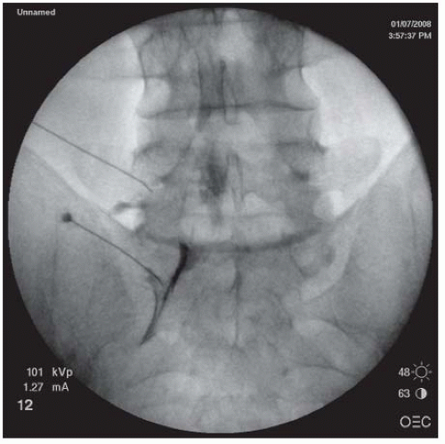
FIGURE 68-4. Left L5-S1 TEI. AP view showing a neurogram pattern of left S1 and L5 nerve roots.


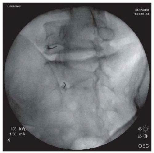
FIGURE 68-5. L5-S1 and S1 TEI. Oblique view showing needle just under the 6 o’clock position of the L5 pedicle and outer lateral quadrant of S1 for L5 and S1 TEIs respectively.

A thoracic TEI is performed with the patient in a prone position. The fluoroscope should be directed in a similar fashion as for lumbar TEI. A critical part of the injection is the correct identification of a clear rectangular-shaped clear space window under the fluoroscope. The upper and lower borders of the rectangle are the lower edge of the lamina of the same vertebral segment and the upper edge of the inferior vertebral endplate of the same segment. The lateral and medial borders of the rectangle are the medial edge of the rib head and the pars interarticularis of the same segment, respectively. After proper skin preparation and local anesthesia, the spinal needle is inserted and directed toward the lower edge of the Scottie dog eye using the same technique as in the lumbar TEI. Caution should be exercised not to direct the needle outside the clear rectangle window. If the needle strays too far laterally outside the rectangular window, it can penetrate the pleura, resulting in a pneumothorax. Needle placement too medially outside the rectangular window can result in spinal cord injury. The final needle position should be in the posterior half of the neuroforamen in the lateral view and the 6 o’clock position of the pedicle in an AP view. The contrast and corticosteroid are injected in a similar fashion as when performing a lumbar TEI.


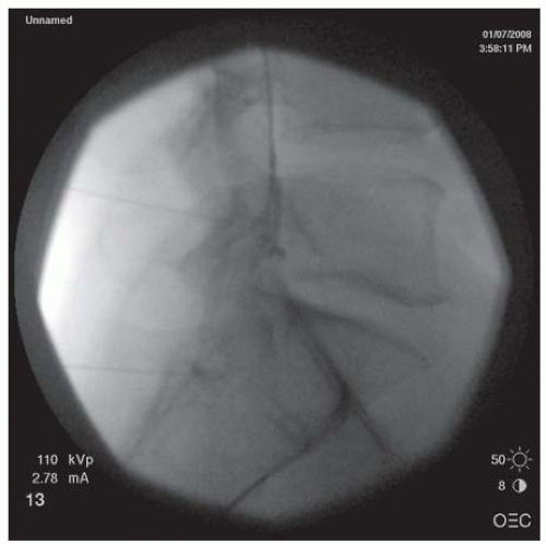
FIGURE 68-6. Lumbar L5-S1 and S1 TEIs. Lateral view demonstrating contrast in the ventral epidural space. Theoretically, the transforaminal approach should achieve more anterior flow of the injectate than would be typical for an interlaminar approach.

A cervical TEI is performed, with the patient in a supine position and the head turned to the contralateral side. A peripheral intravenous line should be placed, and vital signs, as well as oxygen saturation, should be monitored. The C-arm fluoroscope is rotated ipsilaterally and angulated either cephalically or caudally to maximally visualize the targeted neuroforamen (Fig. 68-7). After aseptic skin preparation and draping, the skin entry site is anesthetized with 1% lidocaine. A 22- or 25-gauge, 3.5-in. spinal needle is then inserted at the injection site and directed down and parallel to the fluoroscopy beam until the needle contacts the superior articular process forming the
posterior wall of the neuroforamen. At this point, the needle tip is withdrawn and directed slightly anteriorly to “walk off ” the superior articular process and slip into the neuroforamen. The C-arm is turned to the AP view to assess the needle depth. The needle should be advanced in millimeter-by-millimeter increments in the AP view to ensure that the needle is not advanced past the center of the lateral mass (Fig. 68-8). Overzealous advancement of the needle into the inner half of the lateral mass can potentially lead to penetration of the dura into the subarachnoid space or into the spinal cord. The desired final needle location is the posterior wall of the targeted neuroforamen in the oblique view and the lateral half of the lateral mass in the AP view. After negative aspiration of the cerebrospinal fluid (CSF) or blood, 0.5 to 1 mL of contrast is injected under real-time imaging to exclude a vascular pattern. The needle should be repositioned if there is either blood flashback in the needle hub or a vascular pattern upon contrast injection. If the patient complains of paresthesias or radicular pain, the needle also needs to be repositioned. With satisfactory needle position, the injected nonionic water soluble contrast often outlines the exiting spinal nerve and fills the neuroforamen with epidural spreading or an epidurogram. After the satisfactory position, 1.0 mL of a test dose of 1% lidocaine is injected, and the patient is monitored for 2 minutes for any changes in vital signs or consciousness or neurological deficits in the extremities that would indicate an intravascular injection. For patients without abnormal signs, 40 mg of methylprednisolone, 6 mg of betamethasone sodium phosphate, or 10.25 mg of nonparticulate dexamethasone in a total volume of less than 2 mL per neuroforamen may then be injected (25,26,35). To prevent inadvertent arterial embolism into the spinal cord and brain stem, a nonparticulate soluble corticosteroid such as dexamethasone is recommended for cervical transforaminal ESI.


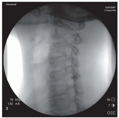
FIGURE 68-7. Oblique view of transforaminal cervical epidural injections showing needles in the posterior walls of the C4/5 and C5/6 neuroforamina.


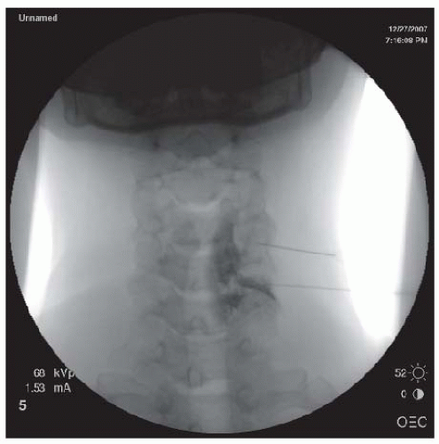
FIGURE 68-8. AP view of transforaminal cervical epidural injection demonstrating left C6 neurogram.


CAUDAL LUMBAR EPIDURAL INJECTIONS

Caudal lumbar epidural injections are performed by inserting a needle through the sacral hiatus into the epidural space at the sacral canal (Figs. 68-9 and 68-10). The patient is placed in a prone position. The legs are slightly abducted and feet turned inward to separate the gluteal fold to facilitate palpation of the sacral cornu. The sacral skin is prepped and draped in a sterile manner. AP imaging can be used to visualize the location of the
sacral hiatus. Alternatively, lateral imaging is used to view the bone defect, consistent with the opening of the sacral hiatus. The skin and the tissues overlying the sacral hiatus are anesthetized with 1% lidocaine. A 22- or 25-gauge spinal needle of appropriate length or a Tuohy epidural needle is inserted into the sacral hiatus. Loss of resistance can sometimes be felt upon needle penetration through the sacral ligament. Several milliliters of the contrast are injected in order to produce a sacral epidurogram to note the level that the contrast reaches. In the lateral view, a typical epidural contrast spread within the sacral canal resembles “smoke up a chimney” (Fig. 68-9), and in the AP view, it often looks like a “Christmas Tree” (Fig. 68-11). If a vascular pattern is observed, the needle should be withdrawn and redirected. Upon proper positioning, a mixture of 10 to 20 mL of solution containing 80 to 125 mg of preservative-free methylprednisolone or other equivalent doses of corticosteroid, preservative-free normal saline, and preservative-free 1% lidocaine is slowly injected into the epidural space through the spinal or epidural needle.


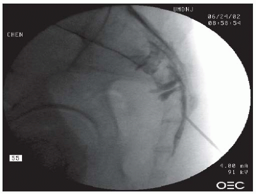
FIGURE 68-9. Caudal epidural injection. Needle tip in the sacral canal. Note the “smoke up the chimney” pattern of contrast in the epidural space of the sacral canal.


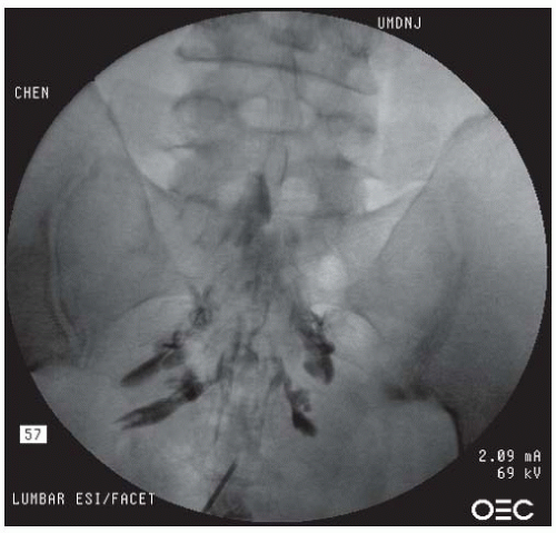
FIGURE 68-10. Caudal epidural injection. AP view demonstrating contrast in a “Christmas tree” pattern within the epidural space.


FIGURE 68-11. Right L5 TEI. Injecting 0.5 mL of contrast outlined the right L5 spinal nerve. However, contrast also spread through the epidural space and outlined the right S1 nerve root as well.


TIMING, FREQUENCY, DOSE, AND VOLUME OF EPIDURAL INJECTIONS


Comment

Optimal timing of ESIs is unknown, although there is evidence of better benefit if ESIs are performed within 3 months of radicular pain onset (36,37). The general consensus is that most patients with radicular symptoms should undergo a few weeks of treatment including oral medications, physical therapy or manual medicine, and relative rest from activities that exacerbate their pain, before undergoing ESIs (3,23,26). If a patient does not have success with such a program, or if the therapy cannot progress because the patient’s pain is too severe, an ESI is indicated for pain control. In contrast, ESIs can be considered earlier in patients with severe radicular pain not responding to even opioid medication or with pain that is significantly interfering with a patient’s sleep and/or function (26,28). Early ESIs also carry the theoretical benefit of controlling inflammation at an early stage (5,7,38) and possibly preventing permanent neural damage such as nerve fibrosis from prolonged inflammation (8). A study demonstrated that an ESI has higher efficacy (>75% pain relief) for patients with radicular pain within 3 months duration, whereas less benefit was found in patients with sciatica longer than 7 months (39). Another study compared epidural injections with bupivacaine alone versus injections of bupivacaine with methylprednisolone in patients with lumbar radicular pain longer than 6 months in duration. At 3-month follow-up, both treatments reduced pain but there was no additional benefit with corticosteroids (40).

The time interval between epidural injections should vary depending upon the steroid preparation used. Because injected methylprednisolone is reported to remain in situ for about 2 weeks (41), the clinician should probably consider waiting for about 2 weeks before fully assessing a patient’s response or administering a repeat injection.

Studies have suggested that the total maximum methylprednisolone dose should be about 3 mg/kg of body weight because excessive salt and water retention can occur at doses above this due to the mineralocorticoid properties of corticosteroids. In general, it is felt that up to three to four ESIs within a year may be performed if clinically indicated (23). Some clinicians schedule and proceed with a series of three ESIs regardless of the clinical response to the first preceding injection(s). Although the efficacy of this approach is unclear, as there are no medical outcome studies to support or refute such a regimen, it may be best to reassess the response to a
given injection at the time of an intervening office visit before proceeding with another injection. Using this approach, the clinician can determine if another injection is still needed and can more readily alter their planned injection technique, rather than trying to make this assessment at the time of the scheduled injection itself.


TABLE 68.1 Volume of Injectate for Epidural Injections


Interlaminar ESI (mL)


Transforaminal ESI (mL)


Caudal ESI (mL)


Lumbar


4-10


2-3


10-20


Cervical and thoracic


3-4


1.5-2


N/A


ESI, epidural steroid injection.


Data from reference 14, 16, 17, 20.


Recommended injection volumes and the corticosteroid doses are dictated mainly by the approach used as shown in Tables 68-1 and 68-2 (23,25,26,29). The epidural steroid can be injected in a preservative-free diluent such as lidocaine (1% to 2%) or normal saline (25,28,29). In the cervical spine, it is recommended that local anesthetics and steroids be injected separately to prevent a potential embolus of poorly dissolved steroid particles within a local anesthetic diluent. A recent study demonstrated that a nonparticulate dexamethasone has similar efficacy compared with particulate triamcinolone, and carries the lowest potential risk of embolization with inadvertent intravascular injection when used in ESIs (41).


TABLE 68.2 Dosage of Corticosteroids for Epidural Injections


Interlaminar ESI (mg)


Transforaminal ESI (mg)


Caudal ESI (mg)


Lumbar


Methylprednisolone


Triamcinolone


Diacetate (Aristocort)


Triamcinolone


Acetonide (Kenalog)


Betamethasone


Dexamethasone


80-120


25-50


40-80


6-18


10-15


40-80


25


40


6


10-15


80-120


25-50


40-80


6-12


10-15


Cervical and thoracic


Methylprednisolone


Triamcinolone


Diacetate (Aristocort)


Triamcinolone


Acetonide (Kenalog)


Betamethasone


Dexamethasone


80-120


25-50


40-80


6-18


10-15


40-80


25


40


6


10-15


N/A


N/A


ESI, epidural steroid injection.


Data from references 16, 17, and 45.


FLUOROSCOPIC GUIDANCE AND CONTRAST

Fluoroscopic guidance and contrast enhancement are essential for accuracy when performing epidural injections (43). Published data show that even in experienced hands, epidural injections without fluoroscopic and contrast-enhanced guidance (i.e., “blind injections”) often result in inaccurate placement (Table 68-3) (43). These misplacements include the needle being inadvertently positioned into the subarachnoid, intravascular (Table 68-4), or subcutaneous regions (caudal approach) or fascial plane superficial to the ligamentum flavum for interlaminar ESI. Misplacement into the subarachnoid or intravascular regions has major potential safety implications, particularly for those injections that include local anesthetics as part of the injectate. Use of detection of flash back of blood in the needle hub to gauge the intravascular placement of needle is not a reliable substitute for looking for a vascular pattern after contrast injection (44). Although injection accuracy should also logically affect efficacy, there is very limited data on the efficacy of fluoroscopic-guided ESIs compared with blind ones. One such study demonstrated that fluoroscopic-guided transforaminal ESIs provided better pain relief than blind interlaminar ESIs (45). ESIs using fluoroscopic guidance have also been shown to reduce procedure-related complications compared to non-image-guided injections (46, 47, 48).

As a result of these factors, it is recommended that ESIs be performed under fluoroscopic guidance and with radiographic contrast, documenting appropriate needle placement in order to improve their accuracy, and by extension their safety and efficacy (49).


TABLE 68.3 Incorrect Needle Placement Associated with “Blind” ESI


Lumbar Interlaminar


Caudal


Lumbar Transforaminal


Cervical Transforaminal


Misplacement


17%-25%


12%-38%


N/A


N/A


Data from Renfrew DL, Moore TE, Kathol MH. Correct placement of epidural steroid injections: fluoroscopic guidance and contrast administration. AJNR Am J Neuroradiol. 1991;12(5):1003-1007.


Efficacy of Epidural Injections

Recent studies have demonstrated good efficacy of lumbar ESIs when proper needle placement is confirmed by using fluoroscopic guidance and radiographic contrast (50,51). A meta-analysis of 12 published randomized controlled trials concluded that ESIs are effective (52). In a systematic review of randomized trials on lumbar epidural injections, Abdi et al. concluded that there was moderate evidence that caudal and TEIs are effective in providing long-term (>6 weeks) pain relief and limited evidence for the effectiveness of lumbar interlaminar ESIs (30). Other studies have suggested that 60% to 75% of patients receive some relief after ESIs (53,54). Benefits include relief of radicular pain and low back pain (generally relieving leg pain more than back pain), improved quality of life, reduction of analgesic consumption, improved maintenance of work status, and a decreased need for hospitalization and surgery in many patients (27,50, 51, 52, 53, 54, 55, 56). One study showed no difference in analgesic use in patients with sciatica who had received three ESIs (58). Another study reported that patients were more likely to start taking opioids and more likely to receive surgery after receiving multiple (>3) injections than patients receiving fewer injections (59). However, the population of patients receiving multiple steroid injections was more likely to have had more advanced disease such as spinal stenosis.

A prospective cohort study was conducted on cervical TEIs for both neck pain and radicular pain from herniated discs or spondylosis. Twenty-one such patients awaiting surgery received cervical TESIs 2 times, at 2-week interval with 12 months follow-up. All patients had reduction in neck and radicular pain, and five of these patients cancelled the surgery (59). In contrast, a prospective randomized study involving 20 patients with cervical radicular pain confirmed by selective nerve root block (SNRB) and with magnetic resonance imaging (MRI) evidence of corresponding segmental pathology demonstrated that there was no difference in radicular pain reduction between steroid/local anesthetic and saline/local anesthetic groups at 3-week follow-up (60). A limitation of this study, however, was that it only involved small numbers of patients and that it is unknown whether saline/local anesthetic is a true control.


TABLE 68.4 Incidence of Intravascular Uptake (IVU) Associated with ESI


Lumbar Interlaminar


Caudal


Lumbar Transforaminal


Cervical Transforaminal


IVU


1.9%


10.9%


11.2%


19.4%


Data from Carette S, Leclaire R, Marcoux S, et al. Epidural corticosteroid injections for sciatica due to herniated nucleus pulposus. N Engl J Med. 1997;336:1634-1640.


There are more studies in support of ESIs for low back pain (7,8,42,53, 54, 55) than there are negative studies (56). Problems with some of these supportive studies, however, include the fact that most of these studies did not use fluoroscopy and radiographic contrast to document accurate placement of the injected substance into the epidural space. Furthermore, many of these injections were not performed at the presumed level of pathology, even though this has been demonstrated to be critical to the success of ESIs (61). These methodologic problems are likely contributing factors to the mixed assessment that ESIs have received. A review of six prospective randomized clinical trials of fluoroscopic-guided transforaminal ESIs, selective nerve blocks, or periradicular nerve injections concluded that there is moderate (level III) evidence that TESIs are safe and effective in reducing radicular pain. However, more prospective, randomized, placebo-controlled studies using sham procedures are needed to provide more conclusive evidence for the efficacy of TESIs in treating lumbar radicular symptoms (62). A recent review article concluded that with proper patient selection, ESIs are a reasonable alternative to surgery for short-term pain relief, reduced medication use, and increased patient activities while awaiting natural recovery (63).

Aside from technical considerations, response to ESIs has been shown to be related to several other factors such as the type and quantity of steroid preparation used, volume of injectate, underlying pathophysiology, and the duration of symptoms (23,26,28). In general, radicular pain or radiculopathy induced by herniated nucleus pulposus appears to respond better to corticosteroid injection than that induced by spinal
stenosis. There is essentially no literature that correlates the type of disc herniation with the response of ESIs. It is the authors’ collective experience and observations that patients with large lumbar disc herniations obliterating the neuroforamen or extraforaminal herniations often have less benefit from ESIs. One study demonstrated that radiculopathy induced by the combination of spinal stenosis and disc herniation has less favorable outcome with ESI. In lumbar spinal stenosis, the efficacy of ESI correlated with the degrees and the levels of stenosis categorized by MRI (64). Patients with single-level lumbar spinal stenosis generally respond better than those with multilevel lumbar spinal stenosis. ESIs provide better efficacy in reducing pain and opioid consumption for patients with mild to moderate rather than severe stenosis. But a prospective cohort study with 12-month follow-up in patients with severe degenerative lumbar spinal stenosis found that fluoroscopic-guided and contrast-enhanced caudal ESIs reduced bilateral radicular pain and improved standing and walking tolerance (65). In contrast to radiculopathy due to herniated discs and/ or spinal stenosis, radiculopathy caused by epidural scar tissues or trauma such as nerve root stretch injury often responds poorly to ESI.

A recent prospective, randomized study on lumbar TESIs demonstrated positive efficacy in treating radicular low back pain. The success rate for TESI is 84%, compared to 48% with trigger point injection, at 1.4 years of follow-up (66). Another prospective, randomized controlled clinical trial compared perineural (transforaminal) epidural injection with conventional posterior (interlaminar) epidural injection with steroid, and perivertebral injection with local anesthetic as a control group (27). The result demonstrated that perineural injection was the most effective approach. Both perineural and conventional epidural injection with steroid were better than that with saline alone (27).

Uncontrolled studies have generally reported favorable outcome of cervical epidural injections for cervical radiculopathy with structural abnormalities such as cervical disc herniation (66,67) and spondylosis (68). However, the prospective, randomized, blinded and controlled clinical trials on the outcome of cervical and thoracic epidural injections have not been reported yet in the peer-reviewed literature.

At the time of this writing, there have been no prospective randomized trials on thoracic ESIs that have been published in the peer-reviewed literature.

Patients should be educated that ESI alone may not be the only solution to give them long-term benefits. ESI is just one of many nonoperative treatments used to treat low back pain or radicular symptoms. Other treatments may include short-term bed rest; medications (e.g., analgesics, muscle relaxants); a properly designed program of physical therapy; and management of any psychological, financial, marital, and work-related problems. A comprehensive treatment approach is likely to produce better outcomes for patients with low back pain than any single modality used in isolation (23,26,51). Recently published research on the outcome of ESIs has supported this notion of multifaceted treatment (50,69).


Recent Advances and Investigations on the Management of Radicular Pain

A study was performed on lumbar TEIs for radiculopathy using autologous conditioned serum (ACS) containing enriched IL-1 antagonist. The ACS group showed statistical superiority over both triamcinolone groups (5 and 10 mg) with regard to the VAS score for pain from week 12 to the final evaluation at week 22, statistical superiority at week 22 compared to the triamcinolone 5 mg group, and no significant difference compared to the 10 mg triamcinolone group. This is an exciting finding, as autologous blood is not associated with the same side effect concerns associated with corticosteroids, and theoretically can be used more frequently than corticosteroids. Additional studies are needed to confirm this finding (70).

An animal study examined the potential benefits from anti-TNF-[α] therapy in reducing neurotoxic effects induced by the nucleus pulposus on neuronal tissues (71). Two openlabel human clinical trials, one using intravenous infliximab (a monoclonal antibody against TNF-[a]) and the other using (etanercept) a soluble TNF-[a] receptor antagonist, in patients with sciatica from disc herniation demonstrated significant efficacy in pain reduction (72,73). Although these basic science and human studies initially implied potential clinical use of anti-TNF-α medication as a treatment for patients with radiculopathy due to disc herniation, there were disappointing long-term findings related to the evaluation of the efficacy of an anti-TNF-[α] treatment versus a placebo injection in disc herniation-induced sciatica in a randomized controlled setting. Specifically, 3-month results showed no difference in the patient-reported symptoms or in the more objective outcomes (SLR, days on sick leave, discectomies) between intravenous infliximab 5 mg/kg and placebo (74). The 1-year results also confirmed the earlier findings (75). Clearly, further studies using multiple intravenous infusion of the anti-TNF-α agents or epidural injection of the similar substances are necessary to clarify any efficacy of anti-TNF-α treatment in radicular pain.


Safety and Complications of Epidural Injections

A retrospective cohort study reviewing the immediate complications of 2,217 patients who received selective lumbar nerve root blocks under fluoroscope, reported a 5.5% minor complication rate (76). When performed by a skilled, experienced clinician within an appropriate setting and on carefully selected patients, the chance of a significant complication from an ESI is remote (23,25,26,77

Only gold members can continue reading. Log In or Register to continue

Stay updated, free articles. Join our Telegram channel

May 25, 2016 | Posted by in PHYSICAL MEDICINE & REHABILITATION | Comments Off on Spinal Injection Procedures

Full access? Get Clinical Tree

Get Clinical Tree app for offline access