Scoliosis and Other Spinal Deformities


Scoliosis and Other Spinal Deformities


Scott M. Paul


DEFINITION

One of the unique features that distinguishes humans from the animal kingdom is our bipedal nature. Quadrupedal ambulation is more stable, with built-in redundancies. However, by standing on our hind legs, we are able to see above many other vertebrates. Our arms and hands are freed to manipulate tools. The unique balance between flexibility and stability in the human spine helped to realize this evolutionary innovation. It is a wonder that this balance is maintained for a lifetime of wear and tear for the vast majority of the world’s population. When that balance is altered, the result is deformity of the spine in one or more of its planes of movement. These conditions have likely been with man since bipedal mobility began. Hippocrates stated that “there are many variations of curvatures of the spine, even in persons who are in good health, for it takes place from natural conformations and from habit, and the spine is liable to be bent from old age and pains” (1).

We have advanced in the effectiveness of the treatment of spinal curvatures since the time of the ancient Greeks and Romans, when various types of traction devices and truncal wraps reportedly were advocated. However, Hippocrates’ description of the pathophysiology of spinal curvatures is fairly accurate. In this chapter, we will discuss the evaluation and treatment of people disabled by spinal deformity, including those whom Hippocrates might have described as in good health (idiopathic), those with natural conformations (congenital), those with curves due to “habit” (neuromuscular), and those who are bent from old age and pains (degenerative and traumatic).

Colloquially, scoliosis is often used as a term to include all spinal deformities. The term was coined by Galen in the second century A.C.E (1). It is derived from the Greek word “skolios” (2), meaning bent, twisted, or curved. As such, it could include any spinal curvature. However, Galen defined it as the (abnormal) curve of the spine in the coronal (aka frontal) plane (Fig. 34-1). Sagittal plane pathology results from excess or reversal of the spine’s natural sagittal curves. Kyphosis, the word Galen applied to sagittal curves with a posterior vertex (concave anteriorly), derives from the Greek word “kuphos” (2), meaning bent forward or humped. Within a range of degrees, it is normal in the thoracic spine. Lordosis is the word used to describe a sagittal curve with an anterior vertex (concave posteriorly). It derives from “lordos” (2), meaning excessive posterior bending. Within a range of degrees, it is normal in the cervical and the lumbar spine (Fig. 34-2).


WHAT IS A PATHOLOGICAL CURVE?

Although the presence of any fixed frontal curve could be considered outside of the design parameters of the spine and abnormal, it is generally accepted that some degree of frontal curve may be “within normal limits.” The Scoliosis Research Society (SRS) has defined a medically significant frontal plane curve (scoliosis) as any curve which is greater or equal to 10 degrees, with or without a rotatory component (3). The definition of a pathological sagittal plane curve is less settled. The spine is “balanced” in the sagittal plane along a plumb line dropped from the center of C7 to the sacral promontory (4). This balance can be influenced by the position of the head and neck, as well as the pelvis and lower limbs. This dynamic balance may help explain some of the outstanding controversy about pathological sagittal curves. Numerous authors (1, 5) give the normal range of thoracic kyphosis to be 20 to 40 degrees, others (1) give the range as 30 to 50 degrees. Fon et al. (6) have shown that kyphosis increases about 4 to 5 degrees over childhood to the end of adolescence. Kyphosis is also known to increase with aging. The SRS has agreed that normal thoracic kyphosis ranges from 20 to 50 degrees. The range of normal for lumbar lordosis is even wider. Jackson and McManus (7) found the mean lordosis to be 59 degrees. Vaz et al. (8) found a mean lumbar lordosis of 46.5 degrees, with a range from 26 to 76 degrees. The SRS has agreed (4) that normal lumbar lordosis can range from 31 to 79 degrees, depending on measurement technique. However, in assessing any curve detected in a clinical situation, it is important to neither simply rely on curve measurements which are within the “normal” range nor to consider the curve amplitude as the only factor in assessing the patient and his/her complaints and impairments.


EPIDEMIOLOGY

As will be discussed later, spinal deformities can impair not only the musculoskeletal system but also many other organs and systems, often in people already coping with other impairments from chronic disabling conditions. Scoliosis
and the other spinal deformities are not uncommon. The prevalence of idiopathic scoliosis is reported to be between 0.3% and 2% of the population (9). The incidence of degenerative scoliosis is reported as 6% in people over 50 years of age and 36% in persons over 50 with osteoporosis (10). Scheuermann’s disease, a common cause of pathological thoracic kyphosis, has a prevalence of between 0.4% and 8.3% of the population. Up to a 100% incidence of spinal deformity can be found in people with many neuromuscular conditions (see section on “Neuromuscular Scoliosis”).


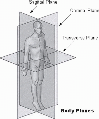
FIGURE 34-1. The planes of the body. (Courtesy of National Cancer Institute’s SEER Program web-based training module.)


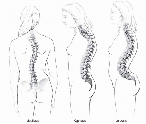
FIGURE 34-2. There are three basic types of pathological spinal curves. (Courtesy of Donald Bliss and Alan Hoofring of the Medical Arts and Publications Branch, NIH.)

A physiatrist cannot avoid scoliosis by subspecializing. Spinal deformity is seen in all ages and almost every type of disabling condition (Table 34-1). Physiatrists who do not treat children cannot avoid childhood onset spinal deformities; they will encounter adults who had spinal deformities as children and need to help them address late sequelae of their deformities.

Hopefully, you have now gained an appreciation for the definition of scoliosis and its importance as an issue in the care of persons with disability and/or pain. We will now proceed to develop a construct to use to help better understand these conditions and effectively care for the persons with these disabilities
and their families. By persevering through the remainder of this chapter, you will have the chance to


TABLE 34.1 Types of Scoliosis and Typical Ages of Onset


Pathophysiological Type of Scoliosis


Typical Age of Onset


Idiopathic


Infancy to adolescence


Neuromuscular


Childhood to adolescence


Bone or ligamentous dysfunction


Childhood


Traumatic/posttraumatic


All ages


Infectious or neoplastic


All ages


Degenerative


Middle age to seniors


  • Regain sufficient memory of the anatomy and physiology underlying the balance of the spine in order to appreciate the factors contributing to the various spinal deformity pathologies and to have a basis to develop a rational treatment plan


  • Understand how to obtain the crucial historical, clinical, and radiographic data to form an educated diagnosis and prognosis


  • Appreciate the impact of spinal deformity on a person’s quality of life


  • Understand the pathophysiology and etiologies of spinal deformity


  • Understand the roles of physiatric modalities in the (re) habilitation of persons with spinal deformity


  • Understand the role of surgical treatment


  • Consider the issues involved in postsurgical rehabilitation


SPINAL ANATOMY AND PHYSIOLOGY


Three Columns for the Price of One (Bony Functional Anatomy)

To achieve the elegant balance of flexibility and stability that the human spine offers, the design requires 7 cervical, 12 thoracic, 5 lumbar, and a fused set of sacral and vestigial coccygeal vertebrae. The stability comes from the fact that the spinal column is actually three columns in one. The anterior column is composed of the anterior longitudinal ligament and the anterior portion of the vertebral body. The middle column is made up of the posterior wall of the vertebral body and the posterior longitudinal ligament. The posterior column is formed by the posterior bony arch, which consists of the transverse processes, facets, laminae, and spinous processes. Each vertebra has the potential for six degrees of freedom (Fig. 34-3): translation in all three axes of movement and rotation around each axis. However, all vertebrae are not created equal. Movement at different levels of the spine is limited in some planes. In essence, in the function of the spine follows its form (with apologies to Louis Sullivan). The cervical vertebrae have the greatest freedom including free flexion, extension, lateral rotation, and lateral flexion (this is fortunate, since this freedom affords the eyes and ears a wider field of function). This mobility is due to larger disks, concave lower and convex upper vertebral body surfaces, and transversely aligned facet joints. Thoracic vertebrae have restricted flexion and extension and limited rotation but freer lateral flexion due to their attachments to the rib cage, smaller disks, flatter vertebral body surfaces, frontally aligned facet joints, and overlapped spinous processes. The lumbar spine has good flexion and extension and free lateral flexion due to large disks, posteriorly directed spinous processes, and sagittally directed facet joints. However, there is only limited lateral lumbar rotation due to facet alignment (Fig. 34-4).


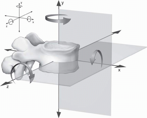
FIGURE 34-3. Six degrees of freedom are possible at each vertebra. The vertebra can move in each of the planes and rotate around each of the axes. (Courtesy of Donald Bliss and Alan Hoofring of the Medical Arts and Publications Branch, NIH.)


Ties That Bind (Elastic Tissues)

The intervertebral disks form one fourth of the total length of the spinal column (11). Since they are a viscoelastic tissue, they serve as shock absorbers and take part of the load applied to the spinal column. They also redistribute the load applied on the vertebral bodies, moving more of the force to the stronger, cortical bone (arranged in parallel with the length of the spine) at the vertebral end plates than the thinner layer of cortex sandwiched between softer cancellous (and therefore weaker) bone in the center of the vertebral body. The disks also help connect the vertebra. And, as implied above, they participate in the articulation of the spine.

A number of ligaments also help to link the spine together. The anterior longitudinal ligament is a thick, strong ligament that stretches from the occiput to the sacrum along the anterior surface of the vertebral body. It attaches to the vertebral end plates and is confluent with the anterior portion of the annulus fibrosis of the disk. At the posterior aspect of
the vertebral body, there is a similar ligament, the posterior longitudinal ligament. However, it has an hourglass shape and is thinner than the anterior ligament. There are also three ligaments which connect portions of the posterior column of the spine. The supraspinous ligament stretches across the tips of the spinous processes while the interspinous ligaments stretch from one spinous process to the next. The ligamenta flava (flava is Latin for yellow and the ligament is that color due to the high proportion of elastic tissue within it) connect the laminae of adjacent vertebrae from the anterior surface of the proximal lamina to the posterior surface of the distal lamina. They also blend in with the capsule of the zygopophyseal joints.


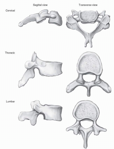
FIGURE 34-4. Typical vertebrae. (Courtesy of Donald Bliss and Alan Hoofring of the Medical Arts and Publications Branch, NIH.)


Spinal Articulations

The spine has two primary joints. The intervertebral joints (as noted above) are formed by the surfaces of the vertebral bodies with the disks. These joints (with the restrictions noted above in the discussion of vertebral anatomy)
allow limited movement in all six directions, although each individual joint only moves to a small degree. The zygopophyseal joints are the only true synovial joints in the spine and are found at the junction of the pedicle with the lamina. The form of their alignment contributes significantly to the functional movement at different levels. The transverse alignment in the neck permits good flexibility in all planes. In the thoracic spine, the facets are aligned frontally which, along with the linkage to the rib cage, limits flexion and extension. The lumbar facets are arranged sagittally, limiting lateral rotation.


Dynamic Balance

It would be possible to place a spine consisting of only its bony and elastic tissues on a base and have the spinal column statically balance in an upright position. However, if the point of the flexibility of the spine is to give humans the freedom to move on two legs, then the spine requires a complex system of dynamic control. The neuromuscular system provides this balance, actuated via muscles intrinsic and extrinsic to the spinal column. The paraspinal muscles are a series of layers of longitudinal muscles attached from the occiput to the sacrum. They are partially distinguishable by their fiber direction and length but “from a functional standpoint, there is little reason to subdivide the back musculature in any detail since the muscles work together in large groups” (11). They serve primarily as extensors to resist the pull of gravity in sitting and standing. In addition, the superficial layers will produce ipsilateral rotation when firing unilaterally, while the deeper groups will produce contralateral rotation with unilateral firing. The primary lateral flexors of the trunk are the quadrati lumborum and oblique abdominal muscles. The primary flexors are the psoas, assisted by the recti abdominus.


CLINICAL ASSESSMENT OF THE PATIENTS WITH SPINAL DEFORMITY


History

Like every other medical condition, most of the diagnosis can be made just by taking a good clinical, functional history and performing a physical examination. “As each symptom is given, it should be pursued with a dogged tenacity until all the details are known” (12). We have mentioned that the majority of patients with scoliosis have an idiopathic condition. It is critical to first rule out spinal deformity associated with another disabling condition. Much of this will be accomplished with good history taking which can then direct and focus the physical and radiological examinations.

The first clue to the etiology of the deformity will be the age of onset. If the scoliosis presents at or soon after birth, it is more likely to be associated with a congenital bony deformity or inherited disease involving the neuromuscular system. If the scoliosis begins in late childhood or adolescence, it is likely to be idiopathic. Spinal deformity that begins in young adulthood is most likely due to trauma or infection; in middle age or later, it is likely to be associated with degenerative disk or joint disease and/or osteoporosis.

It is also important to determine if the onset of the curve is associated with any acute problem such as trauma or infection. The degree of curvature and how fast the size of the curve increases also offer insights into etiology. An underlying neuromuscular or ligamentous condition may promote quicker progression. Children within their adolescent growth spurt may have greater progression due to the rate of overall growth. Progression in an older adult may be an indicator of poor bone health.

Details relating to the nature of the curve itself are insufficient to advance the development of a good diagnosis and rational treatment plan. It is vital to inquire about associated complaints, signs, and symptoms. A thorough review of systems should be performed. However, certain areas of inquiry are more likely to yield clues. Although pain is associated with spinal deformity in adults, it is uncommon in children and adolescents (13) and obligates the physiatrist to search for underlying pathologies including neoplasm or infection. Similar to other pain-evoking conditions, details about the quality, intensity, and temporal and spatial parameters of the pain will help distinguish among bony, soft-tissue-based, and neuromuscular-based pain. Cardiopulmonary dysfunction is the most serious morbidity of spinal deformity. Therefore, questions about fatigability, shortness of breath, palpitations, and decrease in endurance must be included in the history. Any suggestion of cardiopulmonary dysfunction should lead to a more rapid and aggressive workup.

It is important to inquire about the symptoms of central nervous system dysfunction. Problems with loss of balance or falls may indicate a coordination problem such as is seen in cerebellar syndromes. Complaints of muscle spasms may indicate spasticity. Conversely, complaints of weakness or sensory loss may indicate peripheral nervous and/or muscle pathology. Reports of frequent joint sprains may indicate the presence of a condition associated with ligamentous laxity. In children and adolescents, it is important to obtain a history of developmental milestones. Indication of delay in any of the developmental spheres—social/behavioral, communication and cognition, fine motor, gross motor—may provide clues to the presence of an underlying syndrome. Since spinal deformity may be initiated or exacerbated by conditions affecting other areas of the body, the physiatrist should ask about conditions that may affect the lower limbs, such as a history of hip dislocation or leg length discrepancy, and even conditions associated with asymmetry of the upper limbs and head.

Last, but certainly not least, a thorough social and functional history must be obtained. Further clues into etiology may be gleaned. In addition, the insights obtained will identify the barriers to successful functional outcome to the treatment plan and ensure that the goals of the rehabilitation team are in concert with the goals of the patient and his or her family. In some cases, it may be beneficial to use a standardized questionnaire to follow the impact of the person’s spinal deformity on the function and related aspects of quality of life. There
is a validated instrument focused specifically on persons with spinal deformity (14, 15, 16). Alternatively, one of the many generic “disability/burden of care” or “health-related quality of life” instruments may be used (see Chapters 11 and 18).


Physiatric Clinical Examination

Although school screening programs for idiopathic scoliosis may only look at a child’s spine, it is not sufficient for a physiatrist to limit the assessment of spinal deformity to the spine. A comprehensive physiatric examination must first be done. This should include assessment of pain, strength, endurance, sensation, balance (in sitting and standing), coordination, range of motion, reflexes, mobility, and dressing skills. Leg length discrepancy should be assessed, which is not always easy to determine, and corrected.

Once the general examination has been completed, a more focused assessment of the spine must be performed. You do not have Superman’s x-ray version; clothing must be removed so that the spine can be observed directly. The surface anatomy should be viewed in both prone and side lying. If the patient can sit, then the spine must be seen in sitting. If he or she can stand, even with support, then the spine must be viewed in standing. If there is concern about leg length discrepancy and/or pelvic asymmetry, the spine should also be viewed in standing with leveling blocks under the foot on the “short” side and the depth of the correction recorded. Any difference in the shape or degree of curve in different positions should be noted, it is a sign of the degree of flexibility of the curve. The spine should be viewed from both the frontal and sagittal planes. The spinous processes should be palpated to better appreciate the curves’ dimensions, including shape in both frontal and sagittal planes and presence of rotation. The curve should be described by its length (approximately which vertebrae start and end the curve), its shape (e.g., “C” or “S” shaped), and the direction of the vertex of the curve. Truncal range of motion in all planes—forward flexion, rearward extension, R & L lateral flexion, and R & L lateral rotation—should be viewed with special attention to asymmetry and spinal levels where movement is occurring. While the patient is flexing forward, the examiner should look at the shape of the rib cage for prominence or humping (Adam’s forward bending test (17), which is the test used in school screenings) (Fig. 34-5), which may be a sign of a rotatory component to the curve. The shape of the rib cage should be viewed from all angles to look for deformity. Rib cage deformity may not only be a sign of rotation of the vertebrae but also suggest underlying pathologies such as osteogenesis imperfecta. One must look not only at rib prominence but also for deformities such as pectus carinatum or excavatum. Symmetry at the top and bottom of the trunk should be assessed in sitting and standing. Shoulder
symmetry should be viewed at the level of the acromioclavicular joint (13). Pelvic symmetry can be viewed either at the brim of the pelvis at the iliac crests or at the posterior superior iliac spines, which is seen on the surface of the buttocks as the two sacral “dimples.”


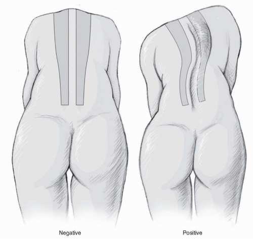
FIGURE 34-5. Adam’s forward bending test. (Courtesy of Donald Bliss and Alan Hoofring of the Medical Arts and Publications Branch, NIH.)

Measurement of symmetry of the appendicular skeleton and head should be done to rule out asymmetries. Head size and alignment should be viewed since reports have associated these issues with scoliosis (18). There are reported associations between upper limb deficiency and scoliosis (19, 20, 21), and, anecdotally, it has been associated with neonatal brachioplexopathy (Erb’s palsy). Therefore, upper limb asymmetries should be assessed. Upper limb length should be measured in a consistent manner. One method is to measure from the acromioclavicular joint to the radial styloid. Lower limb discrepancy produces functional scoliosis in standing and it may contribute to the development of fixed scoliosis, as well (22). Lower limb length is commonly measured from the anterior superior iliac spine at the pelvis down to the medial malleolus. The physiatrist must also look for functional asymmetries that could result from joint contractures or deformities (especially at the hips and knees) or tendon contractures (especially tendons of two joint muscles such as the hamstrings and gastrocnemius). Functional asymmetry may also result from asymmetric neurological function including hemiparesis.


RADIOGRAPHIC ASSESSMENT OF THE SPINE


Measuring the Curve Angle

The standard technique for measuring the angle of a spinal curve is called the Cobb angle (23). Initially developed as a frontal plane measurement, the technique can also be used for assessing sagittal curves. We will review the method for assessing scoliosis, but the rules can be applied similarly in lordosis and kyphosis. The film from which the curve is viewed should, if possible, include the entire spine on one film and be taken posterior-anterior (to reduce the dose of radiation to the breast) (13). Even if the patient is only suspected of having a frontal curve, a lateral film must also be obtained to better assess the amount of rotation in the curve. If at all possible, the films should be done in standing. If the patient is unable to stand, then sitting is preferred to a supine film. The P-A film should be taken with the arms at the side; the lateral film should be done with the arms at 90 degrees of abduction to keep the upper limb from obscuring the spine. Although it is common to shield the perineum when films are done, the initial films should be done without gonadal shield in order to fully visualize the hips, pelvis, and proximal femurs (13). In children and adolescents, a sense of skeletal maturity can be gleaned by looking for Risser’s lines at the crest of the pelvis.

To perform the Cobb measurement, the examiner should first note the upper border, lower border, and vertex of the curve. The curve is classified by the position of the vertex into thoracic, thoracolumbar (vertex at the thoracolumbar junction), or lumbar. The upper and lower borders are identified by the “end vertebrae.” They are the first and last vertebrae that tilt into the concavity of the curve (13). To define the angle, a line is drawn on the radiograph parallel to the upper end plate of the upper end vertebra. A second angle is drawn parallel to the lower end plate of the lower end vertebra. In most cases, these lines will not intersect on the film and, therefore, the angle cannot be measured directly. Instead, using principles learned in high school geometry, a line perpendicular (90 degree angle) to each of the above lines is drawn. The angle formed by the intersection of the perpendicular lines is equal to the angle made by the upper and lower end vertebrae and is measured and reported as the Cobb angle (Fig. 34-6).

More and more facilities are converting to filmless, computer-based radiological information systems. Many of these systems offer tools to directly measure the Cobb angle on the screen. The semimanual methods still require the clinician to place the parallel lines at the top and bottom of the curves. Studies have shown these methods to have precision and
reliability similar to the film-based method (24, 25, 26). More recently, some systems have introduced fully automated Cobb angle calculations. However, it appears that this technology still requires further refinement to achieve adequate accuracy for curves of all magnitudes (27).


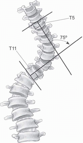
FIGURE 34-6. Measuring the Cobb angle. (Courtesy of Donald Bliss and Alan Hoofring of the Medical Arts and Publications Branch, NIH.)


Assessing Rotation

As mentioned above, the spine has six degrees of freedom, including rotation along the axis of the spinal cord. Many spinal curves are associated with rotation of the vertebrae. Curves with rotation may be more difficult to treat and may cause greater disturbance to the rib cage with resultant adverse effects on respiration. Therefore, it is important to assess the presence and degree of rotation associated with the curve, as well as its rate of progression. Radiographically, the position of the pedicles provides the best sense of degree of rotation on a plain x-ray. The examiner should note the symmetry and alignment of the pedicles in the P-A film. If asymmetry is present, the pedicle that is more prominent will indicate rotation away from that side of the vertebra. The degree of rotation can be graded using the system of Nash and Moe (13) (Table 34-2). Automated computerized systems to assess rotation are also under development (28).


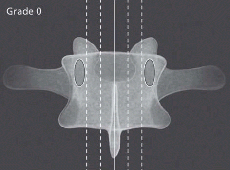
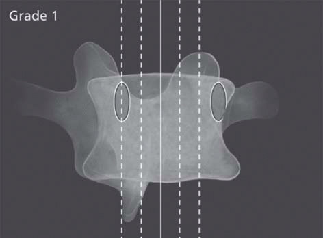
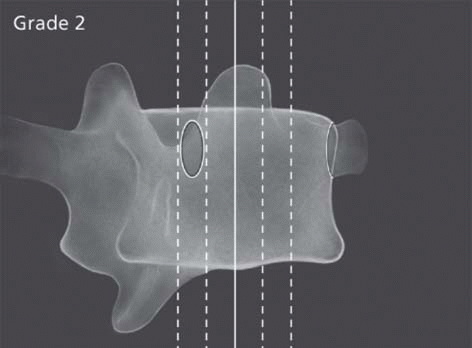
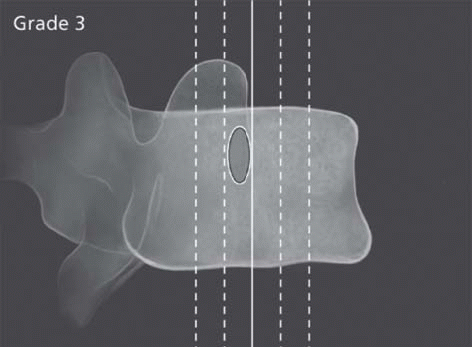
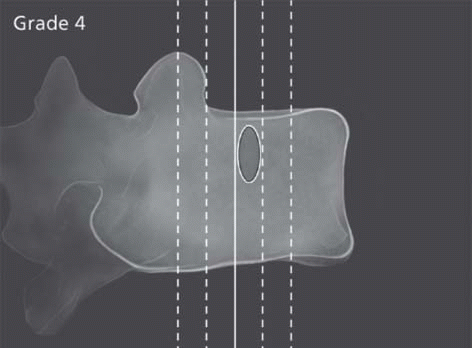
TABLE 34.2 Moe-Nash Classification of Spinal Rotation


Nash and Moe Grade


Description


Figure


Grade 0


Pedicles are seen symmetrically positioned at the lateral border of the vertebral bodies


image


Grade I


Slight asymmetry


image


Grade II


One pedicle is almost out of view


image


Grade III


Only one pedicle is seen, positioned at the center of the vertebral body


image


Grade IV


Only one pedicle is seen, positioned lateral to the center of the vertebral body


image


New computer-assisted techniques are also under development to supply additional information about the three-dimensional characteristics of the curve using the data supplied by digitized plain films (29). Such information may be useful in assessing the results of nonoperative and for planning operative treatments. Despite the additional radiation dosage,
three-dimensional CT may be useful, especially in congenital disorders (30, 31). MRI might offer similar information without radiation exposure (although still with the need for sedation in groups including young children and adults with claustrophobia); however, at the current state of technology, recommendations for its use are still limited to ruling out potential etiologies or neurological abnormalities (32). It is also important to remember that neither CT nor MRI is able to assess the impact of gravity on a curve, since both studies are done in a supine position.


Assessing Vertebral Integrity

It should be obvious by now that among the etiologies of spinal curves are conditions intrinsic to the bone. Most of these will become evident upon careful inspection of the radiographs obtained to determine the magnitude of curvature. The spinal elements should be inspected for signs of hemivertebra, unsegmented bars, and/or bifid spinous processes (see Fig. 34-10 later in this chapter). The ribs should be viewed to look for asymmetry or changes in the natural curve (Fig. 34-7). The pelvis should be examined for signs of asymmetry (Fig. 34-8). The hip joint should be examined for signs of subluxation. Femurs should be assessed for any asymmetry and separate femur films ordered if there is any suspicion of pathology.


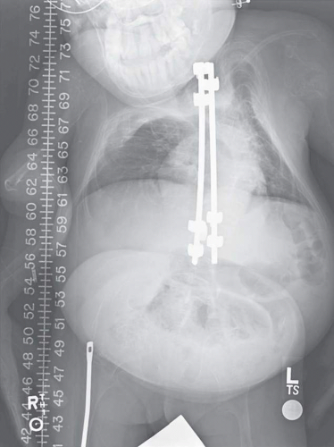
FIGURE 34-7. Rib asymmetry associated with spinal deformity in an adolescent girl with osteogenesis imperfecta. (Courtesy of Department of Imaging Sciences, Warren G. Magnuson Clinical Center, NIH.)


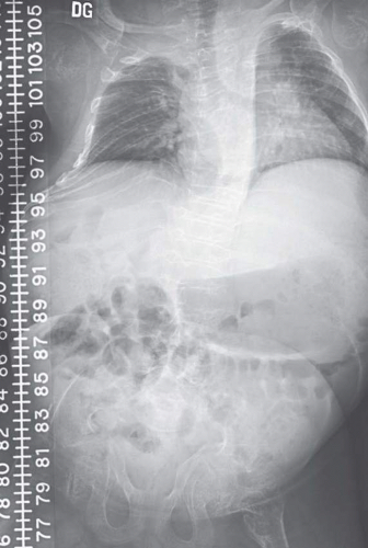
FIGURE 34-8. Pelvic asymmetry (and rib deformity and osteopenia) associated with spinal deformity in a child with osteogenesis imperfecta. (Courtesy of Department of Imaging Sciences, Warren G. Magnuson Clinical Center, NIH.)


THE IMPACT OF SCOLIOSIS ON LIFE FUNCTION (SECONDARY MORBIDITIES)

The reason that this chapter is found in Physical Medicine and Rehabilitation: Principles and Practice is because spinal deformity is not just a cosmetic problem; it impacts on many aspects of quality of life. The types of functional morbidities and their intensity may vary with curves of different etiologies; however, certain problems may be found in almost all types of spinal deformity. In this section, we will review the areas of most common functional concern in persons with spinal curves.


Pain

Pain may be the presenting symptom of spinal deformity; it is in 40% to 90% of adult scoliosis (33). In thoracic kyphosis caused by Scheuermann’s disease, it is an early symptom in 20% to 60% of patients. However, before attributing pain to idiopathic or degenerative etiologies, it is vital to remember that pain may be the sentinel of a critical, even fatal illness. Pain may be the presenting symptom in infectious processes such as epidural abscess or disciitis (34). Rarely, one can have a spontaneous epidural hematoma of the spine. In these cases, due to nerve irritation, the pain may be radicular in character. Of greatest concern is to rule out pain due to tumor. In benign
tumors, the delay between the onset of symptoms and the diagnosis can be up to 19 months. In malignant tumors, it can be up to 4 months between symptom onset and diagnosis. It is important to recall that tumor-related pain is often worse at night and is disassociated from any obvious initiating or exacerbating factors (35).

Chronic pain plays a role in many spinal curvature pathologies. As implied above, it is common in degenerative spine conditions with associated curvature. More than 50% of patients with Scheuermann’s disease have pain later in their course, especially if L1 and L2 are involved (36). There appears to be an increased risk of pain in adults who had scoliosis in childhood or adolescence (37). Pain is often a problem in persons with achondroplasia and is felt to be associated with the mechanical disadvantage of the lordosis on sagittal spinal alignment. Back pain is also found in osteogenesis imperfecta. The pain in osteogenesis imperfecta may also be related to the mechanical disadvantage of the lordosis.


Mobility

As noted above, normal spine function is a delicate dynamic balance between rigidity and flexibility. It is not surprising, then, that balance may be affected by spinal deformity. When the patient has an underlying neuromuscular condition, the spine asymmetry is likely to exacerbate the difficulties with balance control. What is more unanticipated is that balance is disturbed in idiopathic scoliosis as well. Reports have shown increased amounts of sagittal and lateral sway in standing, along with increased sway radius, leading to decreased stability in standing (38). This may result in increased losses of balance and falls as well as increased and asymmetric stress on lower limb joints, creating the potential for a vicious cycle. Recent reports suggest that there may be subtle underlying neurological deficits in persons with idiopathic scoliosis manifesting as balance difficulties (39, 40).


Cardiopulmonary Function

The most critical of all of the potential complications associated with spinal deformity is compromise to the cardiopulmonary system. There is a common pathway toward cardiopulmonary dysfunction. It begins with restrictive lung disease resulting from deformity of the bellows mechanism formed by the rib cage, thoracic spine, and diaphragm. On pulmonary function testing, this is demonstrated by greater decreases in vital capacity than residual lung volume (41). In untreated idiopathic scoliosis, it has been shown that once curves achieve an angle of 70 degrees there will be a mild decrease in vital capacity. By the time a curve reaches 90 to 100 degrees, the patient with have dyspnea on effort. With curves greater than 100 degrees, alveolar hyperventilation, CO2 retention, pulmonary hypertension, and right-sided heart failure are seen (37, 41). At this point, the patient may be irrevocably set on a downward slide of illness leading to death due to pneumonia and/or heart failure. It is likely that these problems will present with much smaller curves when there is also an underlying neuromuscular disease or syndrome affecting other organ systems. Prevention of serious cardiopulmonary decline is one of the major indications for surgical correction. Close monitoring of pulmonary function tests is an essential part of ongoing follow-up of any patient with spinal deformity, especially if progression is noted. It is quite important to remember that there is a “window of opportunity” in progressive curves and that surgery in such curves must be done before pulmonary function has deteriorated to such a degree that the risk of inability to wean from mechanical ventilation after surgery is greater than the potential benefits of the surgery.


Emotional and Behavioral Health

A physiatrist is obligated to approach his/her patient holistically. As with any other disability, the emotional impact of spinal deformity should be addressed. Spinal deformity has traditionally had tragic, negative connotations in Western society as evidenced by Shakespeare’s Richard III and Victor Hugo’s Quasimodo, The Hunchback of Notre Dame. Our modern society often appears obsessed with physical beauty and it seems as if these societal pressures are greater on women and girls. It is important, then, to be mindful of the potential impact that spinal deformity may play on the patient’s body image and emotional health. Studies of patients with idiopathic scoliosis bear out this concern, with findings showing poorer body image, greater unhappiness, lower self-esteem, increased participation in high-risk behaviors, and greater incidence of depression (42, 43, 44). Questions relating to the emotional and behavioral status of a patient with spinal deformity should be a regular part of the physiatrist’s ongoing follow-up of the patient. There should be no hesitation to enlist the help of psychological support professionals such as rehabilitation psychologists and social workers to address the emotional needs of the patients and their families.


PATHOPHYSIOLOGY OF SCOLIOSIS

Spinal stability is derived from the design of the bony, elastic, and muscular elements of the spine and how they are controlled by the nervous system. The bony anatomy has a fair degree of intrinsic stability engineered into it, but must allow movement. Stability is reinforced by the elastic structures but, by their very nature, they will allow movement. The motor control system is designed to adjust to the movements engineered into the bone and elastic tissue system. Understanding the anatomy enables one to determine factors contributing to spinal curvature and better formulate a plan of treatment.

One useful division is to separate factors intrinsic to the spinal column from extrinsic factors. In looking at the intrinsic factors, one should consider how aspects of each of the elements of the spine—bony, elastic, and neuromuscular—may be affected. Once one has identified which systems are involved, the type of involvement should be understood. Is there a failure in the system or is the system overactive? Then, consider the etiology of the impairment. Is it congenital or
acquired? If it is acquired, is the etiology traumatic, infectious, neoplastic, autoimmune, or degenerative? Understanding the biology of the dysfunction is important, that is, is the problem due to a metabolic defect in the tissue or is the tissue reacting to an outside influence. The nature of how these systems interact should also be considered. For example, does the elastic pathology cause secondary bony deformity?


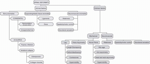
FIGURE 34-9. Pathological factors contributing to spinal deformities. (Courtesy of Donald Bliss and Alan Hoofring of the Medical Arts and Publications Branch, NIH.)

Factors extrinsic to the spine can influence the spine in one of two ways. They can directly impact on the mechanical function of the spine by applying asymmetric forces to the bony and elastic tissue complex of the spine. Disorders resulting in misalignment of the appendicular skeleton, rib cage, and/or head can also apply abnormal forces to the spine. The spine can also be indirectly influenced by abnormal, especially asymmetric, function of the neuromuscular system extrinsic to the spine itself, even if spinal neuromuscular system is intact. It should also be remembered that this extrinsic neuromuscular dysfunction can directly affect the spine or it can indirectly influence the spine by causing extrinsic mechanical failures such as joint contractures or dislocations.

Figure 34-9 is a graphical representation of how all these factors may be organized. However, the relationship between the all of the factors—intrinsic and extrinsic—is even more complex than it appears because of the potential for interactions between each and every factor. Table 34-3 is a list of pathological conditions associated with spinal deformity grouped by category, based on the taxonomy developed by the SRS (9).


SCOLIOSIS DUE TO BONY DYSFUNCTION


Congenital

A number of disorders of vertebral development can occur. These anomalies are present at birth, but they may not be detected immediately. The curvature may be subtle and not noticed until the child begins sitting or standing. Often, clinical evidence of curvature may not be apparent until the child grows and one part of the vertebral column grows faster than another, thus revealing the asymmetry associated with the anomaly. Winter (45) has developed a classification system for these anomalies based on the underlying embryological dysfunction. The dysfunction may be a failure of segmentation, a failure of formation, or a combination of both. The archetype of segmentation failure is a bloc vertebra, formed at two or more spinal levels. This is less likely to cause a curve than a unilateral failure to segment, known as a unilateral bar. The location of the bar will determine the type of pathological curve. A lateral bar will result in scoliosis, a posterior bar will produce kyphosis, and an anterior bar will result in lordosis.

The defect in formation that will result in curvature is usually called a hemivertebra. In reality, any amount of partial defect is possible. The partial vertebra may be “incarcerated,” meaning that the vertebrae above and below it have altered their shape to compensate for the abnormal shape of the partial vertebra, therefore limiting the degree of curvature. A nonincarcerated hemivertebra is more likely to result in curvature because its neighboring vertebrae have not adapted to the hemivertebra’s abnormal shape. Figure 34-10 shows representations of the basic types of deformities according to the Winter classification.

Although congenital bony anomalies may be isolated, it is incumbent to first rule out associated genetic disorders. From 25% to 40% of children with congenital bony anomalies also have other problems including genitourinary anomalies, cardiac anomalies, Klippel-Feil syndrome, and spondylothoracic dysplasia. However, the majority of curves due to congenital bony anomalies are not severe. Only 38% show severe progression, whereas 47% show only mild to moderate
progression and 15% show no progression (46). The character of the curve will help determine likelihood of success of a given treatment approach. Long, flexible curves may respond to orthotic management. Short, stiff curves will require surgery if they progress.


TABLE 34.3 Classification of Scoliosis Based on the SRS Taxonomy


Only gold members can continue reading. Log In or Register to continue

Stay updated, free articles. Join our Telegram channel

May 25, 2016 | Posted by in PHYSICAL MEDICINE & REHABILITATION | Comments Off on Scoliosis and Other Spinal Deformities

Full access? Get Clinical Tree

Get Clinical Tree app for offline access

Idiopathic


Infantile (0-3 y old)


Resolving


Progressive


Juvenile (4 y—puberty onset)


Adolescent (puberty onset—epiphyseal closure)


Adult (after epiphyseal closure)


Neuromuscular


Neuropathic