Total Hip Arthroplasty after Prior Hip Preservation Surgery
Perry Schoenecker
Lisa Berglund
John C. Clohisy
A number of corrective osteotomies of the acetabulum and proximal femur have been described and utilized with varied success to slow or prevent the development of degenerative joint disease in patients with hip deformities such as dysplasia, femoroacetabular impingement (FAI), slipped capital femoral epiphysis (SCFE), and Legg–Calvé–Perthes disease (1,2,3,4,5,6,7,8,9,10,11,12,13,14,15,16,17,18,19). The severity of deformity, age of the patient, and presence of or stage of pre-existing arthritis will often affect the outcome of these procedures. Regardless of the technique or patient selection, a subset of patients will go on to develop end-stage arthritis necessitating conversion to total hip arthroplasty (THA) (20,21,22,23,24,25,26).
Performing THA in patients who have undergone a prior proximal femoral and/or acetabular osteotomy presents unique sets of challenges. In patients who have had a successful joint preservation procedure(s) with pain relief and increased function for a number of years, it is likely that the joint preservation procedure(s) was successful at improving the joint mechanics while avoiding complications of hardware irritation, osteotomy nonunion, avascular necrosis of the acetabulum and/or femur, nerve injury, and infection. Many authors believe that a THA in this situation may be technically less difficult with enhanced results compared to a THA performed in a patient with un-treated deformity (20,21,22,23). Although the structural anatomy may be improved, issues related to retained hardware, prior incisions/approaches, and altered anatomy must be add-ressed (23,24). In the setting of an acute or semiacute failure of a hip preservation procedure, performing THA is clearly more complicated with the outcome at increased risk for complication.
Preoperative Planning
History
When planning for a THA in a patient who has had prior hip preservation surgery it helps to approach the case in a systematic fashion. A thorough history is always obtained. It is clearly advantageous to know in detail what procedure was performed, the details of the prior operative approach, and the operative indications. If at all possible, we obtain the patient’s records, in particular the operative reports and radiographs (both pre- and postoperative). If hardware remains, it is useful to know the manufacturer and size of the implants to facilitate removal if necessary. It is also critical to know if the patient had complications postoperatively such as prolonged wound drainage or infection requiring debridement and/or antibiotics (raising the level of suspicion that a chronic low-grade infection may be present).
Physical Examination
Beyond the standard physical examination of a patient being evaluated for THA, several issues require careful attention. The skin is examined for scars and prior incisions. The soft tissues are evaluated for mobility and for clues to potential wound healing issues. When concern exists regarding compromised soft tissues, a plastic surgery consultation is obtained. Hip range of motion is assessed and recorded. A markedly stiff hip may require an extended surgical approach such as a trochanteric osteotomy. Abductor function is also carefully assessed as damage to the abductors may compromise the function and outcomes of subsequent THA in terms of a high risk of instability and/or a persistent limp even if the procedure is otherwise successful (27). A detailed neurologic examination is performed and recorded. Prior surgeries can either have been associated with neurologic damage which must be documented preoperatively or cause scarring of tissue around neurovascular structures decreasing their flexibility and making intraoperative identification more challenging. Patients with high hip centers, even if corrected from a hip preservation procedure will often have altered neurovascular anatomy and be at higher risk for neurologic injury. In particular, the femoral nerve may perform a hairpin turn, reversing course after exiting the pelvis and proceeding cranial and lateral just distal to the native hip center before turning distal again. This will place the nerve at risk for traction injury from anterior and medial retraction during preparation of the acetabulum (28,29).
Last, perceived or true leg-length inequality is commonly present after hip preservation surgery and must be carefully assessed and documented. Patients are queried as to if one leg feels longer and if they have been using a shoe lift. The leg lengths are then assessed both clinically (both true and perceived) and radiographically (with a standing leg-length film). Measurements are obtained with a tape measure from the anterior superior iliac spine to the lateral malleolus and then assessed by having the patient stand on progressively thicker measured blocks until the patient feels level. Preoperative education as to the limits of leg-length correction and to the risks of postoperative leg-length inequality is critical to prevent patient disappointment.
Radiographic Assessment
Standardized plain radiographs consisting of an anteroposterior (AP) pelvis, AP and lateral hip, and a shoot through lateral are obtained on all patients. The presence of hardware in the acetabulum and/or femur is noted, especially if its location will interfere with femoral or acetabular component placement. We have found preoperative radiographic templating to be extremely useful in planning for these potentially challenging cases as specialized implants may be required that may not normally be available unless specifically ordered.
On the acetabular side, we template the component placement at or near the anatomic hip center. This allows for estimation of component size and coverage while alerting the surgeon to the possible need for acetabular augmentation with either structural bone graft or metallic augments (30,31,32). An estimation of acetabular component size may also effect bearing surface or acetabular component selection as smaller component sizes may limit femoral head size which in turn may affect the risk of dislocation postoperatively (33). Templating the femoral side will elucidate potential issues such as existing hardware, a narrow femoral canal, excessive anteversion, and/or proximal femoral deformity which may require specialized implants or a corrective osteotomy (34). The amount of limb-length correction and offset restoration can then be estimated. In some situations a CT scan will be obtained to better define the anatomy of the hip including identifying any abnormal acetabular or femoral version or anatomy (35). A CT scan is also useful to evaluate osteotomy union as well as localize hardware that may be encountered.
Preoperative Infection Work-Up
Patients who have had any prior hip surgery that have gone on to develop arthritis warranting THA must be ruled out for infection. Given that there are no established guidelines for the detection of chronic indolent infection in the patient who has undergone hip preservation surgery, we approach these patients in a similar fashion as those with a failed THA awaiting revision (36,37). Serum erythrocyte sedimentation rate (ESR) and C-reactive protein (CRP) are drawn. If both the ESR and CRP are within normal limits, the index of suspicion for infection is low. If the ESR and CRP are elevated, the hip is aspirated preoperatively with fluoroscopic guidance and the synovial fluid sent for cultures, synovial fluid white blood cell (WBC) count, and differential. A synovial fluid WBC greater than 3,000 WBC/μL with elevated segmental neutrophils (>60%) is suggestive of infection especially in the setting of an elevated ESR and CRP. If the synovial fluid WBC count and differential are elevated and cultures negative, a repeat aspiration is sent including aerobic, anaerobic, fungal, and acid-fast bacilli cultures. If no fluid is obtained, and clinical suspicion for infection is low, then an intraoperative aspiration is performed (after exposure of the deep tissues but before entering the hip capsule) and the fluid sent for a stat intraoperative synovial fluid WBC count and differential with the synovial WBC counts typically available within 30 minutes and the differential within 1 hour. Finally, capsular tissue can also be sent for an intraoperative frozen section.
In cases where infection has not been definitively ruled out preoperatively, the patient and the surgical team will be prepared for the possibility of performing hardware removal, resection of the femoral head, and reaming of the acetabulum with placement of an antibiotic-loaded spacer. This is followed by a second-stage reconstruction after a 6-week course of intravenous antibiotics dictated by intraoperative culture results and an infectious disease consultation (38). This two-stage approach to the treatment of septic arthritis is preferred over simple hardware removal given its ability to more thoroughly debride any chronic osteomyelitis while obtaining high antibiotic levels from the impregnated spacer. Using this technique, multiple authors have reported excellent outcomes with low rates of infection (39,40,41,42).
Skin Incisions
The incisions used for the hip preservation procedure may not be easily accessible when the patient is positioned for a THA. If the preservation surgery was performed many years earlier, the scars from the index surgery may be faint and possibly not in line with the hardware if the patient’s body habitus has changed. In general, if the prior incision can be incorporated, we choose to do so. However, if the incision will hamper overall exposure or is remote (more than 20 years old) it can be safely ignored as the soft tissue envelope of the hip is more forgiving than would be other areas such as the knee. In general, approximately 6 cm should be left between parallel incisions and sharp corners or angles of less than 60 degrees should be avoided between prior incisions.
Hardware Removal
Easily accessed hardware can be removed with little morbidity but often complete removal of hardware can be challenging and is unnecessary. If hardware removal is required, fluoroscopy and hardware removal tools should be made available for the case. Alternatively, hardware can be addressed with partial removal utilizing a metal cutting burr if the hardware directly interferes with the preparing and placement of THA components.
Hardware in the proximal femur can be particularly challenging to address, especially if incarcerated as can occur with proximal femoral osteotomies performed during childhood. Removing hardware from the proximal femur creates stress risers. In general, to avoid an iatrogenic fracture,
hardware removal should commence only AFTER the femoral head has been dislocated from the acetabulum. The femoral reconstruction should also obtain fixation distal to any diaphyseal cortical defects created from the hardware removal and consideration can be given to protecting the area (particularly if a prior plate was removed) with a strut allograft (Fig. 54.1) (43,44). If complete removal of hardware is required, particularly if intraosseous, an extended trochanteric osteotomy (ETO) may be useful. The ETO is also a useful way of dealing with proximal angular deformity that may be present (34). Alternatively, patients with extensive retained hardware and without large leg-length discrepancies may be a candidate for hip resurfacing (45). However, issues related to the metal-on-metal bearing surface, small native femoral head/acetabulum, leg-length discrepancy, and degree of proximal deformity may preclude use of this technology in these situations (46,47,48,49,50).
hardware removal should commence only AFTER the femoral head has been dislocated from the acetabulum. The femoral reconstruction should also obtain fixation distal to any diaphyseal cortical defects created from the hardware removal and consideration can be given to protecting the area (particularly if a prior plate was removed) with a strut allograft (Fig. 54.1) (43,44). If complete removal of hardware is required, particularly if intraosseous, an extended trochanteric osteotomy (ETO) may be useful. The ETO is also a useful way of dealing with proximal angular deformity that may be present (34). Alternatively, patients with extensive retained hardware and without large leg-length discrepancies may be a candidate for hip resurfacing (45). However, issues related to the metal-on-metal bearing surface, small native femoral head/acetabulum, leg-length discrepancy, and degree of proximal deformity may preclude use of this technology in these situations (46,47,48,49,50).
 Figure 54.1. A: Radiographs of a 44-year-old man with advanced left hip degeneration approximately 10 years after a valgus-producing proximal femoral osteotomy for a malunited femoral neck fracture. B: Postoperative radiographs demonstrate placement and ingrowth of a fully porous cylindrical diaphyseal engaging stem. Note incorporation of an allograft strut to protect the area of prior plate and screw holes. The stem bypasses cortical defects created by screw removal. |
Acetabular Component Choice and Preparation
The vast majority of the time the acetabulum can be reconstructed with a cementless hemispherical component. Although standard titanium shells work well in most situations, the more porous metals utilized commonly in revisions may offer some benefit if implant stability is a concern (51). Once reaming has commenced, great care should be taken to avoid damage to the anterior and posterior columns and the use of trial components is extremely useful in these complex situations as is an intraoperative radiograph to confirm appropriate position of the trial or implant. If there is any question regarding the stability of the trial (and hence the component itself), a multihole shell is recommended to allow for adjunctive fixation with multiple screws.
Although the required amount of host bone/component contact and/or component coverage remains a matter of debate, absolute stability of the implanted component is required for bony ingrowth to occur (52). Care should be taken to appropriately position the component in the optimal amount of abduction and anteversion to maximize stability while resisting the urge to maximize bony coverage at the expense of malposition. Intraoperative radiographs can be useful to confirm appropriate position. Medialization can often improve acetabular component coverage (53,54). We will accept up to 1 cm of elevation of the hip center if this simplifies the procedure by avoiding the need for augmentation.
Femoral Component Choice and Preparation
If possible, the surgeon should template with the femoral component that they normally use as optimal results in difficult cases will most reliably be achieved with the tools
that the surgeon is most comfortable with. Unfortunately, in many of these cases, distorted anatomy may dictate the use of another type of stem. In general, cementless stems are favored in this younger, more active cohort (55). Further, the presence of prior hardware makes creating a perfect cement mantle more challenging although the use of a small cemented hip dysplasia stem may be advantageous in certain situations where the canal is very small and the ability to place the stem in the appropriate amount of anteversion regardless of proximal femoral anatomy is required (26).
that the surgeon is most comfortable with. Unfortunately, in many of these cases, distorted anatomy may dictate the use of another type of stem. In general, cementless stems are favored in this younger, more active cohort (55). Further, the presence of prior hardware makes creating a perfect cement mantle more challenging although the use of a small cemented hip dysplasia stem may be advantageous in certain situations where the canal is very small and the ability to place the stem in the appropriate amount of anteversion regardless of proximal femoral anatomy is required (26).
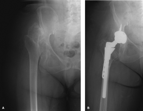 Figure 54.2. A: Preoperative radiograph of a 55-year-old woman with right hip degenerative joint disease and a high congenital hip dislocation. B: Postoperative radiographs demonstrate the use of a modular femoral component that allows proximal sleeve fixation of the metaphysis with ability independently adjust femoral version. |
Given the high frequency of versional abnormalities, a modular stem can be helpful in some of these complex situations. The potential benefits of modular stems must be carefully weighed against the potential risks of fretting, corrosion, metal debris and ion generation, and failure or fracture of the prosthesis at the modular junction (56,57,58,59,60,61,62). These potential complications are of particular concern in the relatively young and active patients requiring THA after failed hip preservation procedures.
Related posts:

Stay updated, free articles. Join our Telegram channel

Full access? Get Clinical Tree


