Mini-open Approach for Femoroacetabular Impingement
James P. Cashman
Javad Parvizi
Clinical Presentation, Diagnosis, and Evaluation for Surgery
Symptoms
Hip pain in young adults can represent a diagnostic challenge. Important features to elicit from the history include developmental dysplasia, trauma, or predisposing factors of avascular necrosis. Patients presenting with femoroacetabular impingement (FAI) present with a variety of clinical signs and symptoms that help differentiate them from patients with other hip pathology. Symptoms of impingement can be insidious in onset in active young and middle-aged adults. Groin pain can be associated with activity and without necessarily having a prior history of trauma. FAI often presents in active young adults as groin pain of slow onset, usually noticed by the patient after an episode of minor trauma. The pain may be felt as the patient starts to walk after rising from a sitting position or it may be a dull groin ache while the patient has the hip in a flexed position. These patients report an inability to perform activities such as high hip flexion or prolonged sitting. They may complain about painful clicking, locking, or instability, possibly associated with a labral tear. The pain is intermittent and may be exacerbated by excessive demand on the hip such as athletic activities or a normal activity of daily living such as walking. The pain may also present after sitting for a prolonged period (1). Based on the presence of unnoticed radiographic changes, these patients sometimes are subjected to an extensive diagnostic workup and even inappropriate surgical procedures.
Patients with cam-type impingement most often have the onset of symptoms during their 30s, although the symptoms may occur earlier. Men are more commonly affected and, although the deformity is often bilateral, patients usually have symptoms on only one side. Pincer-type impingement is more common in females and often presents as groin pain after activity in patients in their 40s (2). Some patients have a history of extensive diagnostic work-ups and perhaps even inappropriate surgical treatments such as repair of inguinal hernia, laparoscopy, and other abdominal procedures (3,4).
Slow onset of initially intermittent groin pain after a minor trauma, which gradually exacerbates by athletic activity or prolonged walking, is the typical presentation. Mechanical symptoms such as locking, catching, or clicking are common with labral tears, but these are nonspecific for the disorder. Chondral lesions, labral lesions, or both may be the cause of pain. Prolonged sitting or driving also elicits pain, although results of routine radiographic studies may be normal.
Signs and Clinical Tests
Patients with FAI can have a reduced range of motion especially flexion, adduction, and internal rotation. There are a number of clinical tests which can be useful in determining a diagnosis of FAI.
The results of the physical examination may be normal, but most patients have a slightly antalgic gait. Examination of the hip often reveals limitation of motion, particularly in internal rotation and passive flexion of the hip of more than 90 degrees while adducted. Typically, the patient has <10 degrees of internal rotation with the hip in 90 degrees of flexion (5). A study by Kubiak-Langer et al. (6) showed that the range of motion of the hips in patients with FAI is decreased in flexion, internal rotation, and abduction. Flexion and adduction leads to the approximation of the femoral neck and the acetabular rim with recreation of the pain, particularly when there is a chondral lesion. The impingement test is positive if one elicits pain on adduction of a flexed, internally rotated hip with the patient supine (7). The impingement sign is positive in most cases (8).
Flexion, abduction, and external rotation (FABER) provocation test can be helpful also. With the hip in flexion, abduction, and external rotation, abutment of the labrum and cartilage also can occur (9). The test is positive if it elicits similar pain as complained by the patient or if the distance between the lateral knee and the examination table differs between the symptomatic and contralateral hip. An additional test is the posterior inferior impingement test (10). With the hip in hyper extension, passively by hanging the leg
over the end of the bed, the affected hip is passively externally rotated. The test is positive if it elicits similar pain as complained by the patient.
over the end of the bed, the affected hip is passively externally rotated. The test is positive if it elicits similar pain as complained by the patient.
All other provocative maneuvers such as classic Trendelenburg test, Thomas test, anterior apprehension test, posterior impingement test, and the bicycle test are suggestive but nonspecific. A positive impingement test has been correlated closely to acetabular rim lesions as visualized on specific modern MRI arthrograms of the hip (11). Wyss et al. (12) found a strong correlation between a lack of internal rotation of the flexed hip and a lack of space between the acetabular rim and the femoral head–neck junction on magnetic resonance imaging.
Imaging
The diagnosis of FAI can be confirmed with imaging. Standard anterior–posterior pelvic, frog-leg lateral, and cross-table lateral radiographs should be obtained (Figs. 42.1 and 42.2). In certain situations a false-profile radiograph can assess anterior femoral head coverage.
Alterations of the proximal femoral anatomy, such as head–neck offset and bump formation can be observed in addition to acetabular and labral pathology. A pistol grip deformity of the femoral head is often seen in cam-type impingement. In this situation the superior-lateral head–neck junction is convex instead of concave. A high fovea can also indicate asphericity of the femoral head that is not able to be appreciated on the AP films. The alpha angle is a useful radiographic measurement for quantifying the head–neck junction deformity. This angle is most accurate when measured on MRI; however, it can also be measured using plain films. The Dunn view (AP of hip in neutral rotation, 45-degree hip flexion, and 20-degree abduction) is the most sensitive x-ray view for detecting femoral head–neck asphericity. The angle is formed by a line drawn from the center of the femoral head through the center of the femoral neck, and a line from the center of the femoral head to the femoral head/neck junction, found by the point by which the femoral neck diverges from a circle drawn around the femoral head. At present, the upper end of normal is an alpha angle of 50 to 55 degrees.
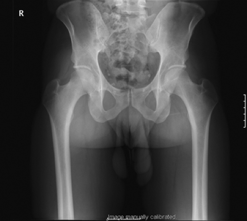 Figure 42.1. AP radiograph can be used to assess the presence of cam/pincer lesions, the presence of acetabular retroversion, and acetabular dysplasia. |
 Figure 42.2. Lateral radiograph demonstrating a cam lesion on the femoral neck. |
Coxa profunda is present when the floor of the acetabular fossa is in line with the ilioischial line; protrusio is present when the medial most femoral head overlaps the ilioischial line. The crossover sign is a sensitive and specific indicator of native acetabular version. On an AP pelvis radiograph, the outlines of the edges of the anterior and posterior walls of the acetabulum should meet superiorly and laterally. In cases of acetabular retroversion, this crossover of the anterior and posterior acetabular wall outlines is more distal. Changes in the acetabular rim may also be noted. A “double line” is seen in labral ossification. An os acetabuli may also be an indicator of pathology.
Magnetic resonance arthrography is the gold standard for detecting labral pathology. An MR contrast agent is injected intra-articularly, thus allowing visualization of labral, chondral, and bony lesions of the hip (Fig. 42.3). A triad of MR arthrographic findings has been described in patients with cam-type FAI (13). The triad consists of an abnormal alpha angle, an anterior-superior acetabular cartilage lesion, and an anterior-superior acetabular labral tear. In that study, 90% of patients with clinical impingement had the triad of MR arthrographic findings. In pincer-type impingement, the cartilage lesions are often seen along the posterior aspect of the acetabulum because of a contrecoup type of injury as the femur abnormally touches the acetabular rim. The associated labral degeneration and tears are most common in the anterosuperior labrum. Tears caused by the pincer-type mechanism extend perpendicular to the surface of the labrum and, in more severe cases, to the subchondral bone, in which case the labrum may become calcified. Endochondral ossification within the labrum is also typically seen.
 Figure 42.3. MR arthrogram demonstrating a labral tear on the superolateral margin of the acetabulum (arrow). |
Specific Indications for Mini-Open Approach
Over the last decade, the surgical treatment of FAI has evolved as surgical techniques through arthroscopy, open surgical dislocation, and combined approaches have been developed. Surgical indications have evolved in line with improved understanding of this clinical entity. The aim of surgical treatment is to improve the head–neck offset in the presence of a cam lesion and to perform acetabular rim resection when pincer impingement is evident, either by open surgery or arthroscopically.
Appropriate management of patients with FAI commences with a trial of conservative treatment, which may include activity modification, restriction of athletic activities, and reduction of excessive motion and demand on the hip. A trial of nonsteroidal anti-inflammatory medications may be appropriate to relieve pain of acute onset. Prolonged pain treatment may mask the symptoms of the underlying destructive process. Physical therapy, with emphasis on improving range of motion or stretching, is not productive; rather, it is counterproductive. Although conservative management is likely to be temporarily successful in some patients, the young age of these patients and their high activity levels and athletic ambitions usually jeopardize patient compliance. Conservative nonsurgical therapy should be exhausted before any decision is made for surgical intervention. Furthermore, the finding of labral tear in these patients should not per se be an indication for surgical intervention.
When conservative measures do not control the patient’s symptoms or when functional limitations remain unsatisfactory, a surgical referral is appropriate. Previously, it was believed that the labrum had little functional importance and that the appropriate treatment for symptomatic labral tears was excision (14). The philosophy has changed because of the evidence supporting the acetabular labrum’s role in preventing premature arthritis, enhancing stability of the hip joint, and participation in nociception and proprioception (15). In regard to current treatment, the literature to date has focused on arthroscopic debridement of labral tears and surgical repair of associated structural problems. In an effort to prevent subsequent joint degeneration after labral injury, open (16) and arthroscopic (17) approaches have been employed.
Repair is particularly important for peripheral labral tears, which have the blood supply to heal. In managing labral tears, the surgeon focuses on preserving healthy labral tissue to maintain its role as a secondary joint stabilizer and to minimize potential arthrosis. Fraying from labral tears is debrided with motorized shavers and radiofrequency probes. Intrasubstance labral tears and detachment of labral tears off the acetabular rim are repaired by placing an absorbable suture through the defect and retrieving the suture through the capsule. Adjacent cartilage lesions should also be debrided and stabilized with the use of shavers and radiofrequency probes to minimize further propagation. Chondral lesions can be managed with microfracture techniques to stimulate fibrocartilage.
The anterior mini-open approach can be used for the treatment of FAI. One study found that the area of femoral head and the portion of the acetabular rim that could be exposed were adequate for treatment of the relevant pathology. However, the area of acetabular rim accessible varied significantly according to the position of the anterior inferior iliac spine (18). In one review comparing surgical disclocation with hip arthroscopy and mini-open assisted arthroscopy, all three surgical approaches led to consistent improvements in patient outcomes (19


Related posts:

Stay updated, free articles. Join our Telegram channel
Full access? Get Clinical Tree
