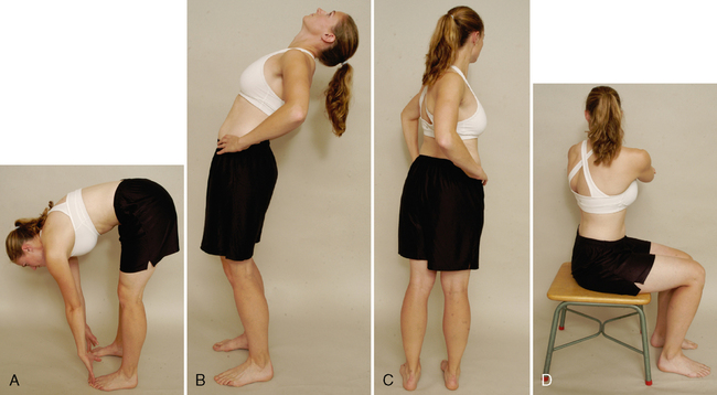
CHAPTER 7 Method 1. Because the ROM at each vertebra is difficult to measure, the examiner can use a tape measure to derive an indication of overall movement. The examiner first measures the length of the spine from the C7 spinous process to the T12 spinous process with the patient in the normal standing posture. The patient then is asked to bend forward, and the spine is again measured. Method 2. If the examiner wishes, the spine may be measured from the C7 to the S1 spinous processes with the patient in the normal standing position. The patient then is asked to bend forward, and the spine is again measured. In this case, the examiner is measuring movement in the lumbar spine as well as in the thoracic spine. INDICATIONS OF A POSITIVE TEST The normal ROM of forward flexion (forward bending) in the thoracic spine is 20° to 45°. • An alternative test method involves having the patient bend forward and try to touch the toes while keeping the knees straight. The examiner then measures from the fingertips to the floor and records the distance. With this method, the examiner must keep in mind that, in addition to the thoracic spine movement, movement also may occur in the lumbar spine and hips; in fact, movement could occur totally in the hips. • Each of the methods described is indirect. Measurement of the ROM at each vertebral segment requires a series of radiographs. The examiner can decide which method to use. It is of primary importance, however, to note on the patient’s chart how the measuring was done and which reference points were used. • While the patient is flexed forward, the examiner can observe the spine from the “skyline” view. With nonstructural scoliosis, the scoliotic curve disappears on forward flexion; with structural scoliosis, it remains. With the skyline view, the examiner is looking for a hump on one side (convex side of the curve) and a hollow on the other side (concave side of the curve). This “hump and hollow” sequence is caused by vertebral rotation in idiopathic scoliosis, which pushes the ribs and muscles out on one side and causes the paravertebral valley on the opposite side. The vertebral rotation is most evident in the flexed position. • When the patient flexes forward, the thoracic spine should curve forward in a smooth, even manner with no rotation or side flexion. The examiner should look for any apparent tightness or sharp angulation, such as a gibbus (hump) when the movement is performed. If the patient has an excessive kyphosis to begin with, very little forward flexion movement occurs in the thoracic spine. • McKenzie1 advocates testing flexion while the patient is sitting to reduce pelvic and hip movements. While sitting, the patient slouches forward, flexing the thoracic spine. The patient can put the hands around the neck to apply overpressure at the end of flexion. If symptoms arise from forward flexion on the spine with the neck flexed by the hands, the examiner should repeat the movement with the neck slightly extended and the hands removed. This can help differentiate between cervical and thoracic pain. • Because extension occurs over 12 vertebrae, the movement between the individual vertebrae is difficult to detect visually. • McKenzie1 advocates having the patient place the hands in the small of the back to add stability while performing the backward movement or to do extension while the patient is sitting or in the prone-lying (sphinx) position. • As the patient extends, the thoracic curve should curve backward or at least straighten in a smooth, even manner with no rotation or side flexion. • Lee2 advocates asking the patient to fully forward-flex the arms during extension to facilitate extension. • The examiner should look for any apparent tightness or angulation when the movement is performed. • If the patient shows excessive kyphosis, the kyphotic curvature remains on extension; that is, the thoracic spine remains flexed, whether the movement is tested while the patient is standing or lying prone. • The test also may be done with the patient in the prone-lying position. If this position is used, the normal kyphotic posture should flatten. If it does not, the patient has a structural kyphosis.
THORACIC SPINE

SELECTED MOVEMENTS
![]()
Stay updated, free articles. Join our Telegram channel

Full access? Get Clinical Tree


THORACIC SPINE


