The Symptomatic Residual Slipped Capital Femoral Epiphysis and Legg–Calvé–Perthes Hip
Eduardo N. Novais
Michael B. Millis
Introduction
Slipped capital femoral epiphysis (SCFE) and Legg–Calvé–Perthes Disease (Perthes) are pediatric hip disorders that may heal with permanent deformity of the proximal femur. More importantly, since the classic work of Murray (1), Stulberg et al. (2), Solomon (3,4), and Harris (5) deformities of the hip secondary to acetabular dysplasia, Perthes, and SCFE have been recognized as the most common cause of symptomatic osteoarthritis (OA) of the hip (1,2,4,6,7). Although, the pathophysiology of OA development secondary to Perthes and SCFE is not completely understood, the conflict between the abnormal femoral head–neck junction and the acetabular rim described in femoroacetabular impingement (FAI) potentially explains the articular cartilage and labral damage reported in adolescents and young adults with symptomatic healed Perthes and SCFE (8,9,10,11,12,13,14). Secondary acetabular deformity may also develop in response to the misshapen proximal femur as the child finishes growing. Therefore, both Perthes and SCFE can lead to two distinct types of mechanical failure of the hip joint: FAI and hip instability because of secondary acetabular dysplasia. Having said that, a number of important questions regarding natural history, pathophysiology, and treatment of Perthes and SCFE remain unanswered.
Theoretically, the goal of treatment of these diseases during childhood is to preserve the femoral head sphericity and the head–neck offset avoiding the development of FAI. Once these deformities are established and FAI is present, the treatment should be focused on reestablishing the normal anatomy of the proximal femoral head–neck junction and acetabular rim. However, because hip OA is a multifactorial disease that is caused by a combination of biologic and mechanical factors, the deformity may have a minimally symptomatic course during adolescence and early adulthood, encouraging procrastination and limited intervention. In this chapter the pathophysiology of residual deformities on the healed Perthes and SCFE hip, the association with early development of OA, its clinical presentation and treatment options will be discussed.
Pathophysiology of Perthes and Scfe—from Disease to Residual Deformities of the Hip
The Perthes Hip
Legg-Calvé-Perthes disease is a pediatric form of osteonecrosis of the femoral head usually affecting children between the ages of 4 and 9 with a male predominance of approximately 5:1 (15). In the United States, the estimate incidence is of 5.1 per 100,000 children (16). Although, Perthes disease was described about 100 years ago (17,18,19), and its etiology has not been completely understood, recent, advanced imaging clinical studies have recognized disruption of femoral head blood supply as the leading event leading to ischemic necrosis (20,21,22). Experimental studies of avascular necrosis (AVN) in animals have confirmed the interruption of the blood supply to the femoral epiphysis as the single most important event on the pathogenesis of the disease process (23). The ischemic necrosis is thought to disturb the entire proximal femur including the metaphysis, growth plate, epiphysis, and the articular cartilage. Four radiographic stages of the disease are typically recognized: initial or necrotic phase, fragmentation, reossification, and residual phase (24). Recent work from Kim et al. (25,26,27,28) using a piglet model of ischemic necrosis have showed that the ischemia leaded to altered mechanical properties of the necrotic femoral head and neck. These changes in the presence of mechanical load of the hip joint may ultimately lead to the subchondral fracture, collapse, and proximal femoral deformity seen later in the disease process. Although the pathophysiology of the necrotic and fragmentation stage of Perthes is becoming clearer by the aforementioned studies, the reossification (remodeling) and residual stages are not
completely understood. It is widely accepted that the remodeling potential and prognosis of the affected femoral head is directly affected by the patient’s age (29). As the child grows, reossification and remodeling the femoral head changes its shape and increases its size resulting in a wide spectrum of residual deformities. Femoral head deformity may be oligosymptomatic and tolerated in the childhood and early adolescent years but usually will lead to the development of degenerative changes throughout adulthood.
completely understood. It is widely accepted that the remodeling potential and prognosis of the affected femoral head is directly affected by the patient’s age (29). As the child grows, reossification and remodeling the femoral head changes its shape and increases its size resulting in a wide spectrum of residual deformities. Femoral head deformity may be oligosymptomatic and tolerated in the childhood and early adolescent years but usually will lead to the development of degenerative changes throughout adulthood.
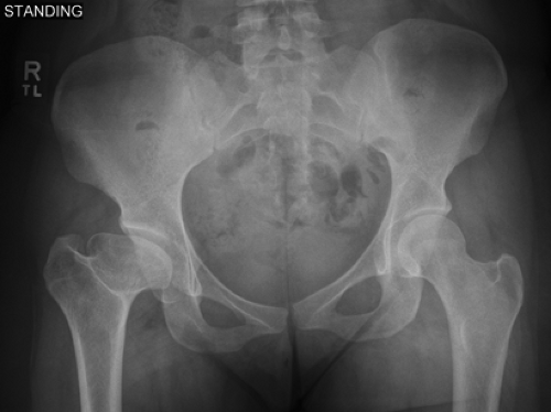
Once the disease has passed the necrosis, fragmentation, and reossification stages, the residual abnormal proximal femoral anatomy is thought to be the most important factor for the long-term prognosis of Perthes (30). Although in a young child Perthes may heal and have very mild residual changes, residual proximal femoral deformities secondary to the healed Perthes may lead to FAI and the associated early articular cartilage degeneration (8,12,31,32). The most common residual deformity is an enlargement of the involved epiphysis (coxa magna) with the more severe cases also having short and broad femoral neck (coxa brevis) with the unopposed growth of the greater trochanter resulting in a reduced articulotrochanteric distance (cranial to the center of rotation of the femoral head) that clinically manifests as abductor weakness and fatigue due to abductor lever insufficiency (Fig. 20.1). The femoral neck–shaft angle is usually preserved, however, the high-riding greater trochanter represents a functional coxa vara deformity (12). The affected limb may be shorter and lower extremity length inequality may also affect the gait and mechanics of the hip. Osteochondritic lesions in the femoral head, although rare, may also be a source of mechanical dysfunction represented clinically by complains of locking and catching. Misalignment in the axial plane has also been described in Perthes. In residual Perthes the articular surface is frequently in the posteromedial superior portion of the femoral head, adjacent to an anterolateral inferior portion of the femoral head that protrudes from the acetabulum. The anterolateral portion is considered the false head that often blocks internal rotation of the hip and is represented radiographically by the sagging rope sign. The posteromedial portion is considered the true head and is the remnant of the original articulating surface. This segment (true head) is retroverted relative to the anterolateral segment and results in a deformity described by Kim and Wenger (33) as functional retroversion that manifests clinically by an externally rotated gait. In addition to proximal femoral deformity, the residual abnormal shaped femoral head may induce secondary adaptive changes of the acetabulum that further contribute to mechanical dysfunction of the hip joint. The acetabulum may become dysplastic and in the presence of an aspherical large femoral head potentially lead to anterior and anterolateral undercoverage of the femoral head with hip instability as the final mechanical failure. Acetabular retroversion has been described in about 40% of adults with Perthes (34). Sankar and Flynn (35) demonstrated that children with Perthes initially have normal acetabular version but overtime acetabular retroversion is likely to develop in cases with more severe deformity of the femoral head. Stulberg et al. (30) studied patients with Perthes disease and identified characteristic pattern of involvement during the active stages of the disease with five specific categories after long-term clinical and radiographic courses. Class I was described as a completely normal hip joint; class II a spherical femoral head (same concentric circle on anteroposterior and frog-leg lateral radiographs) but with one or more of the following abnormal characteristics of the femoral head, neck, or acetabulum: larger-than-normal (although spherical) femoral head (coxa magna); shorter-than-normal femoral neck; or abnormally steep acetabulum. Class I and II hips were thought to have spherical congruency and in these categories arthritis did not develop. Class III was described as a nonspherical (ovoid femoral head) but not flat femoral head. The abnormalities described on class II hips may also be present. In class IV there was a flat femoral head and abnormalities of the femoral head, femoral neck, and acetabulum as described in class II. Class III and IV were described as hips with aspherical congruency associated with mild-to-moderate arthritis in late adulthood. Finally, in class V a flat femoral head and a normal femoral neck and normal acetabulum. These hips were described as aspherical congruency and severe arthritis developed before the age of 50. In Perthes a hip may be unstable in upright activities and yet still impinge when the hip is flexed because of the abnormal shaped femoral head.
The Scfe Hip
SCFE is the most common hip disorder presenting in adolescence characterized by abnormal shearing failure through the proximal femoral growth plate in which there is displacement of the femoral metaphysis anteriorly and cranially with relative posterior and inferior positioning of the femoral head. It is estimated to affect 2 per 100,000 with a male to female rate of 2:1 (36,37). The mean age at diagnosis is during 13.5 years for boys and 12 years for girls. SCFE can affect children younger than 10 years; however, in this scenario a hormone dysfunction (mainly thyroid) needs to be ruled out (38). At presentation, SCFE tends to involve
only one hip; however, the rate of subsequent contralateral slip is high. The overall prevalence of bilateral involvement ranges from 21% to 80% (39). Classically SCFE has been classifying according to duration of symptoms in acute (less than 3 weeks), chronic (more than 3 weeks), and acute on chronic (prodromal pain for more than 3 weeks with a new event leading to an acute exacerbation of the symptoms). It has also been classified according to the degree of displacement of the epiphysis measured by the femoral epiphysis–shaft angle described by Southwick (40) as mild (less than 30 degrees), moderate (30 to 60 degrees), and severe (more than 60 degrees). The most used and perhaps the single most important classification scheme was described by Loder et al. (41) and divides SCFE into stable (patients that are able to bear weight even with crutches) and unstable (patients unable to ambulate or get up even with crutches). In their series, Loder et al. (41) reported a rate of 47% (14 of 30 patients) of AVN of the femoral head for patients with unstable SCFE compared to 0% (0 of 25 patients) with stable SCFE. Following its description the instability of the physis has been widely used and accepted as the most important prognostic factor for occurrence of AVN following SCFE (42,43,44).
only one hip; however, the rate of subsequent contralateral slip is high. The overall prevalence of bilateral involvement ranges from 21% to 80% (39). Classically SCFE has been classifying according to duration of symptoms in acute (less than 3 weeks), chronic (more than 3 weeks), and acute on chronic (prodromal pain for more than 3 weeks with a new event leading to an acute exacerbation of the symptoms). It has also been classified according to the degree of displacement of the epiphysis measured by the femoral epiphysis–shaft angle described by Southwick (40) as mild (less than 30 degrees), moderate (30 to 60 degrees), and severe (more than 60 degrees). The most used and perhaps the single most important classification scheme was described by Loder et al. (41) and divides SCFE into stable (patients that are able to bear weight even with crutches) and unstable (patients unable to ambulate or get up even with crutches). In their series, Loder et al. (41) reported a rate of 47% (14 of 30 patients) of AVN of the femoral head for patients with unstable SCFE compared to 0% (0 of 25 patients) with stable SCFE. Following its description the instability of the physis has been widely used and accepted as the most important prognostic factor for occurrence of AVN following SCFE (42,43,44).
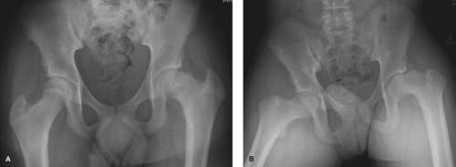
The primary goal of treatment in SCFE is to stabilize the physis and prevent further displacement, while avoiding the complication of AVN. For years in situ fixation has been the standard initial treatment of stable SCFE independently of the severity of displacement (45,46,47,48,49). In situ fixation usually leads to closure of the physis and short-term reliable improvement in function. However, in situ fixation does not allow for restoration of the normal concave offset between the femoral epiphysis and the metaphysis (11). Residual deformity with prominent metaphysis has been shown to cause FAI and lead to mechanical damage to the acetabular cartilage and labrum (9,50,51,52) (Fig. 20.2). Remodeling of the femoral head–neck junction after in situ fixation has been reported previously (53,54). Recently, however, it has been postulated that the remodeling process will occur with impingement between the anterolateral prominence in the femoral head–neck junction and the acetabular rim leading to articular cartilage and labral damage (9,50). Besides proximal femoral residual deformity, SCFE also has associated acetabular deformities (34,55,56).
Femoroacetabular Impingement as the Common Cause of Mechanical Failure in the Healed Perthes and Scfe Hip
Mechanical dysfunction is a known risk factor in the etiology of OA of the hip joint (57,58). Symptomatic hip OA is the major adult consequence of Perthes and SCFE (1,2,4,6,7,30). Hip OA has traditionally been categorized as primary (idiopathic) or secondary (caused by recognized structural abnormalities that are often of congenital or developmental origin). In the early 50s more than half of end-stage hip OA was believed to be primary or idiopathic (59). In the 60s, Murray (1) described the “tilt deformity” of the proximal femur as a possible cause of hip OA. In the 70s, Stulberg et al. (2) examined the radiographs of 75 patients with idiopathic OA of the hip and found that 39% had acetabular dysplasia as determined by four measurements of acetabular configuration. An additional 40% of the patients were found to have the so-called “pistol-grip” deformity of the proximal femur. Subsequent work from Solomon (3,4) and Harris (5) clearly demonstrated that the vast majority of cases of hip OA previously thought to be idiopathic are associated with a developmental hip deformity that may or may not have been recognized prior to skeletal maturity. In 1986, Aronson (6) reported that 76% of hips that underwent total hip arthroplasty (THA) had associated diagnoses of pediatric hip disease: 43% developmental dysplasia of the hip, 22% SCFE, and 11% LCPD. The pathologic mechanics of the hip joint secondary to Perthes and SCFE have been studied in the
past; however, it was not until the work from Ganz et al. (58,60,61,62,63) that FAI was recognized as the main etiologic mechanical factor for early cartilage damage and progression to hip OA.
past; however, it was not until the work from Ganz et al. (58,60,61,62,63) that FAI was recognized as the main etiologic mechanical factor for early cartilage damage and progression to hip OA.
The Perthes Hip
The abnormal hip morphology in the symptomatic healed Perthes hip leading to FAI has been recently described (8,12). Novais et al. categorized residual deformities secondary to Perthes according to its location following observations during surgical dislocation of the hip. Proximal femoral deformity was classified as intra-articular, extra-articular, or in both locations. Intra-articular deformities are related to the aspherical femoral head, enlarged femoral head (coxa magna), flat femoral head (coxa plana), or a combination of these deformities, and ultimately result in cam impingement. The cam impingement is due to the aspherical femoral head entering a relatively spherical acetabulum that fails to accommodate the enlarged anterior segment of the femoral head. The large femoral head may create a cam-induced pincer impingement resulting in linear abutment of the femoral head on the labrum causing primary labral injury. Kim and Wenger (33) described the phenomenon of functional retroversion in which the articulating surface of the femoral head is out of line with the femoral neck. The anterolateral portion is protruding and seen on an anteroposterior radiograph of the hip as the sagging rope sign and often blocks internal rotation of the flexed hip, whereas the posteromedial portion is the articulating portion of the femoral head. This segment is retroverted relative to the anterolateral segment and results in a “functional retroversion” manifested clinically in an externally rotated gait (64). Extra-articular impingement may be secondary to greater trochanteric overgrowth. Residual short and broad femoral neck (coxa brevis) and high-riding trochanter may result in anterior and/or posterior trochanteric impingement. Secondary acetabular morphology in healed Perthes may contribute to abnormal hip mechanics and include acetabular dysplasia and acetabular retroversion (32,34,35,65,66


Stay updated, free articles. Join our Telegram channel

Full access? Get Clinical Tree


