Introduction
The peripheral nervous system provides the link between the central nervous system and all parts of the body. The nerves of the peripheral nervous system transmit information to and from the brain and the spinal cord.
Sensory information, originating in a variety of receptors all over the body, is transmitted to the spinal cord and the brain by the peripheral nervous system. The receptors in the sense organs and the skin monitor changes in the external environment, while those in blood vessels, glands and organs of the body respond to the internal environment. During movement, the proprioceptors in the muscles and the joints are activated by the changing position of the body. All this information is carried to the central nervous system in the peripheral nerves.
Motor commands originating in the brain and spinal cord are transmitted by the peripheral nervous system to the skeletal muscles to execute or modify movement. By the same route, the activity in the organs and glands is regulated to maintain a constant internal environment.
All the nerves of this system contain axons of sensory and motor neurones bound together by connective tissue. There are two functional categories of axons found in the peripheral nerves. The somatic component consists of all the sensory and motor axons associated with activity in the muscles, the joints and the skin. The visceral component is all the axons carrying nerve impulses to the glands, organs and blood vessels. The visceral nerve fibres are part of the autonomic nervous system.
Damage to the nerves of the peripheral nervous system at any point from their origin in the central nervous system to their terminations inside the muscles will result in loss of both muscle function and sensation in the skin. Trophic changes, such as flushing and dryness of the skin, will also occur if the visceral fibres are damaged.
The nerves of the peripheral nervous system are arranged in a bilateral system of paired nerves. The cranial nerves leave the brain, and the spinal nerves leave the spinal cord.
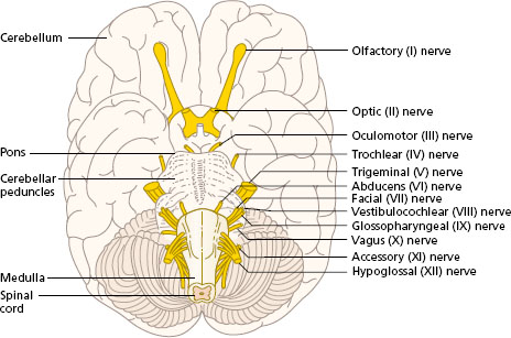
Twelve pairs of cranial nerves, the cell bodies of which are located in the brain, can be seen most clearly in a ventral view of the brain (Figure 4.1). The pairs of cranial nerves appear at irregular intervals as a result of the folding of the embryonic neural tube in the development of the brain. The cranial nerves are summarised in Appendix II, Table A2.1.
The spinal nerves consist of the 31 pairs of nerves leaving the spinal cord. Each pair of spinal nerves emerges from the vertebral canal between adjacent vertebrae at the intervertebral foramina. The latter can be seen in the lateral view of a thoracic vertebra in Appendix I. The lower end of the spinal cord in adults lies at the level of the disc between the first and second lumbar vertebrae. The lower spinal nerves therefore lie in the spinal canal below this level before emerging at their corresponding level. This sheath of lumbar and sacral nerves is known as the cauda equina (horse’s tail).
Figure 4.1 Cranial nerves seen in a ventral view of the brain.
Spinal nerves
Each spinal nerve begins at the spinal cord with two roots: the anterior (ventral) root, and the posterior (dorsal) root. Each root consists of a series of rootlets which eventually join. Diagrams of the formation of a spinal nerve represent each root as a single trunk for clarity.
The anterior root consists of axons that grow out from multipolar nerve cells in the spinal cord. These axons are all motor (efferent), carrying impulses away from the cord. Those originating in the anterior horn are lower motor neurones supplying muscles. Visceral motor fibres of the autonomic nervous system are also found in the anterior roots. The cell bodies of these autonomic neurones lie in the lateral grey matter of certain segments of the spinal cord (described later).
The posterior root develops in a different way. A ridge of cells on each side of the neural tube in the embryo forms a pair of ganglia (cells) for each segment of the spinal cord. Fibres grow centrally from each ganglion into the spinal cord, and also laterally to lie alongside the fibres originating in the anterior root. The fibres of the posterior root are all sensory (afferent), carrying information from the receptors in the skin, the muscles and the joints. The cell bodies lie in the posterior root ganglion, isolated from the hundreds of synaptic connections possible for the cell bodies of neurones in the grey matter of the spinal cord. Axons of the sensory neurones enter the spinal cord, branch to segments of the cord above or below, or turn into the posterior white matter to reach the brain stem before synapsing.
The spinal nerve is the common nerve trunk formed by the anterior (motor) root and the posterior (sensory) root joining, distal to the posterior root ganglion. Spinal nerves are mixed; each contains motor and sensory nerve fibres. Alternative terms are efferent and afferent respectively.
Each spinal nerve contains all the somatic and visceral nerve fibres that supply the corresponding body segment. The thoracic spinal nerves follow the basic plan described. The other spinal nerves show considerable mixing, branching and joining. This regrouping of nerve fibres forms a plexus.
There are four major plexuses formed by the anterior primary rami of the spinal nerves:
- cervical plexus (C1–C4) to the muscles of the neck;
- brachial plexus (C5–T1) to the muscles of the upper limb;
- lumbar plexus (L1–L4) to the muscles of the thigh;
- sacral plexus (L4–S4) to the muscles of the leg and foot.
Figure 4.2 shows the four plexuses formed by the spinal nerves. The lumbar and the sacral plexus can be considered together as the lumbosacral plexus supplying the whole of the lower limb.
As a result of the formation of a plexus, some nerve fibres from one spinal nerve may eventually lie alongside those from a different spinal nerve in one peripheral nerve.
Figure 4.2 Spinal nerves emerging from the vertebral column and formation of the cervical, brachial, lumbar and sacral plexuses.
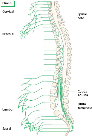
Figure 4.3 Area of skin supplied by the posterior branches of the spinal nerves.
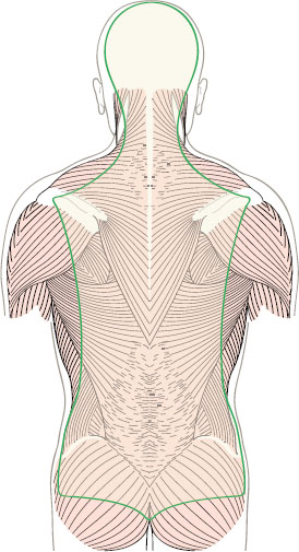
The first branch of each spinal nerve after it emerges from the vertebral column supplies the deep muscles of the back and the skin covering them (Figure 4.3). This branch is known as the posterior primary ramus. Visceral nerve fibres of the autonomic nervous system lying in the spinal nerve connect with sympathetic ganglia, which lie on the sides of the bodies of the vertebrae, by grey and white rami. Look at Chapter 3, Figure 3.20 to see the position of the posterior primary ramus of a spinal nerve and the rami of the sympathetic ganglion. The autonomic fibres will be considered later in this chapter.
Dermatomes and myotomes
A dermatome is an area of skin supplied by all the sensory nerves fibres of one spinal nerve. An example of a dermatome is a band of skin around the trunk innervated by the sensory nerve fibres of the second pair of thoracic nerves. A map of the dermatomes of all the spinal nerves is shown in Figure 4.4. In the trunk, the dermatomes form a series of bands, one for each spinal nerve from T2 to L1 in order. There is some overlap, and each dermatome may receive nerve fibres from three or four spinal nerves. In the limbs, the arrangement of dermatomes is more complicated. Each limb develops from a bud, which grows out in the embryo, and some dermatomes are carried to the ends of the limb. C7 and C8 are carried in this way to the hand, while L4, L5 and S1 reach the skin of the foot. From the diagram, it can be seen that damage to the spinal nerves in the upper part of the neck (C4, C5) will give loss of sensation around the shoulder, while severance of lower roots (C7, C8) will affect sensation in the hand.
Figure 4.4 Dermatomes. The cutaneous distribution of the spinal nerves in (a) anterior and (b) posterior view.
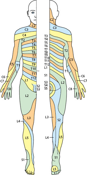
Figure 4.5 Formation of a peripheral nerve (musculocutaneous) from two spinal segments (C5 and C6).
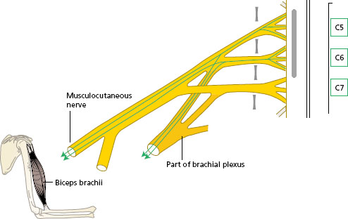
A myotome is all the muscles supplied by one spinal segment and its pair of spinal nerves. For example, nerve fibres from the first thoracic nerve (T1) are distributed to a long finger flexor muscle in the forearm and some of the intrinsic muscles of the hand. Each individual muscle, however, receives fibres from two or three spinal nerves (Figure 4.5), so that injury to one spinal segment may have only a limited effect on one particular muscle.
The segmental origin of the nerves supplying the muscle groups of the limbs is given in Appendix II, Table A2.2.
Peripheral nerves
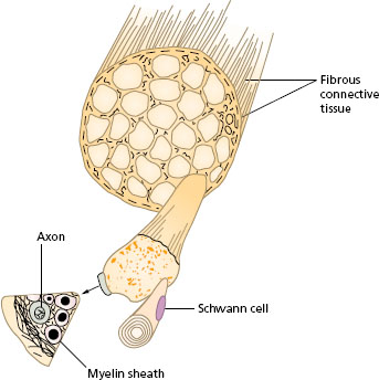
Branches of the spinal nerves, and the plexuses formed from them, are distributed to all the parts of the body. These branches are known as peripheral nerves. The structure of a peripheral nerve is shown in Figure 4.6. Half of the total bulk of a nerve is connective tissue, surrounding both the nerve and also the bundles of axons within the nerve. Each nerve has its own blood supply, which branches along the length of the nerve in both directions.
Figure 4.6 Transverse section of a peripheral nerve showing the axons and connective tissue.