PART I: THE BRAIN
Introduction to the form and structure
At first glance the brain seems to be composed only of the two cerebral hemispheres (Figure 3.1). Although they are the largest feature of the brain, they conceal many other important areas. The two symmetrical hemispheres have a folded surface with their inner aspects lying close together in the midline. Underneath the posterior end of each cerebral hemisphere is the cerebellum, which also has two hemispheres that are joined together in the midline. Part of the pons is visible anterior to the cerebellum, and below the pons is the cone-shaped medulla oblongata. The medulla leads down into the spinal cord at the foramen magnum (‘large hole’) in the base of the skull.
Development of the brain
A look at the development of the brain shown in Figure 3.2a will help in the understanding of the position and form of the adult brain areas.
The forebrain first grows laterally and backwards. It then folds forwards on itself and takes on the appearance of a hand wearing a boxing glove with the thumb touching the palm when viewed from the side. Hidden by the extensive growth of the cerebral hemispheres, the forebrain also develops less rapidly to form the basal ganglia. The wall of the remaining part of the forebrain thickens to form the thalamus and hypothalamus, collectively known as the diencephalon or ‘between brain’. The structures in the diencephalon provide important links between the cerebral hemispheres and other parts of the central nervous system for both sensory and motor activity.
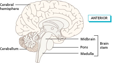
The midbrain continues in the same position during development, increasing in total size, but obscured in the external view of the brain by the lower temporal lobes of the cerebral hemispheres. In the adult, the midbrain looks like the ‘waist’, area with the expanded forebrain above and hindbrain below. Find the midbrain in the sagittal section of the brain (Figure 3.3). The mid-brain provides routes for pathways carrying impulses up or down to various levels of the central nervous system and is also important for integration of information from the eyes and ears.
Figure 3.1 External appearance of the brain, lateral view of the left side.
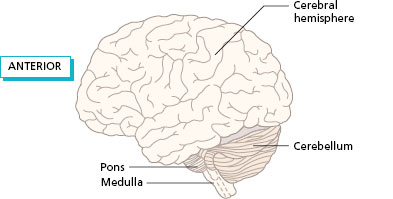
Figure 3.2 (a) Development of the brain showing folding of the forebrain; (b) adult brain viewed from the left showing the position of the cavities.
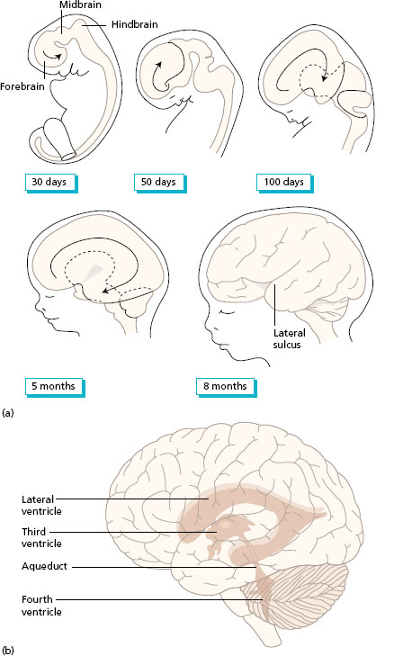
Figure 3.3 Median sagittal section of the brain.
The hindbrain develops into the pons and medulla oblongata. The cerebellum, also part of the hindbrain, grows out from the pons to lie under the posterior lobes of the cerebral hemisphere in the adult brain. Three fibre tracts link the cerebellum to the midbrain, pons and medulla.
The brain stem is the term used to describe the long cylindrical base of the brain which links the diencephalon to the spinal cord below. The brain stem is composed of the midbrain, pons and medulla (Figure 3.3). If the outgrowths of the cerebral hemispheres and cerebellum are removed from the brain, the complete brain stem can be seen with the diencephalon above.
The developing brain retains an internal cavity which forms the ventricular system containing cerebrospinal fluid. The cavity within each cerebral hemisphere follows the shape of the clenched hand and is known as the lateral ventricle. The cavity in the centre of the diencephalon is a thin slit between the two thalami, called the third ventricle. The central cavity of the midbrain is a narrow canal called the cerebral aqueduct which leads down into the fourth ventricle, the cavity of the hindbrain. The fourth ventricle lies behind the pons and upper part of the medulla, with the cerebellum forming the roof of the cavity. Figure 3.2b shows the cavities of the brain in position in the adult brain.
Cerebrospinal fluid
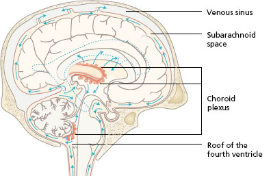
The central nervous system is surrounded and protected by the bones of the skull and the vertebral column. Further protection is provided by the cerebrospinal fluid, which is found in all the cavities of the brain and in the central canal of the spinal cord. The same fluid is also found surrounding the brain and spinal cord, in between two of the three layers of protective connective tissue known as the meninges; these will be described later. The main function of the fluid is to act as a shock absorber. It also carries nutrients and other essential substances to the nerve tissue. Figure 3.4 shows a sagittal section of the brain and part of the spinal cord to illustrate the way in which the cerebrospinal fluid circulates through the central cavities and around the outside of the central nervous system. The fluid is secreted from special patches of blood capillaries called choroid plexuses situated in each of the ventricles of the brain. The ventricles are found in the areas of greatest growth and expansion during development. Cerebrospinal fluid is formed by a process of filtration from the capillaries of each choroid plexus at the rate of 500 ml/day. Follow the arrows in Figure 3.4 to see how the fluid flows downwards in the brain and then through openings in the roof of the fourth ventricle into the space between the coverings of the brain. The absorption of cerebrospinal fluid into the blood takes place mainly in the venous sinus between the two cerebral hemispheres, known as the superior sagittal sinus.
Figure 3.4 Circulation of cerebrospinal fluid seen in a sagittal section of the brain.
Organisation of neurones into grey and white matter
The brain is composed of neurones (see Chapter 1) and their supporting cells, the neuroglia. Surprisingly, half the volume of the central nervous system is made up of neuroglia, which are special support cells found in between the neurones, and of capillaries which supply the high oxygen demands of nerve tissue. The neuroglia act as transporting and insulating cells, and also co-operate in the function of the neurones. Sections through the brain reveal areas of light and darker shade, the white and the grey matter, respectively. The overall pink colour of living brain tissue reflects the abundant blood supply.
The grey matter is all the cell bodies and dendrites of the neurones which form the core of the central nervous system. In the brain the core is not continuous, but the cells are collected together to form many nuclei of grey matter. For example, the thalamus is a nucleus of grey matter where ascending pathways from the spinal cord synapse on the way to many areas of the cerebral hemisphere. Grey matter also forms the outer layer or cortex of the cerebral hemispheres and the cerebellum. The cell bodies of the cortical neurones are laid down in layers in an organised way. The cortical grey matter is folded and each raised part, seen on the surface, is known as a gyrus. Each depression in between the gyri is called a sulcus (Figure 3.5a). A very deep sulcus is sometimes called a fissure.
Figure 3.5 (a) Grey matter: cortex with gyrus, sulcus and deep nucleus; (b) white matter: projection, association and commissural fibres.
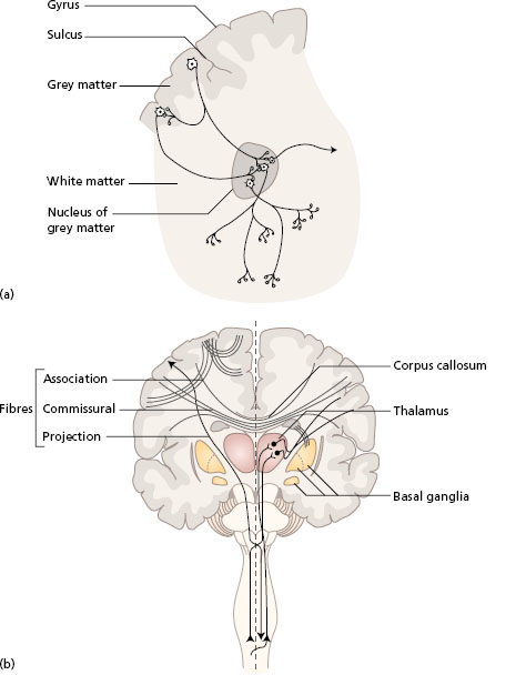
The white matter is the axons of neurones. It is found surrounding the nuclei in the brain stem and forming the core of the cerebellum and the cerebral hemispheres. The white matter is mainly organised into bundles of axons lying in particular directions (Figure 3.5b). Commissural fibres connect the right and left cerebral hemispheres. The main bridge between the two lies above the diencephalon and is known as the corpus callosum. It contains an estimated one million nerve fibres. Association fibres link one gyrus to another gyrus in the same hemisphere. Projection fibres convey information between the surface grey matter and both the lower centres of the brain stem and the spinal cord. Each of the projection fibres carries impulses in one direction only, either upwards or downwards.
It is important to build up a three-dimensional picture of the shape, position and relations of the areas of the brain. Diagrams of sections taken through the brain at different levels can be compared with slices in various directions of a Swiss (jelly) roll or a piece of marble. Each slice shows one particular colour in a different way, but the shapes can be put together to determine the three-dimensional shape inside. This task is not easy, but can be achieved with practice.
The location and overall functions of each of the main brain areas will now be considered in the following order: cerebral hemispheres (frontal, parietal, temporal and occipital lobes), basal ganglia, thalamus, hypothalamus and limbic system, brain stem and cerebellum.
Cerebral hemispheres
The great expansion of the cerebral hemispheres (or cerebrum) to envelop nearly all other brain areas distinguishes the primates, especially humans, from other animals. It is, therefore, not surprising that the surface of the hemispheres (cerebral cortex) has been studied extensively for over two centuries. The microscopists of the mid-nineteenth century noted variations in the basic cellular architecture in different regions of the cerebral cortex. The result of these studies was a detailed mapping into 52 areas numbered by Brodman (1909) and used in practice to this day for purposes of description. Meanwhile, evidence from brain damage was accumulating to suggest that different areas of the cerebral cortex have particular functions. In 1861 Broca had identified a particular area in the left hemisphere concerned with speech, from the post-mortem examination of a patient with a severe motor speech defect. Evidence from head injuries in soldiers in the trenches in World War I, and studies of the electrical activity of the surface of the brain during surgical intervention led to the identification of distinct motor and sensory areas related to particular parts of the body.
The primary areas identify and localise information from the sense organs, skin and muscles (sensory), or send out motor commands to the muscles for the correct force, timing and speed of movement (motor). Other areas, called association areas, process information from the primary areas at a higher level for recognition and meaning. For example, there is a primary sensory area receiving information from receptors in the skin, muscles and joints. An adjacent association area has links with the primary area and with other areas involved in perception and memory. The integration of all this information leads to the ability to recognise objects held in the hand without vision, known as stereognosis.
In recent years, neuroimaging studies using positron emission tomography (PET) have extended our knowledge of brain function by identifying the active brain areas during the performance of activities using the upper limbs. Studies have shown that the number and the location of active areas vary in different individuals performing the same task, and activity may occur in both the right and left hemispheres during the performance of a single-handed task. Similarly, functional magnetic resonance imaging (fMRI) scans can measure the changes in blood flow as a result of neuronal activity linked to different functions. This technology can assist in mapping different areas of the cortex in response to undertaking different activities. More recently, TMS (transcranial magnetic stimulation) studies involve placing a magnetic coil near a specific area of the person’s head to extrinsically stimulate functional movements, i.e. make someone perform a specific movement. If this is combined with fMRI, the specific location of that function can be very accurately mapped on the cortex.
PET scan studies have shown that the function of one brain area can shift to another area with related function after brain damage. A group of people who had been blinded in early life showed activity in the areas of the brain normally concerned with vision when they performed tactile tasks. Normal subjects doing the same tactile tasks showed no activity in the visual areas. These results confirm the plasticity of neurones in the brain, particularly in early life.
The lobes of the cerebral hemispheres
Each cerebral hemisphere is divided into four lobes, named after the skull bones that cover them. In each hemisphere, the lobes are separated by two deep sulci: the central sulcus and the lateral sulcus (Figure 3.6a).
- The frontal lobe lies anterior to the central sulcus, and above the lateral sulcus.
- The parietal lobe lies behind the central sulcus.
- The occipital lobe is at the posterior end of the hemisphere, above the cerebellum at the base of the skull.
- The temporal lobe lies below the lateral sulcus.
Each lobe continues on to the medial surface of the hemisphere (Figure 3.6b). The median sagittal sulcus separates the right and left frontal, parietal and occipital lobes (Figure 3.6c).
It is important to realise that the surface of the cerebral hemispheres extends from the level of the eyebrows in front, to the base of the skull at the back of the head, and down to the level of the ears at the side. This becomes obvious when a life-sized model of the brain is placed inside the cranial cavity of the skull.
The overall functions of each lobe of the cerebral hemispheres will be described in turn. It is important to stress that the numerous interconnections among the four lobes means that no individual lobe functions alone.
Figure 3.6 Lobes of the cerebral hemispheres seen in: (a) lateral view; (b) medial view; (c) view from above.

Frontal lobe
The frontal lobe is a large part of the cerebral hemisphere found underneath the frontal bone of the skull. The part of the frontal lobe particularly concerned with the performance of movement lies more posteriorly in the lobe, leading up to the central sulcus. The larger anterior part of the lobe, which lies above the orbit of the eyes (supraorbital area), is involved in planning and problem-solving aspects of both movement and behaviour. This part of the frontal lobe is also called the prefrontal lobe.
The band of grey matter lying immediately in front of the central sulcus (precentral gyrus) is the primary motor area, which is concerned with the generation of movement in the whole of the opposite side of the body (Figure 3.7a). Adjacent to this area are the premotor area and the supplementary motor area which are discussed in more detail below.
The cell bodies of the neurones in the motor cortex project not to individual muscles, but to functional groups of muscles. Direct links to the small muscles of the hands, the feet and the face are particularly important, and damage to the primary motor area often results in loss of precision movements.
There is representation of half of the body in an ‘upside-down’ position in each primary motor cortex. The head is represented in the lower cortex on the lateral side, then the upper limb and trunk above, and finally the lower leg and feet in the cortex on the medial surface of the lobe. In Figure 3.8, a vertical section through the cerebral hemisphere at the level of the primary motor area is shown (frontal section). Note that the sizes of the body parts are not in normal proportions. The body parts that move with the greatest degree of precision have larger areas of representation, so that the face and hand are large, while the trunk and leg are small. A figure constructed with these dimensions has a head like a hippopotamus, the hands of a giant, and the trunk and legs of a dwarf, and is known as the ‘motor homunculus’.
The premotor area lies anterior to the primary motor area on the lateral surface of the lobe (Figure 3.7a). Visual and auditory information from the occipital and temporal lobes, respectively, is integrated in the premotor area to guide movement, more specifically it helps guide body movement by integrating sensory information and controls muscle groups that are closest to the axis of the body (midline). Neurones project from this area to the primary motor area on the same and the opposite side. Projection fibres from the premotor area descend directly to the spinal cord, or indirectly via the primary motor cortex.
The supplementary motor area (SMA) also lies anterior to the primary motor area, but mainly on the medial side of the frontal lobe (Figure 3.7b). Neuroimaging studies have recorded an increase in cerebral blood flow in the supplementary motor area immediately before the execution of complex sequences of movements of the fingers, and when both hands are involved. These studies suggest a role in the planning of movement that is internally generated.
Figure 3.7 Main functional areas of the cerebral hemispheres: (a) lateral view; (b) medial view. MC = primary motor cortex; SMA = supplementary motor area; SC = somatosensory area.
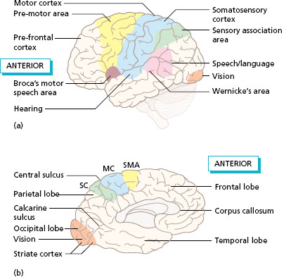
The motor speech area identified by Broca lies in the lower part of the frontal lobe in the lip of the lateral sulcus (see Figure 3.7a). The function of this area, usually only found in the dominant hemisphere, is in the production of fluent speech.
The prefrontal area (supraorbital area) occupies the large anterior area of the frontal lobe and connects with all the other lobes of the cerebral hemispheres, the thalamus, the limbic system and many other brain areas. Interaction with the limbic system is concerned with the emotional aspects of movement. The prefrontal area is also concerned with the planning of goal-directed movement and behaviour and in modifying the plan in response to changes in the environment. These are known as the executive functions.
Parietal lobe
The parietal lobe lies posterior to the frontal lobe and beneath the parietal bone of the skull. The overall function of the parietal lobe is the processing of sensory input from receptors in all parts of the body and also from the special sense organs (eyes and ears). This provides awareness of the position of the parts of the body during movement and spatial awareness of the environment.
Figure 3.8 Primary motor cortex seen in frontal section to show the representation of body parts.
- A lesion in the primary and premotor cortex on one side leads to muscle weakness in the muscles of the opposite side of the body, known as contralateral hemiplegia. Muscle tone may be low (flaccid) or high (spastic). Fine skilled movements of the extremities are particularly affected.
- Lesions in the prefrontal cortex area lead to problems in planning movement and the ongoing review of movement whilst it is being carried out. Loss of insight into movement performance may be a major factor in the rehabilitation process. Frontal lobe lesions are also associated with innapropriate social behaviour and lack of insight. The inability to plan and monitor movement and behaviour resulting from lesions in the prefrontal cortex is known as dysexecutive syndrome.


