Surgical Techniques

Chapter 80 Surgical Techniques


Surgical Techniques


Preoperative Preparation


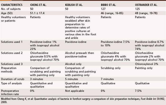
Because of the normal bacterial flora residing on the foot, particularly in the web spaces, which often are moist because of confinement in hosiery and shoes, the extremity to be operated on is scrubbed for 8 to 10 minutes from toes to knee with an antibacterial soap of the surgeon’s choice that the patient’s skin can tolerate. Particular attention is directed to the web spaces. After this cleansing, the extremity is wrapped sterilely with a cloth that is secured with a gauze wrap. This is not removed until the patient is on the operating table, at which time the skin is prepared again with an antiseptic solution. Goucher and Coughlin, however, found no benefit in covering the toes during hindfoot or ankle surgery after skin preparation with chlorhexidine gluconate and isopropyl alcohol. Only two of 40 patients had positive cultures after surgery, and neither developed an infection. Forefoot procedures are worrisome because of the high bacterial counts between the toes. Bacteria resides in colonies between the stratum corneum, in the follicles of the sebaceous glands, and in the hair. Presently, no consensus has been reached for the best skin preparation in foot and ankle surgery, although several studies have compared different types of solutions and preparation methods (Table 80-1). Quantitative analysis of positive cultures have shown significant reductions in heavy bacterial growth when bristled brushes are used with povidone-iodine and isopropyl alcohol.


Prophylactic antibiotics are used routinely in foot and ankle procedures. A first-generation cephalosporin, or an aminoglycoside if the patient is allergic to penicillin or cephalosporin, is the most frequently used antibiotic.


When performing foot or ankle surgery, the surgeon and assistant may sit or stand. Additional draping, secured to the table and the body of the surgeon, ensures a sterile field in the surgeon’s lap when seated. Lighting that reduces shadows and focuses sharply on the foot is necessary. A high-intensity headlight is useful, especially during deep plantar dissections. To reduce injuries to the cutaneous nerve, an assistant should hold the foot motionless. Careful preoperative planning, a thorough knowledge of the pertinent anatomy, good surgical exposure, skill in use of equipment, and elimination of distractions reduce the likelihood of an undesirable outcome.


Tourniquet


The use of a tourniquet allows dissection in a bloodless field, decreasing the likelihood of injury to nerves, vessels, and tendons. Adequate surgery of the foot can be performed without a tourniquet, however; and in selected patients, because of age or a vascular condition, a tourniquet might be contraindicated. Smith and Hing, in a systematic review, reported that patients who had foot and ankle surgery without a tourniquet had less pain, reduced swelling, and a shorter hospital stay than those who had tourniquet-assisted surgery. Also, they suggested a greater incidence of wound infection and deep vein thrombosis in tourniquet-assisted foot and ankle surgery, although they conceded that further study is warranted. If a tourniquet is used, the pressure should be set 100 to 125 mm Hg higher than the systolic blood pressure and inflated after elevation of the leg for 2 minutes or after exsanguination of the extremity from toes to tourniquet with an elastic rubber wrap that is 10 cm wide. Most surgeons consider blood pressure, limb size, or both in determining the amount cuff pressure. For calf and ankle cuffs, pressures most commonly used by surgeons in a survey of members of the American Orthopaedic Foot and Ankle Society (AOFAS) were 201 to 250 mm Hg, although Younger et al. thought that these pressures might be higher than necessary for many patients. We rarely leave the tourniquet inflated more than 90 minutes during procedures on the foot but occasionally have kept it inflated for 120 minutes. If the procedure is prolonged, however, and the surgeon anticipates the need of a tourniquet for longer than 60 to 75 minutes, it is advisable to deflate the tourniquet, elevate the leg for 10 to 15 minutes, and not exceed 30 minutes for a second tourniquet inflation.


The use of an elastic wrap for a tourniquet has proved useful and safe in our hands. When the blood supply to the foot is questionable, a tourniquet is not recommended and a constricting wrap around the ankle especially should be avoided. The use of an elastic rubber tourniquet around the foot and ankle permits most forefoot and midfoot procedures to be performed in a bloodless field for 2 hours, if needed, and the patient experiences little tourniquet discomfort as long as the tourniquet does not reach the musculotendinous junction at the distal third of the leg. Grebing and Coughlin evaluated pressures beneath 4- and 6-inch elastic rubber bandages (Esmarch) using three and four tensioned wraps around the ankle, followed by tucking the remainder of the bandage beneath the proximal end of the wrap. Three wraps with a tuck produced an average pressure of 222 mm Hg (range, 146 to 319 mm Hg), and four wraps around the ankle with a tuck generated an average pressure of 288 mm Hg (range, 202 to 405 mm Hg). The investigators concluded what clinical experience has shown: This technique of exsanguination to improve visibility in the operative field is safe and effective. A survey of 140 members of the AOFAS showed that 73% of surgeons use an elastic bandage for exsanguination. Rudkin et al. audited 1000 patients who had foot and ankle surgery with ankle block anesthesia and an ankle cuff tourniquet and recommended the method as safe and effective; however, they cautioned that patients older than 70 were at greater risk (3.5 times greater than younger patients) of experiencing tourniquet pain with ankle block anesthesia. When a thigh tourniquet is used, the length of procedures done under local anesthesia is markedly shortened because of discomfort caused by the tourniquet. Ankle and upper calf pneumatic tourniquets are safe and convenient for procedures on the foot and ankle.


Instruments


Two trays of instruments, designated as “foot tray—soft tissue” and “foot tray—bone,” are helpful. The instruments in the former should include fine two-tooth retractors and delicate forceps with 1.5 mm between the teeth. A Brown-Adson forceps is helpful for grasping small bone fragments and bulky soft tissue. A No. 15 Bard-Parker blade attached to a multisided handle facilitates quick changes in angle of dissection. Sharp dissection with a scalpel is indicated whenever practical to prevent tearing of the tissue and the edematous reaction that follows indelicate dissection. When blunt dissection is needed, small scissors with gently curved and slightly blunted tips are helpful. Narrow- and wide-neck mini-Hohmann and small double-ended, right-angle retractors allow the assistant’s hand to be out of the field of dissection and yet afford excellent exposure. Mosquito hemostats for small vessel occlusion, Webster needle holders with smooth jaws for grasping fine (4-0 to 5-0) suture, and a 70-degree angled probe complete the foot tray for use in soft tissues.


The foot tray for bony procedures has many of the same instruments but on a larger scale. Heavier blades, forceps, dissecting scissors, retractors, and needle holders may be needed for procedures that include bone and large tendon surgery, as in various midfoot and hindfoot arthrodeses, osteotomies, or tendon advancements, transfers, or transpositions. Thin osteotomes, a small mallet, small curets, heavy-duty two-tooth retractors, a wide dissecting probe, and an Inge retractor with arms that have been thinned and teeth narrowed all facilitate bony procedures.


Two power instruments are helpful in surgery of the foot. One is a Kirschner wire inserter that does not require key release to change wires or to change the length of a wire. Another is a power oscillating saw with thin, narrow blades. Magnification is helpful; we routinely use 2.5× to 3.5× magnification loupes. A camera that is simple and easily used by the operating room personnel is recommended for taking pictures of the procedure before, during, and after surgery.


Anesthesia


As more foot surgery is performed on an outpatient basis, the benefits of regional forefoot or ankle block anesthesia become more obvious. A combination of 1% lidocaine (Xylocaine) and 0.5% bupivacaine (Marcaine), given in a recommended volume per kilogram of weight dose, provides adequate anesthesia for most forefoot surgeries and for a significant portion of hindfoot surgeries. We have anesthesia personnel in the room or operating area to sedate and monitor the patient while the block is performed. Suggested techniques for forefoot and ankle block anesthesia are given.


Jun 5, 2016 | Posted by in ORTHOPEDIC | Comments Off on Surgical Techniques

Full access? Get Clinical Tree

Get Clinical Tree app for offline access