Fig. 1
Chronic dislocations may be associated with erosion of the femoral head by the indirect head of the rectus femoris and/or other deformations resulting in mild to severe loss of sphericity and degenerative changes. There are no absolute guidelines to determine which hips are reconstructible, and direct inspection may be required to assess the degree of damage and decide what to do
Displacement of the femoral head is assessed using the Reimer’s migration index (Fig. 2). This measurement is taken by first drawing a line horizontal through the triradiate cartilage (Hilgenreiner’s line) and then a line vertical at the edge of the lateral margin of the acetabulum (Perkin’s line). The percent of displacement of the capital femoral epiphysis is then calculated as the percentage of the capital femoral epiphysis that lies outside Perkin’s line. If the migration percentage is over 50 % [3], subluxation and dislocation is highly likely [4]. The acetabular index is also evaluated and is felt to be abnormal if >25°. The characteristics of the sourcil should also be noted.
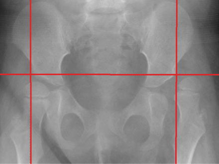
Fig. 2
AP x-ray of the pelvis showing a migration percentage of 20 % on the right and >90 % on the left
Surveillance programs have been set up to screen for dysplasia and institute early treatment with the goal of preventing dislocation and minimizing the magnitude to surgery required to achieve this goal. These surveillance systems involve repeating the physical examination, typically every 6 months, and repeating radiographs at an interval defined by the relative clinical risk of progressive dysplasia .
Surgical treatment strategies for neuromuscular hip dysplasia may be (1) prophylactic, (2) reconstructive, or (3) salvage. Prophylactic procedures are indicated with younger patients who have developed soft tissue contractures but do not have significant subluxation. These patients typically exhibit clinical “at risk” signs (passive abduction in extension <45°) and minimal elevation in the migration percentage, <40–50 %. In such cases, an open adductor release with or without psoas tenotomy may prevent the need for reconstructive surgery in more than 50 % of patients at 9 years follow-up. Even when soft tissue surgery fails to eliminate the need for bony reconstructive surgery, the reconstructive procedures are performed at a later age which should reduce the chance or resubluxation or dislocation with growth and remodeling. Reconstructive surgery is performed when significant bony abnormalities are present (significant subluxation and acetabular dysplasia), ideally in a child older than 4 years of age. Salvage surgery is considered in hips that are felt to be non-reconstructible, in which femoral head deformity is severe or arthritic changes are present, typically in patients with long-standing dislocation and chronic pain. The degree of femoral head deformity beyond which reconstructive surgery should not be considered remains unknown, and there is no scientific evidence to guide this decision.
In this chapter reconstructive surgery will be considered which typically involves a soft tissue release, correction of proximal femoral deformity (excessive anteversion and valgus) by a varus derotational osteotomy (VDRO), and correction of coexisting acetabular dysplasia with a volume-reducing, incomplete pelvic osteotomy. In some cases a medial capsulotomy or an open reduction may also be required as a component of this reconstruction. The first goal of the femoral osteotomy is to restore normal rotational alignment to the proximal femur by correcting excessive femoral anteversion, which will improve abductor mechanics in the ambulatory, and lower extremity positioning in the nonambulator. The second goal is correction of coxa valga, recognizing that the desired end point for neck-shaft angle differs in ambulators versus nonambulators. The goal of the acetabular osteotomy is to restore acetabular morphology and improve coverage of the femoral epiphysis.
Indications for Surgery
Equipment
1.
Blade plate set: Comes with infant, child, adolescent, and adult sizes
2.
Sterile goniometer
3.
K wires
4.
Oscillating saw
5.
Cobb and Crego elevators
6.
Hohman retractors
7.
Lamina spreader
8.
Bone holding forceps or clamps (Verbrugge, Lowman)
9.
Metallic wedges of predetermined angles
Technique of Femoral Varus Derotational Osteotomy
Positioning and Preparation
1.
Either supine or prone positioning may be chosen based on whether a pelvic osteotomy is required and the surgeon’s experience and preference. When a pelvic osteotomy is not performed, typically in the ambulatory population, the procedure can be performed in the prone position. This position is familiar as the clinical assessment of lower extremity rotational alignment is usually carried out in this position. There are several technical advantages, especially with regard to clinically estimating femoral anteversion. For one, it is easier to insert and maneuver the chisel for the blade plate when it is facing upwards, and fine-tuning of the chisel’s trajectory on the lateral image can be made with simple positioning of the leg rather than while an assistant holds up the leg while you are striking the chisel close to the surface of the table. It is also possible to clinically estimate femoral anteversion using a guide pin up the femoral neck (Fig. 3).
A supine position can also be used in nonambulators since they frequently require a pelvic osteotomy. In this position, the patient is placed with a bump under the ipsilateral buttock. If a bilateral procedure is to be performed, then a small square bump under the sacrum is helpful, particularly if performing pelvic-sided procedures (Fig. 4).
2.
If a bilateral procedure is being done, a folded surgical towel can be placed over the genitalia and sealed using an occlusive dressing. The prep may be done over this sealed dressing.
3.
The entire leg is prepped out with care to provide an adequate superior margin, up to the lower ribs, and also an adequate posterior margin.
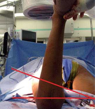
Fig. 3
Intraoperative assessment of femoral anteversion pin placed in center position on the lateral compared to the transepicondylar axis. In this instance the patient is in the prone position, but a similar assessment can be conducted in the supine position by holding the knee flexed to 90°
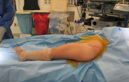
Fig. 4
Positioning of patient for cerebral palsy unilateral hip reconstruction following draping (photograph courtesy of Wudbhav Sankar, MD)
Adductor Tenotomy
1.
An adductor release is routinely performed with the VDRO in the nonambulatory population. This is rarely needed in an ambulatory patient and if required may often be performed as a percutaneous release of the fascia overlying the adductor longus rather than the more extensive open release. In the ambulatory patients, correction of derotation is usually the major aim and little if any varus is required.
2.
A transverse incision is made in the groin crease directly overlying the adductor longus tendon, about two fingerbreadths distal to the superior pubic ramus.
3.
A longitudinal incision is made in the deep fascia overlying the adductor longus tendon, and the subfascial interval developed which exposes the adductor longus and surrounding muscles (Fig. 5).
4.
A right angle retractor is passed around the longus tendon, and the tendon is then released with electrocautery. The anterior neurovascular bundle must be identified on the surface of the adductor brevis and protected. The gracilis is then bluntly identified and released with electrocautery. This muscle is placed under tension by abducting the hip with both the hip and knee extended.
5.
A partial myotomy of the adductor brevis may be performed if additional passive abduction is required.
6.
If a psoas tenotomy is planned at this stage, the interval between the pectineus and the brevis is used and widened laterally. The psoas tendon is identified as it attaches to the lesser trochanter. The sheath is opened up and a right angle is passed around it. It is released with electrocautery, while being careful to protect the medial femoral circumflex artery.
7.
When the desired amount of hip abduction is obtained, the wound is irrigated and close in layers once hemostasis has been achieved.
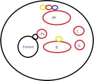
Fig. 5
Cross-sectional schematic of the proximal femur at the level of the lesser trochanter” B adductor brevis, G gracilis, L adductor longus, Pe pectineus, Ps psoas, yellow nerve, blue vein, black bone
Surgical Approach for VDRO
1.
A longitudinal incision is made over the lateral proximal femur extending from the trochanteric flare or slightly above down along the femoral shaft. Sufficient length must be made to accommodate the length of the plate taking into account shortening from the varus with or without removal of any additional bone wedges.
2.
Dissect through the skin subcutaneous tissue down to the level of the fascia lata. The fascia lata is then divided in line with the skin incision. A self-retaining retractor is placed in the fascia lata.
3.
The fascia overlying the vastus lateralis is incised longitudinally approximately 5 mm anterior to the intramuscular septum. The muscle is teased off the posterior fascia until the periosteum is visualized, and then the periosteum is incised to subperiosteally expose the proximal femur. This cuff of remaining posterior fascia is utilized for the repair.
4.
Proximally, the origin of the vastus lateralis is incised transversely (perpendicular to the longitudinal cut in the fascia) along the trochanteric flare and distal to the trochanteric apophysis, to allow the muscle to be reflected anteriorly (Fig. 6). Care is made not to plunge posteriorly with the cautery as the sciatic nerve is very close.
5.
The periosteum is incised to expose the femur subperiosteally. Hohman retractors are placed anteriorly and posteriorly for protection.
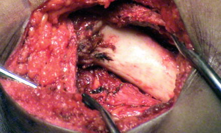
Fig. 6
The lateral approach to the femur. The proximal femur has been subperiosteally exposed. The lateral aspect of the greater trochanter apophysis is visible in the proximal aspect of the wound
Osteotomy Technique (90° Blade Plate)
Determining Rotation and Neck-Shaft Angle
The first component is to estimate or measure the true neck-shaft angle and the degree of anteversion. Recognizing that a more important concern is how to accurately achieve targets for final anteversion and neck-shaft angle, it is advantageous to get baseline measurements for completeness. These measurements can be made either before or after the patient is prepped and draped. With regard to neck-shaft angle, the hip is internally rotated until a true AP is obtained, typically when the length of the femoral neck appears greatest on the AP image. This image can be saved and measured with a goniometer. The degree of anteversion may be been measured using several techniques. Some prefer to obtain a CT scan preoperatively; however, anteversion may be measured intraoperatively. In the first, prior to prepping and draping, the patient is slid down to the end of the table, and the knee is flexed 90° over the edge. The hip is then medially rotated until a true AP is obtained, and the version is then measured as the angle between the tibial axis or shank and the vertical. Alternatively, version may be measured during the procedure.
Related posts:
 Neuromuscular Hip Disorders: Focus on Cerebral Palsy
 Femoral Deformities: Varus, Valgus, Retroversion, and Anteversion
 Surgical Technique: Arthroscopic Labral Management
 Surgical Technique: Endoscopic Gluteus Medius Repair
 Introduction to Static and Dynamic Overload of Hip Pathology
 Physical Examination of the Hip and Pelvis
Stay updated, free articles. Join our Telegram channel

Full access? Get Clinical Tree


