Fig. 1
(a) AP radiograph left hip demonstrating a severe unstable SCFE. (b) Frog lateral radiograph right hip confirming no contralateral SCFE. Note that the left hip was not manipulated. (c) Intraoperative image demonstrating incomplete reduction after positioning patient. (d) AP radiograph left hip (e) Frog lateral radiograph left hip following in situ fixation with two 6.5 mm fully threaded cannulated screws. Note the residual proximal femoral deformity but no AVN
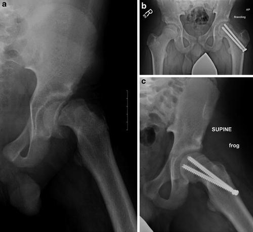
Fig. 2
(a) AP radiograph left hip demonstrating severe unstable SCFE. (b) AP pelvic radiograph (c) Frog lateral radiograph left hip. Note near anatomic alignment with no AVN 7 months following positional reduction and in situ pinning
Early studies of epiphyseal fixation in a bovine shear model demonstrated double-screw fixation to be stiffer than single-screw fixation, but there was no difference when tested under physiologic cyclical loading (no torsional testing performed). As a result, the authors recommended single-screw fixation as the risk of intra-articular placement of the second screw outweighed the mechanical benefit [15, 16]. Subsequent torsional testing in a nonreduced, immature bovine model demonstrated that double-screw fixation resulted in a 25 % increase in axial stiffness when tested under shear and a 312 % increase in torsional stiffness when compared to single-screw fixation [17]. In an immature porcine model of mild to moderate unstable SCFE, single versus double 7.3 mm (16 mm thread length) cannulated screws were tested with a posterior-inferior directed force instead of a pure shear load. Double-screw constructs were 66 % stiffer and 66 % stronger when compared to single-screw fixation. Therefore, a double-screw construct for an unstable SCFE is biomechanically superior to a single-screw construct, but this benefit must be weighed against the potential complications of a second screw, i.e., intra-articular penetration/chondrolysis [18].
Capsular decompression has been recognized as an important step when performing an in situ fixation of an unstable SCFE. Herrera-Soto et al. [19] demonstrated a significantly elevated intracapsular pressure in the effected hip when compare to the normal hip. In addition, there was a further significant increase in pressure after attempted manipulation. Given the risk of the increased intracapsular pressure causing a tamponade effect on the retinacular vessels and causing AVN, a capsular decompression is recommended.
There are multiple decompression techniques that can be utilized. An 18 gauge spinal needle can be placed percutaneously anterolaterally through the proximal thigh into the hip capsule under C-arm guidance. If there is a large traumatic effusion, this can be aspirated to decrease the intra-articular pressure and protect the posterior superior retinacular vessels from intra-articular compression. Alternatively, a large hemostat can be placed through one of the incisions created for screw placement and placed onto the femoral neck and through the capsule, guided by fluoroscopic imaging. The capsule can then be opened and decompressed. With this method, it is difficult to estimate how much fluid is decompressed from the hip. Another alternative is a formal Smith-Peterson anterior approach to the hip for a capsulotomy and decompression under direct visualization.
Author’s Preferred Operative Technique
The patient is brought into the operating room and general anesthesia is induced while the patient is on the stretcher. The patient is not moved to the operating table prior to the induction of the anesthesia to minimize the patient’s discomfort. Once asleep, the ipsilateral foot and ankle are well padded. The patient is then gently transferred to the fracture table. The fracture table is selected so that the affected hip can be maintained still during the procedure while moving the image intensifier for the AP and lateral views of the hip. Pinning the hip on a flattop table would require increased manipulation of the hip, which could be detrimental to the position of the epiphysis and potentially to its blood supply. The affected foot and ankle are placed into the traction boot of the fracture table, but no formal manipulation is formed. The contralateral hip is flexed and abducted and the leg placed on a well-padded leg holder. The effected lower extremity is positioned so the knee cap is pointed toward the ceiling. The image intensifier is then used in the AP and lateral views to confirm that appropriate imaging can be achieved. Any reduction achieved while positioning the patient is accepted, but no further reduction maneuver is performed. The patient is then prepped and draped in the usual sterile fashion.
Under fluoroscopic guidance, a 2.8 mm threaded guide pin is placed percutaneously into the center of the femoral epiphysis on the AP and lateral view of the hip. Commonly, the unstable SCFE has severe posterior displacement, and placing the guide pin in the center of the femoral head would require placing the pin quite anterior on the femoral neck (Fig. 1c). This can result in postsurgical screw impingement [20]. In this case, the guide pin is started more laterally on the femoral neck with the goal of creating the starting point at or lateral to the intertrochanteric line on the AP image. When planning the exact placement of the pin, thought should be given to placement of a second guide pin and a second screw. Two 6.5 mm fully threaded, cannulated screws are recommended. When compared to partially threaded screws, the fully threaded screws have superior purchase in the metaphyseal bone of the proximal femur and are easier to remove if needed. Once fluoroscopy has confirmed appropriate placement of the first guide pin in the center of the head, a second guide pin can be placed, most commonly in the posterior-inferior aspect of the femoral epiphysis. Placement of the second screw anteriorly may make the screw prone to cutting out of the anterior neck because of the limited metaphyseal bone anteriorly, and the screw may be more likely to become intra-articular if there is collapse of the anterior epiphysis with the onset of AVN. After both guide pins are placed in the appropriate position and confirmed under fluoroscopy in AP and lateral views, a 1 cm incision is made around each guide pin. Blunt dissection is taken down to bone along the guide pins. The appropriate screw lengths are measured, and a 5.0 mm cannulated drill is used to over-drill the guide pin up to, but not across, the physis. The appropriate length screw is placed by hand over the guide pin into the epiphysis. The goal is to have at least four threads of the screw across the physis into the epiphysis. The second guide pin is then over-drilled, and the second screw is placed in a similar fashion. Once both screws are secured, fluoroscopic images are obtained through a full arc of motion, from a true lateral to a true AP of the hip, confirming that neither screw is encroaching the subchondral bone of the epiphysis. Because of the residual malposition of the femoral epiphysis, it is frequently helpful to rotate the image intensifier beyond a true AP angle so that the femoral epiphysis can be imaged in an orthogonal manner.
Capsular decompression is then performed by placing a large hemostat through one of the incisions into the capsule under fluoroscopic guidance. Once the hip is decompressed, the guide pins can be safely removed. The wounds are copiously irrigated with normal saline. The deep tissues are closed with a 2-0 Vicryl suture and the skin approximated with a 2-0 Monocryl suture in a subcuticular fashion. Bupivacaine (0.25 %) is injected around the incision for local anesthetic. The incisions are covered with a gauze dressing that can be removed in 48 h.
Related posts:
 Neuromuscular Hip Disorders: Focus on Cerebral Palsy
 Surgical Technique: Bone Graft for Avascular Necrosis of the Hip
 Rehabilitation of Non-Operative Hip Conditions
 Surgical Technique: Open Proximal Hamstring Repair
 Subspine Impingement and Surgical Technique
 Atraumatic Instability and Surgical Technique
Stay updated, free articles. Join our Telegram channel

Full access? Get Clinical Tree


