Surgical Approach to PCL Injury
Craig S. Mauro
John C. Karpie
Christopher D. Harner
INDICATIONS/CONTRAINDICATIONS
Posterior cruciate ligament (PCL) injuries are rare, may be partial or complete, and frequently occur with other ligamentous and soft tissue injuries. There is a relative lack of consensus regarding the optimal management of PCL injuries. Most authors advocate nonoperative management of isolated, partial PCL injuries (grades I and II). Our treatment protocol includes immobilization in full extension with protected weight bearing for 2 weeks. Range of motion exercises are advanced as tolerated and strengthening is focused on the quadriceps muscles through closed-chain exercises. Return to sport after isolated grade I and II PCL injuries is usually 4 to 6 weeks, paying special attention to protect the athlete from injury and potential progression to a grade III injury. We have found functional bracing to be of little benefit after return to sport.
Treatment of isolated grade III injuries is more controversial, as some authors dispute the existence of an isolated grade III injury. We recommend nonoperative treatment with immobilization in full extension for 2 weeks to prevent posterior tibial subluxation. Weight bearing is protected during these 2 weeks, then slowly advanced. Quadriceps sets and straight leg raises are encouraged, while hamstring loading is prohibited until later in the rehabilitation course. After 1 month, range of motion, full weight bearing, and progression to functional activities is instituted. Return to sport is usually delayed for 2 to 4 months in patients with grade III injuries.
Our indications for reconstruction of isolated PCL tears are displaced bony avulsions or chronic grade II/III injuries with persistent instability and pain. We also advocate PCL reconstruction in the setting of combined ligamentous injuries, especially those involving the posterolateral corner. Surgical treatment of isolated grade II or III injuries may be considered for higher-level athletes.
PREOPERATIVE PLANNING
Accurately diagnosing isolated PCL or combined ligamentous knee injuries through a complete history and thorough physical exam is the first step in injury management. Once identified, understanding PCL injuries with respect to their natural history, surgical indications, surgical technique, and postoperative rehabilitation is important in order to achieve a successful outcome. The timing of PCL reconstruction depends on the severity of the injury and the associated concomitant ligamentous injuries. Displaced bony avulsions and knees with multiligamentous injuries should be addressed within the first 3 weeks to provide the opportunity for anatomical repair (4).
The initial history should focus on the mechanism of injury, its severity, and associated injuries. With acute injuries, the patient will often not report feeling a “pop” or “tear,” as is often described with ACL injuries. The history should also focus on assessing the chronicity of the injury, and the pain and instability the patient experiences. Acute injuries usually result from a history of a direct blow to the anterior lower leg. Motor-vehicle accidents and sports-related trauma are the most commonly cited causes of PCL injuries (2). In motor-vehicle trauma, the classic “dashboard injury” occurs when the proximal tibia strikes the dashboard, causing a posteriorly directed force to the proximal tibia with the knee in a flexed position. Athletic injuries usually involve a direct blow to the anterior tibia or a fall onto a flexed knee with the foot in plantar flexion. Noncontact
hyperflexion injuries have been reported to result in partial tearing of the PCL. Hyperextension injuries, which are often combined with varus or valgus forces, often result in combined ligamentous injuries.
hyperflexion injuries have been reported to result in partial tearing of the PCL. Hyperextension injuries, which are often combined with varus or valgus forces, often result in combined ligamentous injuries.
A comprehensive knee examination, including inspection, palpation, range of motion testing, neurovascular examination, and special tests should be performed. Special tests to examine the PCL and posteriolateral corner include posterior drawer test (Fig. 36.1), posterior sag (Godfrey) test (Fig. 36.2), quadriceps active test, reverse pivot shift test, dial test, and posterolateral external rotation test. The posterior drawer test is the most sensitive test to detect a PCL injury. The quadriceps active test must be performed in the office setting as voluntary quadriceps contraction must occur. This test is generally only helpful for higher grade injuries, as the examiner must visualize the posteriorly subluxed tibia reduce anteriorly with quadriceps contraction. The reverse pivot shift test is performed with the patient supine and the knee flexed to 90 degrees. The knee is extended while the foot is externally rotated and a valgus stress is applied to the knee. A palpable reduction of the tibia occurs at 20 to 30 degrees during a positive test. As previously mentioned, PCL injuries frequently occur with other ligamentous and soft tissue injuries. They are most commonly associated with posterolateral corner injuries. A dial test may be performed in the office with the patient prone. The tibiae are externally rotated with the knees at 30 and 90 degrees. A positive test results in asymmetry of >10 degrees of external rotation. Another important test for diagnosing posterolateral corner injuries, especially during the exam under anesthesia, is the posterolateral external rotation test. With the patient supine, the knee is flexed to 90 degrees, and the foot is supported on the table. A posterior and external rotation stress is placed on the tibia. The tibia is palpated for posterolateral subluxation.
Standard AP and lateral radiographs of the knee are an essential part of the diagnostic evaluation. These films should be carefully scrutinized for subtle posterior tibial subluxation, which may be the only radiographic finding in isolated PCL injuries. An avulsion of the tibial insertion of the PCL may be identified on a lateral radiograph. Stress radiographs and contralateral views, although not routine, may be helpful in some situations (7). In the setting of a chronic injury, flexion weight-bearing and long cassette films are also essential to assess for medial and patellofemoral compartmental arthrosis and coronal malalignment. An MRI is important to confirm a PCL injury (Fig. 36.3), determine its location and completeness, and to assess for a concomitant injury, including meniscal and posterolateral corner pathology.
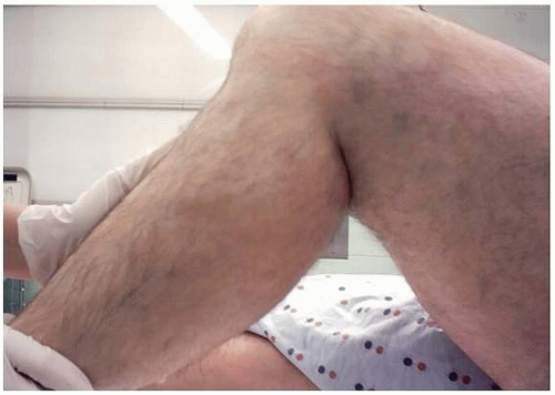 FIGURE 36.1 The posterior drawer exam is performed with the patient supine, the knee flexed to 90 degrees, and the foot supported on the table. The examiner’s thumbs are placed on top of the medial and lateral tibial plateaus and a posterior force is applied to the proximal tibia. Posterior tibial translation compared to contralateral side results in a positive test. Grade I (0-5 mm), grade II (5-10 mm), grade III (>10 mm). |
 FIGURE 36.2 Godfrey test (posterior sag) is performed with the patient supine, the hip and knee flexed to 90 degrees, and the foot supported by the examiner. Abnormal posterior sag of the tibia relative to the femur results in positive test. |
 FIGURE 36.3 T2 weighted sagittal MRI demonstrating a midsubstance tear of the PCL. |
SURGERY
Sciatic and femoral nerve blocks catheters may be placed prior to surgery. However, no anesthetic is introduced until neurologic assessment has occurred upon completion of the case. After induction, an examination under anesthesia (EUA) is performed on both the nonoperative and the operative knees. Data from the contralateral knee may be particularly helpful with combined injuries. A detailed exam to determine the direction and degree of laxity is recorded. Fluoroscopy may be used after the EUA to assess posterior tibial displacement.
Treatment Algorithm
For acute injuries, we employ the single-bundle technique. If there is some component of the native PCL remaining, we spare this tissue and utilize the augmentation technique. This technique can be time consuming and difficult, but preservation of PCL tissue may provide enhanced posterior stability of the knee and may promote graft healing. We perform the double-bundle technique most commonly in the chronic setting when any remaining structures are significantly incompetent. Although some authors advocate the tibial inlay technique for all settings, we typically do not utilize this technique. In the setting of a displaced tibial avulsion or a combined PCL and posterolateral corner injury, we use the techniques described below.
Single-Bundle Technique
The patient is positioned supine on a radiolucent operating room table. We do not use a tourniquet. A side post is placed on the operative side just distal to the greater trochanter to support the proximal leg with the knee in flexion. A padded bump is taped to the operating room table to hold the knee flexed to 90 degrees during the case. After prepping and draping the operative site, a hole is cut in the stockinette for access to the dorsalis pedis pulse throughout the case. A bump is placed between the post and the leg to stabilize the knee in a flexed position while the foot rests on the prepositioned sand bag (Fig. 36.4). The knee is flexed to 90 degrees and the vertical arthroscopy portals are delineated. The anterolateral portal is placed just lateral to the lateral border of the patellar tendon and adjacent to the inferior pole of the patella. The anteromedial portal is positioned 1 cm medial to the medial border of the superior aspect of the patellar tendon. A diagnostic arthroscopy is conducted to determine the extent of injury and evaluate for other cartilage or meniscal pathology. The notch is examined for any remaining intact PCL fibers. If there are remaining fibers, care is taken to preserve these fibers and an augmentation is performed (see Single-Bundle Augmentation technique). Using an arthroscopic electrocautery device and shaver, overlying synovium and ruptured PCL fibers are debrided and the superior interval between the ACL and PCL is defined. An accessory posteromedial portal is created just proximal to the joint line and posterior to the MCL. A 70-degree arthroscope is placed between the PCL remnants and the medial femoral condyle to assess the posterior horn of the medial meniscus and to localize the posteromedial portal with a spinal needle. A switching stick may be placed into the posteromedial portal to facilitate exchange of the arthroscope. The 30-degree arthroscope is used when viewing via the posteromedial portal. A trans-septal portal also
may be created for better visualization of and access to the tibial PCL insertion (8). Preparation and exposure of the tibia is essential for drilling the tunnel safely in the appropriate position. First the 70-degree arthroscope is placed into the anterolateral portal and a commercially available PCL curette is introduced through the anteromedial portal. A lateral fluoroscopic image may be obtained to confirm its position. The 30-degree arthroscope is then introduced through the posteromedial portal. The soft tissue on the posterior aspect of the tibia is carefully elevated centrally and slightly laterally. A shaver may be placed through the anterolateral portal to debride some of the surrounding synovium. The 70-degree arthroscope is returned to the anterolateral portal and the shaver placed in the posteromedial portal to complete the exposure. A commercially available PCL tibial drill guide set to 55 degrees is advanced through the anteromedial portal and placed just distal and lateral to the PCL insertion site, 1.5 cm distal to the articular edge of the posterior plateau along the sloped face of the posterior tibial fossa. The position is checked fluoroscopically with a lateral view and arthroscopically via the posteromedial portal. An incision and dissection through periosteum to bone is made on the anteromedial aspect of the tibia in line with the guide. The PCL guide is set and its position is confirmed with fluoroscopy and arthroscopy (Fig. 36.5). A guide wire is drilled to but not through the posterior cortex. Fluoroscopy is used to confirm the path of the guide wire (Fig. 36.6). With the 30-degree arthroscope in the posteromedial portal, the PCL curette is introduced through the anteromedial portal and is used to protect the posterior knee structures as the guide wire is carefully advanced through the posterior cortex under arthroscopic visualization. A parallel pin guide can be used to make small pin placement corrections if necessary. A cannulated compaction reamer is used to drill the tibial tunnel. The tibial cortex is cautiously perforated by hand reaming under arthroscopic visualization (Fig. 36.7). The tunnel is irrigated and increasing serial dilators are used under arthroscopic visualization up to the graft size.
may be created for better visualization of and access to the tibial PCL insertion (8). Preparation and exposure of the tibia is essential for drilling the tunnel safely in the appropriate position. First the 70-degree arthroscope is placed into the anterolateral portal and a commercially available PCL curette is introduced through the anteromedial portal. A lateral fluoroscopic image may be obtained to confirm its position. The 30-degree arthroscope is then introduced through the posteromedial portal. The soft tissue on the posterior aspect of the tibia is carefully elevated centrally and slightly laterally. A shaver may be placed through the anterolateral portal to debride some of the surrounding synovium. The 70-degree arthroscope is returned to the anterolateral portal and the shaver placed in the posteromedial portal to complete the exposure. A commercially available PCL tibial drill guide set to 55 degrees is advanced through the anteromedial portal and placed just distal and lateral to the PCL insertion site, 1.5 cm distal to the articular edge of the posterior plateau along the sloped face of the posterior tibial fossa. The position is checked fluoroscopically with a lateral view and arthroscopically via the posteromedial portal. An incision and dissection through periosteum to bone is made on the anteromedial aspect of the tibia in line with the guide. The PCL guide is set and its position is confirmed with fluoroscopy and arthroscopy (Fig. 36.5). A guide wire is drilled to but not through the posterior cortex. Fluoroscopy is used to confirm the path of the guide wire (Fig. 36.6). With the 30-degree arthroscope in the posteromedial portal, the PCL curette is introduced through the anteromedial portal and is used to protect the posterior knee structures as the guide wire is carefully advanced through the posterior cortex under arthroscopic visualization. A parallel pin guide can be used to make small pin placement corrections if necessary. A cannulated compaction reamer is used to drill the tibial tunnel. The tibial cortex is cautiously perforated by hand reaming under arthroscopic visualization (Fig. 36.7). The tunnel is irrigated and increasing serial dilators are used under arthroscopic visualization up to the graft size.
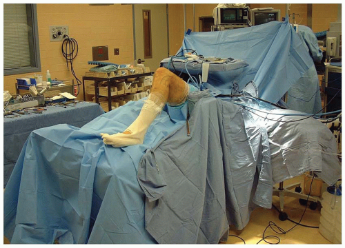 FIGURE 36.4 Operating room setup. |

Related posts:Stay updated, free articles. Join our Telegram channel
Full access? Get Clinical Tree
 Get Clinical Tree app for offline access

|
