Distal Biceps Tendon Rupture in the Athlete
Bernard F. Morrey
Once thought to be an uncommon injury, the avulsion of the distal biceps tendon from the radial tuberosity is being seen in increasing numbers in both competitive and recreational athletes. In general it is of interest that the injury occurs almost exclusively in males, usually those with heavy lifting requirements of work or avocation.
PATHOLOGY
The distal biceps tendon complex may be injured at the musculotendinous junction, by a disruption of the tendon itself in continuity, or a complete or partial tear or avulsion of the tendon from the radial tuberosity (Fig. 5.1). By far the most common lesion is the avulsion from the tuberosity and this is the only lesion that will be dealt with in this chapter.
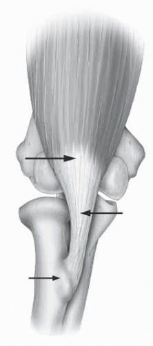 FIGURE 5.1 The biceps mechanism may be injured at the muscle/tendinous junction, intratendinous, or at the tuberosity. |
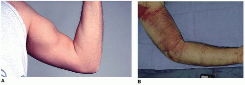 FIGURE 5.2 In most instances, proximal retraction is diagnostic (A). Ecchmosis is uncommon. In this instance, this competitive athlete had been on high-dose aspirin at the time of injury (B). |
Of the tears from the radial tuberosity, approximately 95% are complete ruptures whereas about 5% are partial tears. Both conditions, along with delayed reconstruction, will be addressed in this chapter.
THE DIAGNOSIS
Complete rupture is easy to diagnose in most instances due to retraction of the distal biceps muscle belly with elbow flexion. The history is that of eccentric loading during flexion. Hematoma formation is variable, as is the location of the pain (Fig. 5.2).
Imaging
In recent years, there has been a significant improvement in the ability to diagnose the biceps tendon injury, especially incomplete rupture, with MRI. By placing the arm overhead the course of the biceps tendon may be brought in plane thus allowing a more accurate assessment of the pathology. This position was described by Giuffre and Moss (9) and is termed the “flexion abduction supination ‘FABS’” view (Fig. 5.3). In addition to this improvement in patient position, three dimensional reconstructions are also improving our preoperative understanding of the precise location and extent of the pathology present. This is of particular value in instances of partial rupture.
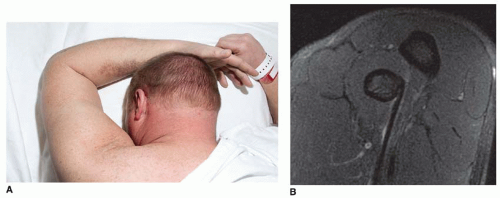 FIGURE 5.3 The FABS view (A) brings the entire tendon and its attachment into profile with MRI (B). |
INDICATIONS/CONTRAINDICATIONS
In my judgment there is little question that in the athlete particularly distal biceps tendon rupture should be repaired as soon as possible (1,3,15,17,19). There have been several studies including our own attempting to estimate and evaluate both subjective and objective dysfunction following the nonrepaired biceps tendon. These studies have generally shown reasonable function under most circumstances with minimal pain. However, with excessive exertion the patients do have pain and lack endurance, hence the need for reconstruction in some patients if the repair is not performed acutely. The average age of patients with the injury is approximately 55 and virtually every report has documented the almost exclusive occurrence in the male. In our practice at Mayo, we have treated only two females from among over 100 with this diagnosis, both with partial ruptures. Usually, the patient is involved with heavy labor or athletic activity, which further emphasizes the need of early definitive treatment.
Delayed reattachment is difficult because the tendon retracts. If this has occurred, reattachment or embedding the biceps tendon into the brachialis is easy but not considered acceptable today. Reconstruction for selected patients has been effective in recent years but this is a difficult surgical procedure (11) and typically is referred to those surgeons with experience with this procedure. The author prefers the achilles allograft for this procedure as described herein.
Contraindications
Reattachment is contraindicated in patients who do not have significant functional impairment. This is not very applicable in the athlete but might be considered in a sedentary patient—but rarely does such an individual sustain this injury. Attempts to reattach this tendon if there has been a delay of over 3 weeks requires careful thought as the tendon typically is retracted into the biceps muscle and the tendon track is scarred. If the delay is prolonged there may not be adequate length to reach to the radial tuberosity (17). Furthermore, the tract of the tendon to the tuberosity will have scarred and become obliterated, making the surgery much more difficult with a higher complication rate (13).
Surgical Considerations
The surgeon has two interrelated technical considerations when addressing these patients. The first is the selection of either a one or two-incision technique. The second is the mode of fixation. In this chapter, we will deal with three types of fixation which with their variations reflect virtually all of the approaches used today: bone tunnel, suture anchor, and endo button.
The surgical approach is clearly the preference of the surgeon. Surgical procedures have been described using a modified Henry approach (7,15) or through a two-incision approach described by Boyd and Anderson (5). The advantage of the anterior Henry approach is that it is felt to be less likely to create ectopic bone. The disadvantage is that it puts the radial nerve at jeopardy (7,15,19). However, it must be emphasized that the two-incision approach currently used is NOT that described by Boyd and Anderson. The advantage of the two-incision technique is that it lessens and virtually eliminates the likelihood of injury to the radial nerve (17). The original Boyd-Anderson approach exposes the ulna, and hence can be associated with ectopic bone (8). Through the years we have employed the Mayo modification of the Boyd-Anderson approach which does NOT expose the ulna, and hence is associated with very little ectopic bone (13).
The author continues to use the two-incision technique with excellent results and minimal complications (13). It is recognized that the one-incision technique is also popular with the thought that it lessens the likelihood of ectopic bone formation. This has not been demonstrated to be the case but it does motivate many to use a single anterior approach. The direct exposure is correlated with the mode of fixation. An anterior approach can be used for the endo button or the suture anchor. For bone tunnels, a two-incision technique is required.
PREOPERATIVE PLANNING
If the injury is more than 4 weeks since onset, be prepared to perform a more detailed dissection in the antecubital space. If the tendon has retracted, direct reattachment to the tuberosity with the elbow flexed up to 90 degrees is preferred. If this is not possible, restoration of length with an achilles tendon allograft is preferred. The patient must be prepared for these eventualities.
TECHNIQUE
Complete Rupture—Immediate Reattachment
Incision
The arm is prepped and draped with the patient supine. A sandbag may be placed under the shoulder to allow the arm to comfortably be brought across the chest. Under a general anesthesia, a single 4-cm transverse incision in the antecubital crease is employed (Fig. 5.4).
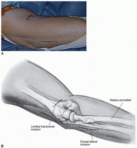 FIGURE 5.4 The two-incision technique employs a simple 4-cm transverse incision in the antecubital space (A) and a 5- to 7-cm incision over the posterior aspect of the proximal forearm (B). |
TENDON PREPARATION.
By digital palpation or limited dissection, the tendon is identified, dissected free of soft tissue, and is delivered from the wound (Fig. 5.5). The end of the tendon tends to be bulbous and is trimmed in order to allow it to fit well into the tuberosity. After the tendon has been trimmed, two 5 Mersilene sutures are placed through the torn portion entering the end of the tendon. A crisscrossed (Bunnell) suture or locking stitch (Krakow) is employed (Fig. 5.6).
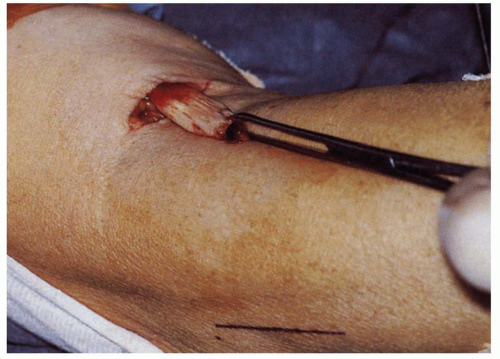 FIGURE 5.5 The tendon is identified by digital palpation and delivered through the skin incision revealing a bulbous degenerative process at the site of disruption. |
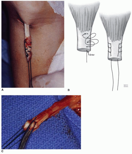 FIGURE 5.6 A-C: Two 5 nonabsorbable sutures are inserted by the crisscross Bunnell or Krakow locking technique. |
Forearm Incision
A curved clamp is then introduced into the tunnel previously occupied by the biceps tendon (Fig. 5.7). It is directed by palpation to and then past the tuberosity between the radius and ulna. Rotation of the forearm confirms proper position of the instrument on the ulnar side of the radius. A lead suture is grasped by the curved hemostat and is advanced until it punctures the muscle and subcutaneous tissues of the dorsal aspect of the forearm. An incision is then made over the site of prominence splitting the common extensor and supinator muscles (Fig. 5.8).
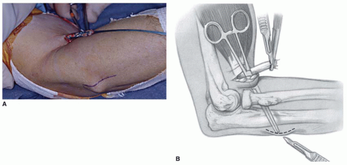 FIGURE 5.7 The curved hemostat is passed between the radial tuberosity and the ulna (A) to emerge through the common extensor muscle mass and tent the skin on the proximal posterolateral aspect of the forearm (B). |
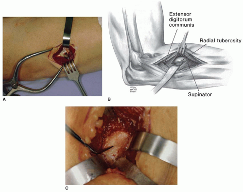 FIGURE 5.8 A-C: An incision is made over this prominence, the muscle is split, the forearm is fully pronated, and the tuberosity is exposed. |
Tuberosity Identity and Preparation
With full forearm pronation the tuberosity is identified and cleaned of soft tissue.
A high-speed burr is used to excavate the cancellous bone from the midportion of the tuberosity (Fig. 5.9). After an adequate orifice of 10 to 12 mm × 7 to 8 mm has been made to receive the tendon, the forearm is partially segmented and three drill holes are placed on the radial side of the tuberosity (Fig. 5.10). Allowing the forearm to supinate slightly brings this margin of the radial tuberosity into better alignment. The holes should be placed in such a way as to leave sufficient bone to avoid osseous rupture or pullout of the sutures (Fig. 5.11).
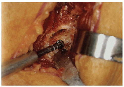 FIGURE 5.9 The greater tuberosity is excavated with a high-speed bur in such a way as to receive the distal biceps tendon. |
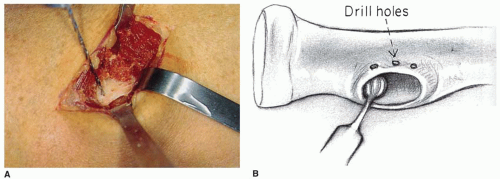 FIGURE 5.10 A,B: The tuberosity is prepared by releasing some of the pronation and three holes are drilled in the radial aspect of the tuberosity. |
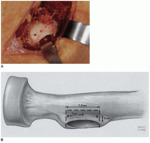 FIGURE 5.11 A,B: Care is taken to provide sufficient space between the holes to provide secure fixation in bone. |
REATTACHMENT OF THE TENDON
The tendon is then brought through the tunnel from the antecubital space (Fig. 5.12) and drawn past the ulnar side of the tuberosity with the lead suture (Fig. 5.13). The sutures are threaded into each of the holes at the margin of the tuberosity. One suture is brought into the proximal and distal, and two into the central hole (Fig. 5.14). The biceps tendon is threaded into the tuberosity; once again this is facilitated by slightly supinating the forearm. With the arm remaining in less than full pronation, the sutures are tied (Fig. 5.15).
Related posts:

Stay updated, free articles. Join our Telegram channel

Full access? Get Clinical Tree


