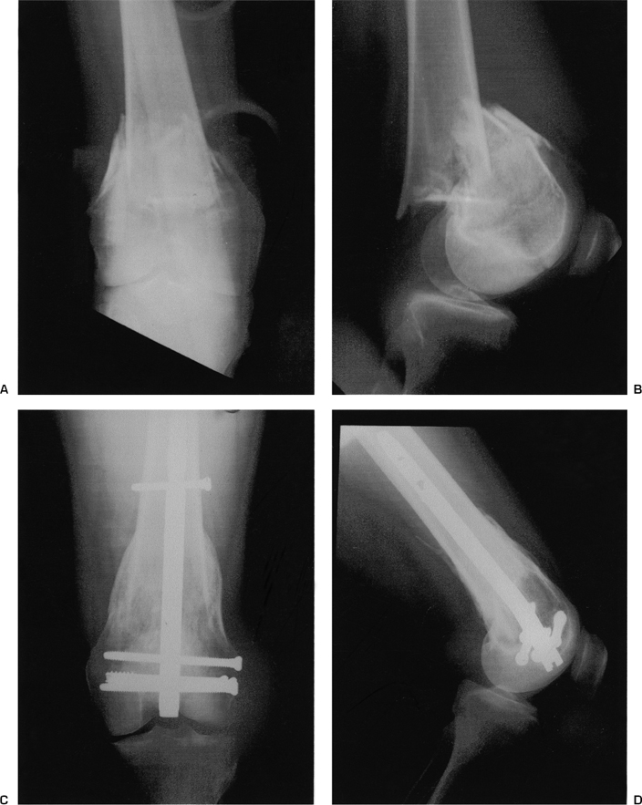
Supracondylar Femur Fracture: Rod
Supracondylar femoral fractures have been classified as type A, extraarticular fractures; type B, unicondylar fractures; and type C, intraarticular fractures.1
Of the various techniques available for fixation of the distal femur, the intramedullary supracondylar nail is a simple solution.
Indications
- Type A and C supracondylar femoral fractures (Fig. 30–1)2–5
- Fractures proximal to a knee prosthesis or distal to hip implants
- Obesity, injury at the trochanteric entrance point, or for positioning problems
Contraindications
- Type B or transcondylar fractures involving the distal 2 to 3 cm of the femur
- Preexisting intramedullary implant in the distal femoral canal
Physical Examination
Knee effusion, ecchymosis, soft tissue damage, pain, tenderness and swelling; also length, axial, or rotational deformity centered in the supracondylar region
Diagnostic Tests
- History and physical exam, complete blood count, urinalysis, prothrombin time, type, and screen; anteroposterior (AP) and lateral views of the distal femur and knee
- Computed tomography scan if fracture pattern is complex
Special Considerations
Open reduction may be indicated for type C fractures and
requires more time and equipment.
Preoperative Planning and Timing of Surgery
- The procedure is delayed until the patient’s general and local problems allow surgery.
- Ten- to 20-lb skeletal traction is used in comminuted or displaced fractures.
- Nail length and width is estimated preoperatively by using implant templates.
Special Instruments
- A full set of conventional, interlocking, bioabsorbable, and cannulated screws. Conical nuts and washers are useful in osteopenic patients.2
- Periarticular bone-reduction clamps
Anesthesia
General or spinal anesthesia
Patient and Equipment Positions
- Patient supine in radiolucent table with a folded drape to flex the knee 30 to 60 degrees
- Image intensifier brought in across the uninjured leg
Surgical Procedure
- Reduction, temporary fixation, and vertical 2- to 3-cm transpatellar tendon incision
- Entrance portal identification and canal opening (Fig. 30–2). A ball-tipped guide is passed, and the canal is reamed 1.5 to 2 mm more than the planned diameter of the nail.
- Guide exchange and manual nail insertion to 2 to 3 mm in the distal femur
- Interlocking guide assembly, interlocking screws placement from lateral to medial and from distal to proximal, and incision closure (Fig. 30–3)
- Control AP and lateral films are obtained before the patient leaves the room.

Stay updated, free articles. Join our Telegram channel

Full access? Get Clinical Tree


