CHAPTER 22 Subscapularis Tears
Arthroscopy has revolutionized the way we treat rotator cuff tears1 and many authors have reported excellent clinical results following all arthroscopic rotator cuff repairs. Arthroscopic repair of the subscapularis tendon, however, remains the final frontier for many arthroscopists because of its difficulty with visualization, the awkward angle of approach, and the complex releases required for mobilization and repair.
Subscapularis tears, however, are commonly missed, particularly partial-thickness articular surface tears and full-thickness upper subscapularis tendon tears. The incidence of subscapularis tears has been reported as high as 27% in a study of 165 arthroscopically evaluated shoulders with rotator cuff, labral, or ligamentous disorders.2 Although subscapularis tears do occur in isolation, they are more commonly seen in combination with other rotator cuff tears. In one study, 90% of subscapularis tears were associated with tears of the supraspinatus tendon.3
ANATOMY AND PATHOANATOMY
Recently, cadaveric studies have clarified the insertional anatomy of the subscapularis tendon.4,5 The shape of the tendinous insertion grossly resembles a human ear, with the medial border almost parallel to the longitudinal axis of the humerus, and the insertion broad proximally and tapered distally. The average footprint measures 25.8 ± 3.2 mm in superior to inferior height, and the maximum width measures 18.1 ± 1.6 mm.4 These anatomic dissections highlight the broad insertional anatomy of the footprint and are helpful when determining the percentage of subscapularis tendon torn during arthroscopic evaluation.
Traditionally, the main function of the subscapularis has been described as an internal rotator and anterior stabilizer of the glenohumeral joint. However, the subscapularis tendon is also an important dynamic stabilizer during normal glenohumeral motion. In the normal shoulder, the transverse plane force couple consists of the subscapularis anteriorly, which is balanced against the infraspinatus and teres minor posteriorly, resulting in a balanced force couple in the transverse plane. In contrast, a disruption of the subscapularis leads to loss of the transverse plane force couple and an unstable fulcrum. When rotator cuff repair restores the anterior (subscapularis) and posterior (infraspinatus, teres minor) forces so that the moments between the two are balanced, function will be restored, even in the presence of a persistent defect in the superior rotator cuff (supraspinatus).1
The precise cause of subscapularis tears has yet to be fully defined, but is likely multifactorial and includes intrinsic degeneration, microtrauma or macrotrauma, impaired vascularity, and subcoracoid impingement. Subcoracoid stenosis (narrowing of the subcoracoid space) and subcoracoid impingement (direct contact of the coracoid against the subscapularis tendon or lesser tuberosity) have been implicated in the pathogenesis of subscapularis tears.6 The coracohumeral distance, as measured by axial computed tomography (CT) and magnetic resonance imaging (MRI), has been reported to be 8.7 to 11.0 mm in normal shoulders.7,8 In one study, the average coracohumeral distance was measured by a single axial cut on a preoperative MRI scan. In 35 patients requiring subscapularis repair, the coracohumeral distance was 5.0 ± 1.7 mm; compared with a control group (N = 35) that had no rotator cuff, subscapularis, or subcoracoid pathology, the coracohumeral distance was 10.0 ± 1.3 mm.9 Whereas these and other studies have confirmed a relationship between a narrowed coracohumeral distance and subscapularis tears, the exact temporal or causal relationship between tearing and a decreased coracohumeral distance and subcoracoid impingement is still unclear.10 Cadaveric studies of subscapularis tendons have suggested that intrinsic tendon degeneration may be an important causative factor in tears of the subscapularis.11,12 These studies have demonstrated that the incidence of histologic changes in the subscapularis tendon is similar to that of the supraspinatus tendon; these are most commonly seen in the superior and deep portion of the tendon insertion. Clinically, this corresponds to the area most commonly affected by partial-thickness tears of the subscapularis.
Full-thickness tears of the subscapularis tendon are much less common than partial-thickness tears of the subscapularis tendon or full-thickness tears of the supraspinatus tendon. One key pathoanatomic finding of subscapularis tendon tears has been called the comma sign, which is a comma-shaped arc of soft tissue located superior to the superolateral border of the torn subscapularis tendon.13 This tissue is composed of the fibers of the medial portion of the coracohumeral ligament, as well as a portion of the superior glenohumeral ligament (the medial biceps sling), which interdigitate with the superior fibers of the subscapularis tendon at its humeral insertion. When the subscapularis tendon is torn, the medial biceps sling tears along with it and stays attached to the superolateral corner of the subscapularis tendon, forming a continuous arc of soft tissue. By identifying the comma sign, this tissue can then be followed inferiorly and medially to the superolateral border of the subscapularis tendon.
HISTORY AND PHYSICAL EXAMINATION
The lift-off test is performed with the affected arm of the patient placed in internal rotation and extension, with the back of the hand on the lumbar region. The test is considered positive if the patient is unable to raise the arm posteriorly off the back.14 Although this test is reliable in patients with complete subscapularis tendon tears, it may not be positive until more than 50% to 75% of the subscapularis tendon is torn. In addition, it is of limited value in patients who cannot bring the affected arm into the starting position because of pain or restricted range of motion. In this case, the belly press test may be used.
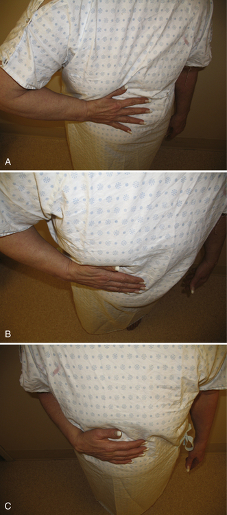
To perform the belly press test,15 the patient presses on the abdomen with the palm of the hand and attempts to keep the arm in maximum internal rotation by maintaining the elbow anterior to the midcoronal plane of the body. This test is considered positive if the elbow drops in a posterior direction behind the midcoronal plane while pressure on the abdomen is exerted by extension of the shoulder. A variation is the Napoleon test, which is similarly performed but, in addition to the elbow dropping posteriorly, the wrist commonly palmar-flexes against the abdomen. The amount of wrist flexion has been correlated with the size of subscapularis tendon tearing. Tears of less than 50% of the subscapularis tendon can have negative Napoleon signs (wrist fully extended), tears involving more than 50% but not the entire tendon have an intermediate result (wrist flexed 30 to 60 degrees), and tears involving the entire subscapularis tendon have a positive Napoleon test (wrist flexed 90 degrees; Fig. 22-1).16
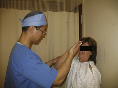
Recently, the bear hug test has been described, whereby the patient places the hand of the affected side on the opposite shoulder, with the fingers extended and the shoulder in a forward-flexed position.17 The examiner then tries to pull the patient’s hand off the shoulder using an external rotational force perpendicular to the plane of the forearm as the patient resists. If the examiner is able to lift the patient’s hand off the shoulder, this is considered a positive examination and is suggestive of at least a partially torn subscapularis (Fig. 22-2). In one study, the bear hug test was more sensitive than the belly press test or the lift-off test and may be particularly helpful for partial thickness, upper subscapularis tendon lesions.
DIAGNOSTIC IMAGING
There are few studies that have specifically evaluated the usefulness of diagnostic imaging for the assessment of the subscapularis tendon. Plain radiographs are usually nonspecific but are helpful in ruling out other types of shoulder pathology. In patients with long-standing subscapularis tendon insufficiency, static anterior subluxation of the humeral head may be apparent on axillary radiographs. The use of ultrasound can provide a relatively inexpensive, noninvasive, dynamic assessment of the rotator cuff but is highly observer-dependent. Ultrasound has been reported to be 100% sensitive and 85% specific for the detection of all rotator cuff lesions.18 In this study, ultrasound correctly identified six of seven tears of the subscapularis tendon. In a larger study of 17 isolated subscapularis tendon tears, ultrasonography correctly demonstrated 86% of full-thickness tears of the subscapularis. However, ultrasonography was not as accurate in demonstrating partial-thickness tears, particularly small upper subscapularis tendon tears.19,20
MR arthrography (MRA) has been reported to be 91% sensitive and 86% specific for detecting tears of the subscapularis tendon.21 However, others have reported less promising results and subscapularis tendon tears, particularly full-thickness, upper subscapularis, or partial-thickness tears, are commonly missed by MRI.22 Failure to include T-2 weighted axial images and the experience of the reader have been suggested as contributing to missed subscapularis tendon tears on MRI.
In addition to tendon tearing, the chronicity of the tear and quality of its muscle may be assessed on sagittal oblique images medial to the glenoid. Severe atrophy and fatty degeneration on preoperative MRI or CT generally correlate with poor tendon quality and limited tendon excursion, and may be a negative prognostic sign regarding the ability to repair the subscapularis tendon.23–25 However, others have argued that the subscapularis tendon is commonly reparable, even in the presence of severe atrophy and fatty degeneration and, although not functional, may act as a tenodesis to provide a stable fulcrum of motion.26
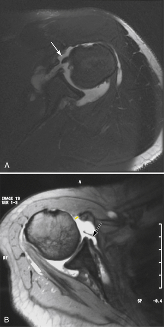
Biceps tendon and rotator interval pathology may be associated with subscapularis tears. The stabilizing reflection pulley of the long head of the biceps tendon may be lost with defects of the superior subscapularis and result in medial subluxation or dislocation of the long head of the biceps tendon (Fig. 22-3A). This may be detected as extravasation of contrast material anterior to the superior border of the subscapularis tendon on axial images. In addition to subscapularis pathology, radiographic evidence of subcoracoid stenosis should be considered. Proximal humeral migration, particularly static anterior subluxation of the humeral head, can contribute to narrowing of the subcoracoid space, especially when imaging studies are obtained with the patient in the supine position (e.g., by MRI).