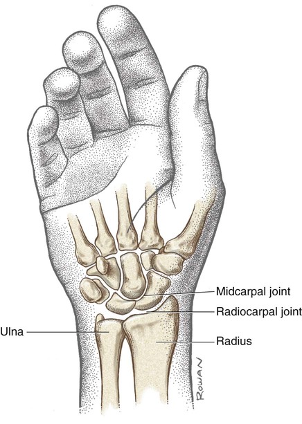
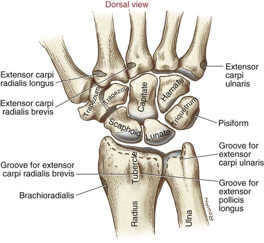
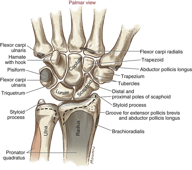
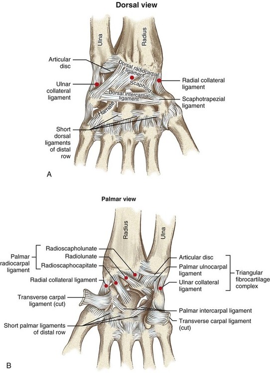
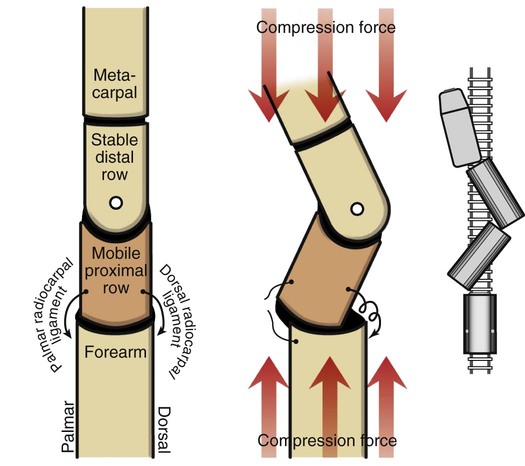
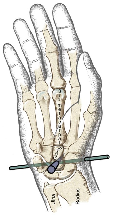
• Identify the bones and primary bony features relevant to the wrist complex. • Describe the supporting structures of the wrist. • Cite the normal ranges of motion for wrist flexion and extension and radial and ulnar deviation. • Describe the planes of motion and axes of rotation for the joints of the wrist. • Cite the proximal and distal attachments and innervation of the primary muscles of the wrist. • Justify the primary actions of the muscles of the wrist. • Describe how compressive forces are transferred from the hand through the wrist. • Explain the function of the wrist extensor muscles when grasping. • List the structures that travel within the carpal tunnel. • Explain the synergistic action between the muscles of the wrist when flexion-extension and radial and ulnar deviation are performed. The wrist contains eight small bones that are located between the distal end of the radius and the hand (Figure 6-1). Although slight, the passive movements that occur within the carpal bones help absorb forces that cross between the hand and the forearm, as when crawling on all four limbs, or when bearing weight through the hands when using crutches or a walker. The distal radius and ulna (Figure 6-2) articulate with the proximal row of carpal bones. The distal forearm is bordered laterally by the radial styloid process and medially by the ulnar styloid process. The radial tubercle, also called Lister’s tubercle, is a small, palpable projection on the dorsal aspect of the distal radius. This ridge of bone helps guide the direction of the tendons of several wrist and thumb extensor muscles. From a radial (lateral) to ulnar direction, the proximal row of carpal bones includes the scaphoid, lunate, triquetrum, and pisiform. The distal row includes the trapezium, trapezoid, capitate, and hamate (see Figures 6-2 and 6-3). The bones within the proximal row are loosely joined. In contrast, strong ligaments tightly bind the bones of the distal row. The natural stability of the distal row provides an important rigid base for articulations with the metacarpal bones. The transverse carpal ligament bridges the palmar side of the carpal bones, helping to form the carpal tunnel (Figure 6-4). The carpel tunnel serves as a passageway that helps protect the median nerve and the tendons of the extrinsic flexor muscles of the digits. Carpal Tunnel Syndrome All the tendons that flex the digits travel with the median nerve and pass through the tightly packed carpal tunnel (see Figure 6-4). Also traveling within the carpal tunnel are several synovial membranes that help reduce friction between tendons and surrounding structures. Hand activities that require prolonged and often extreme wrist positions can irritate these tendons and synovial sheaths. Because of the small size of the carpal tunnel, swelling of the synovial membranes can increase pressure on the median nerve. Carpal tunnel syndrome, which is characterized by pain or paresthesia (tingling), or both, over the sensory distribution of the median nerve, may result. In more extreme cases, muscular weakness and atrophy may occur in the intrinsic muscles around the thumb. As is illustrated in Figure 6-1, the wrist is a double-jointed system, consisting of the radiocarpal and midcarpal joints. Many smaller intercarpal joints also exist between carpal bones. Compared with the large ranges of motion permitted at the radiocarpal and midcarpal joints, motion at the many intercarpal joints is relatively small. The proximal part of the radiocarpal joint consists of the concave surface of the radius and the adjacent articular disc (Figure 6-5). The distal part of the joint consists primarily of the convex articular surfaces of the scaphoid and the lunate. Approximately 80% of the force that crosses the wrist passes between the scaphoid and the lunate, and then to the radius. The large, expanded distal end of the radius is well designed to accept this force. Unfortunately, however, for many persons, a fall onto an outstretched hand fractures the distal end of the radius, as well as the scaphoid. Persons with weakened bones due to osteoporosis are particularly susceptible to these fractures. The ulnar-located carpal bones and the distal ulna are less likely to fracture from such a fall because they are not in the direct path of weight bearing. Furthermore, a relatively wide space exists between the distal ulna and the ulnar carpal bones. This space, formally known as the ulnocarpal space (see Figure 6-1), helps buffer the forces that cross the wrist. The midcarpal joint separates the proximal and distal rows of carpal bones (see Figure 6-5). Although this joint involves several articulations, the most prominent is formed between the head of the capitate and the socket formed by the distal surfaces of the scaphoid and lunate. Note that the scaphoid and the lunate bones are important members of the main two articulations of the wrist. The joints of the wrist are enclosed within a fibrous capsule. The capsule is thickened by extrinsic and intrinsic ligaments. Extrinsic ligaments have their proximal attachments outside the carpal bones but attach distally within the carpal bones. Intrinsic ligaments, in contrast, have both their proximal and distal attachments located within the carpal bones. Table 6-1 lists the main attachments and primary functions of the four primary extrinsic ligaments: radial collateral, ulnar collateral, dorsal radiocarpal, and palmar radiocarpal. Three of the four primary extrinsic ligaments are indicated by red dots in Figure 6-6, A and B, and are summarized along with their individual functions in Table 6-1. The detailed anatomy of the intrinsic ligaments is beyond the scope of the text. As a group, however, the intrinsic ligaments (1) interconnect various carpal bones; (2) help transfer forces between the hand and the forearm; and (3) maintain the natural shapes of radiocarpal and midcarpal joints, thereby minimizing joint stress during movement. Ulnocarpal Complex A complex set of connective tissues, known as the ulnocarpal complex, exists near the ulnar border of the wrist (see Figure 6-6, B). (This group of tissues is often referred to as the triangular fibrocartilage complex, or TFCC). The ulnocarpal complex includes the articular disc (described in Chapter 5 as an important component of the distal radioulnar joint), the ulnar collateral ligament, and the palmar ulnocarpal ligament. This set of tissues fills most of the ulnocarpal space between the distal ulna and the carpal bones (see Figure 6-1). The ulnocarpal space allows the carpal bones to follow the pivoting radius during pronation and supination of the forearm, without interference from the distal end of the ulna. Tears in the articular disc, the central component of the ulnocarpal complex, may result in instability and pain of the wrist and the distal radioulnar joint. Consider that the loosely articulated proximal row of carpal bones is located between two rigid structures: the radius and the distal row of carpal bones. Ligaments weakened by injury or disease often lead to instability of the wrist and even collapse. When compressed strongly from both ends (e.g., from a fall), the proximal row of carpal bones is prone to collapse in a zigzag fashion, much like derailed cars of a freight train (Figure 6-7). An unstable wrist can become painful and is often disabling. Osteokinematics of the wrist involves flexion and extension and ulnar and radial deviation. Except for minimal accessory motions, the wrist does not spin in a circular motion relative to a fixed radius. The bony fit and ligaments of the radiocarpal joint naturally block this twisting motion. As studied in Chapter 5, pronation and supination involve rotation of the forearm, with the hand and wrist “following” the path of the radius. The axis of rotation for wrist movement pierces the head of the capitate (Figure 6-8). The axis runs in a medial-lateral direction for flexion and extension, and in an anterior-posterior direction for radial and ulnar deviation. The firm articulation between the capitate and the base of the third metacarpal bone causes rotation of the capitate to direct the overall path of the entire hand. On average, from a neutral (0-degree) position, the wrist flexes approximately 70 to 80 degrees and extends approximately 60 to 65 degrees, for a total of approximately 130 to 145 degrees (Figure 6-9, A). Total flexion normally exceeds extension by approximately 15 degrees. Extension is normally limited by tension in the thicker palmar radiocarpal ligaments, as well as by contact of the carpal bones with the slightly elongated dorsal side of the distal radius. On average, from a neutral (0-degree) position, the wrist allows approximately 30 to 35 degrees of ulnar deviation and approximately 15 to 20 degrees of radial deviation, for a total of about 45 to 55 degrees of motion (Figure 6-9, B). Maximum ulnar deviation is normally twice that of radial deviation, mostly because of the void created by the ulnocarpal space. Radial deviation is blocked by contact between the styloid process of the radius and the radial side of the carpal bones.
Structure and Function of the Wrist
Osteology
Distal Radius and Ulna
Carpal Bones
Carpal Tunnel
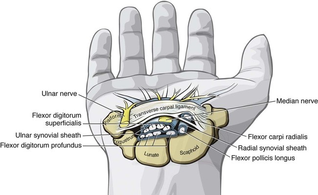
 Clinical insight
Arthrology
Joint Structure
Radiocarpal Joint
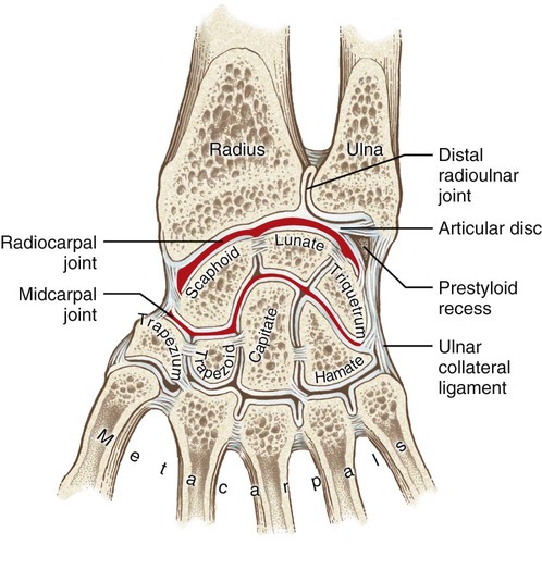
Midcarpal Joint
Ligaments of the Wrist
 Table 6-1
Ligament
Function
Comments
Dorsal radiocarpal ligament
Resists extremes of flexion
Attaches between the radius and the dorsal side of the carpal bones
Radial collateral ligament
Resists extremes of ulnar deviation
Strengthened by muscles such as the abductor pollicis longus and the extensor pollicis brevis
Palmar radiocarpal ligament
Resists extremes of wrist extension
Thickest ligament of the wrist; consists of three parts
Ulnar collateral ligament
Resists extremes of radial deviation
Part of the ulnocarpal complex; helps stabilize the distal radioulnar joint
 Consider this…
Wrist Instability
Kinematics
Osteokinematics
Sagittal Plane: Flexion and Extension
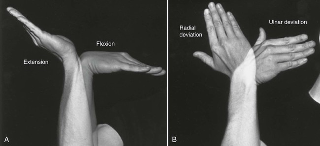
Frontal Plane: Radial and Ulnar Deviation
![]()
Stay updated, free articles. Join our Telegram channel

Full access? Get Clinical Tree


 Objectives
 Key Terms
 Consider this…
 Consider this…