• Describe the components of the axial versus appendicular skeleton. • Define the primary components found in bone. • Describe the five types of bones found in the human skeleton. • Describe the three primary classifications of joints and give an anatomic example of each. • Identify the components of a synovial joint. • Describe the seven different classifications of synovial joints in terms of mobility (degrees of freedom) and stability. • Provide an anatomic example of each of the seven different classifications of synovial joints. • Describe the three primary materials found in connective tissue. • Explain how tendons and ligaments support the structure of a joint. • Explain how muscles help to stabilize a joint. • Describe the effects of immobilization on the connective tissues of a joint. The bones of the skeletal system can be grouped into two categories: the appendicular skeleton and the axial skeleton. The axial skeleton consists of the skull, hyoid bone, sternum, ribs, and vertebral column, including the sacrum and coccyx, forming the central, bony axis of the body. The appendicular skeleton is composed of the bones of the appendages, or extremities. All bones of the upper extremity, including the scapula and clavicle, and all bones in the lower extremity, including the pelvis, are part of the appendicular skeleton. Figure 2-1 differentiates the axial and appendicular skeleton and labels the major bones of the body. Bone provides the rigid framework of the body and equips muscles with a system of levers. This text describes bone as having two primary types of tissue: cortical (compact) bone and cancellous bone (Figure 2-2). Most bones have common structural features important for maintaining their health and integrity. Figure 2-3 illustrates the primary components found in a bone. Many of the cells important for forming and repairing bone are housed within the endosteum. Bones can be classified into five basic categories based on their structure, or shape: long, short, flat, irregular, and sesamoid (Figure 2-4). A synarthrosis is a junction between bones that allows little to no movement. Examples include the sutures of the skull and the distal tibiofibular joint. The primary function of this type of joint is to firmly bind bones together and transmit forces from one bone to another (Figure 2-5). An amphiarthrosis is a type of joint that is formed primarily by fibrocartilage and hyaline cartilage. Although these joints allow limited amounts of motion, they play an important role in shock absorption. For example, the intervertebral body joints of the spine allow relatively little motion, but the thick layers of fibrocartilage that form the intervertebral discs absorb and disperse the large compressive forces often transmitted through this region (Figure 2-6). A diarthrosis is an articulation that contains a fluid-filled joint cavity between two or more bones. Because of the presence of a synovial membrane, diarthrodial joints are frequently referred to as synovial joints. Seven different categories of diarthrodial (synovial) joints exist, each with unique functional abilities; however, all synovial joints contain the seven common elements listed below (Figure 2-7): • Synovial fluid: Provides joint lubrication and nutrition • Articular cartilage: Dissipates and absorbs compressive forces • Articular capsule: Connective tissue that surrounds and binds the joint together • Synovial membrane: Produces synovial fluid • Capsular ligaments: Thickened regions of connective tissue that limit excessive joint motion • Blood vessels: Provide nutrients to the joint • Sensory nerves: Transmit signals regarding pain and proprioception The pivot joint (Figure 2-9) allows rotation about a single longitudinal axis of rotation, similar to the rotation of a doorknob. Examples include the proximal radioulnar joint and the atlantoaxial joint between the first and second cervical vertebrae.
Structure and Function of Joints
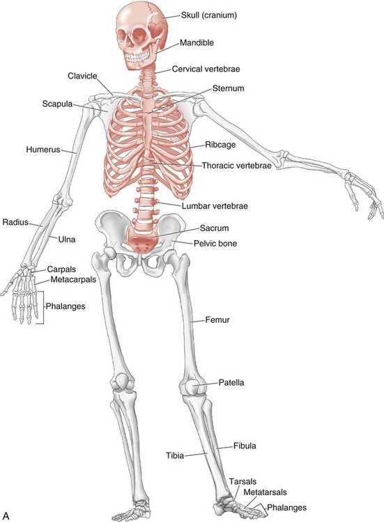
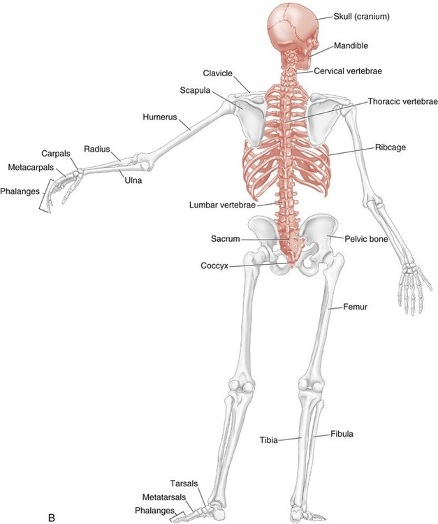
Axial versus Appendicular Skeleton
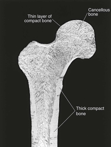
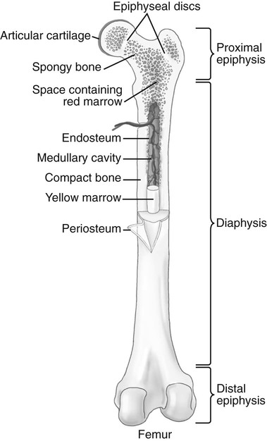
Bone: Anatomy and Function
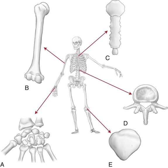
Types of Bones
Classification of Joints
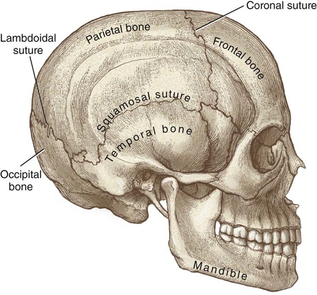
Synarthrosis
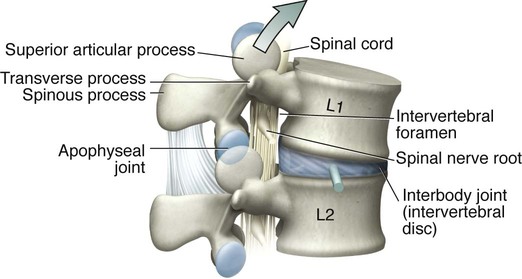
Amphiarthrosis
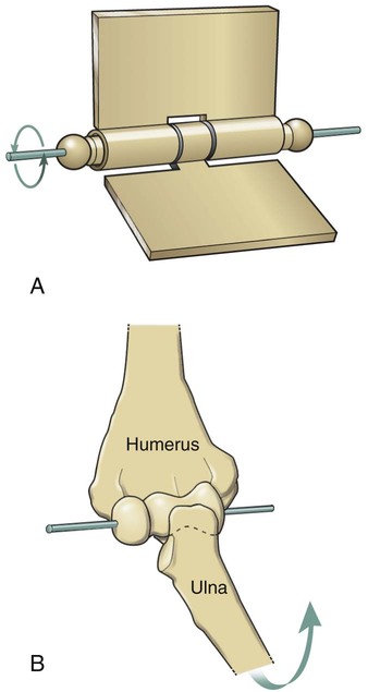
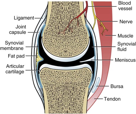
Diarthrosis: The Synovial Joint
Classification of Synovial Joints
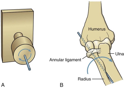
Pivot Joint
![]()
Stay updated, free articles. Join our Telegram channel

Full access? Get Clinical Tree


 Objectives
 Key Terms