Structure and Function of Skeletal Muscle


Structure and Function of Skeletal Muscle


Nearly all physical rehabilitation programs involve stretching, strengthening, or retraining of muscles. As the sole producer of active force in the body, muscle is ultimately responsible for all active motions and therefore plays a fundamental role in kinesiology. Muscles also control and stabilize our posture by their actions at joints. Clinicians therefore often advocate strengthening muscles to stabilize the underlying joints, especially when structures such as ligaments have been weakened by disease or trauma. This chapter provides a basic overview of the structure and function of skeletal muscle and reviews the important features of muscle as it relates to our study of kinesiology.


Fundamental Nature of Muscle


Muscles develop active force after receiving input from the nervous system. Once stimulated, a muscle produces a contractile, or pulling, force. By pulling on bones, muscles create movement. Although not always obvious, it is important to understand that muscles act by pulling, not pushing, regardless of whether the muscle is shortening, lengthening, or remaining a constant length.


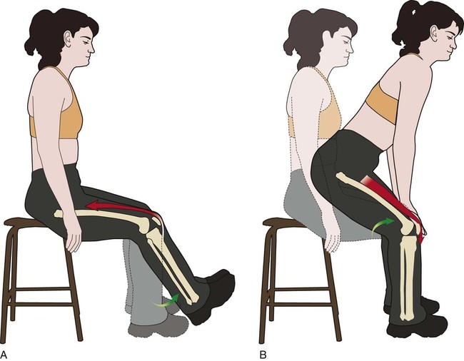
A fundamental principle of kinesiology states that when a muscle contracts, the freest (or least constrained) segment moves. This principle applies whether a muscle is pulling its distal attachment toward its proximal attachment or vice versa (Figure 3-1).


Types of Muscular Activation


An active muscle develops a force in only one of the following three ways:


These muscle activations are referred to as concentric, eccentric, and isometric, respectively.


Eccentric


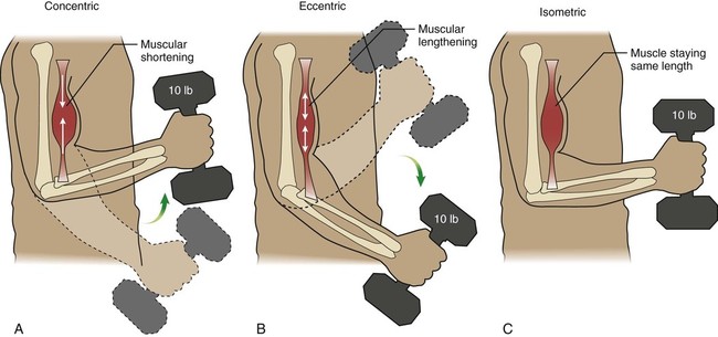
Eccentric activation occurs as a muscle produces an active force—attempts to contract—but is simultaneously pulled to a longer length by a more dominant external force. During eccentric muscular activation, the external torque, often generated by gravity, exceeds the internal torque produced by muscle. Most often, gravity or a held weight is allowed to “win,” effectively lengthening the muscle in a controlled manner. For example, slowly lowering a barbell involves eccentric activation of the elbow flexors. As a consequence, the proximal and distal attachments of the muscle become farther apart (Figure 3-2, B).


Isometric


Isometric activation occurs when a muscle generates an active force while remaining at a constant length (Figure 3-2, C). This occurs when the muscle generates an internal torque equal to the external torque; as a consequence, there is no motion and no change in joint angle.


Muscle Terminology


Specific terminology is commonly used when describing muscles or the actions of muscles. The following paragraphs outline some of these terms and their definitions.


The terms proximal attachment and distal attachment are used throughout this text to describe the relative points of attachment of muscle to bone. The proximal attachment, or origin, of a muscle refers to the point of attachment that is closest to the midline, or core, of the body when in the anatomic position. The distal attachment, or insertion, refers to the muscle’s point of attachment that is farthest from the midline, or core, of the body.


An agonist is a muscle or muscle group that is most directly related to performing a specific movement. For example, the quadriceps (knee extensors) are the agonists for knee extension. An antagonist, on the contrary, is the muscle or muscle group that can oppose the action or actions of the agonist. Usually, the antagonist muscle passively elongates as the agonist actively contracts. For example, when elbow flexion is performed, the biceps are considered the agonists as they perform elbow flexion. The triceps (elbow extensors), which are the antagonists of this action, passively elongate as the elbow is flexed. Therefore, an overly stiff antagonist muscle that fails to elongate can significantly limit the action of an agonist muscle.


A co-contraction occurs when agonist and antagonist muscles are simultaneously activated in a pure or near-isometric fashion. Co-contractions of muscle often stabilize and therefore protect a joint. Similarly, a muscle that fixes or holds a body segment relatively stationary so that another muscle can more effectively perform an action is referred to as a stabilizer.


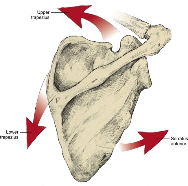
Muscles that work together to perform a particular action are known as synergists; furthermore, most meaningful movements of the body involve the synergistic action of muscles. A force-couple is a type of synergistic action that occurs when two or more muscles produce force in different linear directions but produce torque in the same rotary direction. Figure 3-3 illustrates the force-couple generated by three different shoulder muscles to upwardly rotate the scapula.


Muscles are elastic in nature and therefore are constantly being lengthened or shortened. This change in the length of a muscle is known as its excursion. Typically, a muscle can shorten or elongate only about half of its resting length. For example, a muscle that is 8 inches long at its resting length could contract to roughly 4 inches or could elongate to about 12 inches in length.


Muscular Anatomy


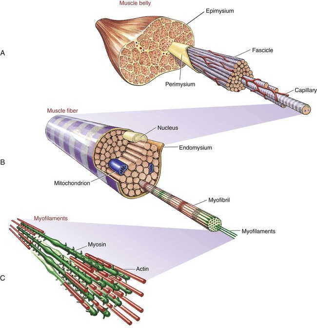
Figure 3-4 illustrates the primary functional components that constitute skeletal muscle, whereas Box 3-1 describes each of these components. A whole muscle consists of three main components, each surrounded by a particular type of connective tissue that supports its function.


The Sarcomere: The Basic Contractile Unit of Muscle
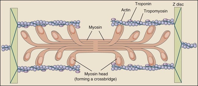

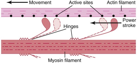
A sarcomere is the basic contractile unit of muscle fiber. Each sarcomere is composed of two main protein filaments—actin and myosin—which are the active structures responsible for muscular contraction. The most popular model that describes muscular contraction is called the sliding filament theory. In this theory, active force is generated as actin filaments slide past the myosin filaments, resulting in contraction of an individual sarcomere.


Figure 3-5 illustrates a sarcomere and emphasizes the physical orientation of the actin and myosin filaments. The thick myosin filament contains numerous heads, which when attached to the thinner actin filaments create actin-myosin cross bridges. In essence, a myosin head is similar to a cocked spring, which on binding with an actin filament flexes and produces a power stroke. The power stroke slides the actin filament past the myosin, resulting in force generation and shortening of an individual sarcomere (Figure 3-6). Because sarcomeres are joined end to end throughout an entire muscle fiber, their simultaneous contraction shortens the entire muscle.


Each myosin filament has numerous heads, and each actin filament has numerous binding sites. This is important because in order for a sarcomere to maximally contract, numerous power strokes must occur. In fact, the force of a muscular contraction is determined largely by the number of actin-myosin cross bridges that are formed. This concept is addressed later in the section on the importance of muscular length.


Form and Function of Muscle


The three following factors help determine the functional potential of a muscle: cross-sectional area, shape, and line of pull.


Cross-Sectional Area


The physiologic cross-sectional area of a muscle describes its thickness—an indirect and relative measure of the number of contractile elements available to generate force. The larger a muscle’s cross-sectional area, the greater is its force producing potential. This simple concept explains why a person with larger muscles can usually generate larger muscular forces.


Stay updated, free articles. Join our Telegram channel

Dec 5, 2016 | Posted by in MANUAL THERAPIST | Comments Off on Structure and Function of Skeletal Muscle

Full access? Get Clinical Tree

Get Clinical Tree app for offline access