Chapter
9
Soft Tissues Are Equally Important!
Bone and joints are the trees, but its roots lie in soft tissues!
What Is Special about Soft Tissues in Foot and Ankle?
Important characteristics of skin and soft tissues in foot and ankle are represented in Fig. 9.1.
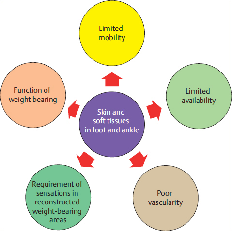
Fig. 9.1 Characteristics of skin and soft tissues in foot and ankle.
Prevention of Iatrogenic Foot and Ankle Soft-Tissue Damage
Some important tips and tricks for the prevention of iatrogenic soft-tissue damage at foot and ankle are enumerated below:
♦ Avoid putting small incisions
♦ Avoid vigorous retraction of skin
♦ Always take full-thickness flaps
♦ At tendo Achilles surgeries, incise skin and sheath together, and never create a plane between both of them
♦ A safe spacing of 4 to 6 cm between two incisions over midfoot and forefoot is mandatory to prevent skin ischemia and necrosis
♦ In an acute trauma setting, wait till wrinkles over the traumatized area are seen
♦ While waiting for wrinkles to appear and edema to settle down, always reduce subcutaneous bony prominences to prevent skin necrosis
♦ While waiting for skin condition to improve, a spanning fixator would be of help for ligamentotaxis and early settlement of edema
♦ Aspirate serous blister and wait for 3 to 5 days before operating through it
♦ Do not incise through blood-filled blisters; a healing time of 1 to 3 weeks before going in for surgery is mandatory
♦ Delicate skin flaps should be handled with blunt skin hooks and should be retracted with bent K-wires put in nearby normal bones
♦ Knots must be put outside the delicate skin flaps
♦ Anticipation of skin closure problems must be envisaged preoperatively and help and guidance of reconstructive plastic surgeon must be sought beforehand
♦ While dealing with compound injuries of foot and ankle, reconstructive plastic surgeon should always be involved at early stage for better outcome
♦ While putting external fixator frame or ring fixator, careful planning is required to not violate future areas of soft-tissue reconstruction
Management of Soft-Tissue Defects
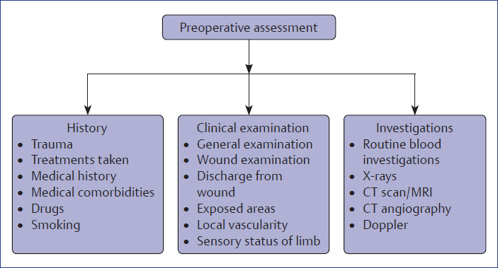
Preoperative Assessment
It includes three steps, which are further separately enumerated in Flowchart 9.1.
Prerequisites
Some of the prerequisites for the management of soft-tissue defects include:
♦ Sequential radical debridement
♦ Definitive or temporary skeletal stabilization
♦ Early soft-tissue coverage (Flowchart 9.2)
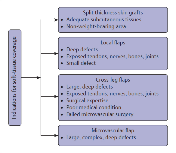
Flowchart 9.2 Soft-tissue coverage.
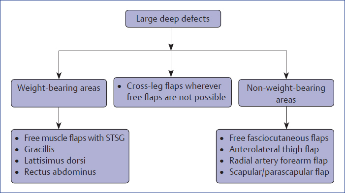
A brief overview of the soft-tissue defects is illustrated in Flowcharts 9.3 to 9.5.
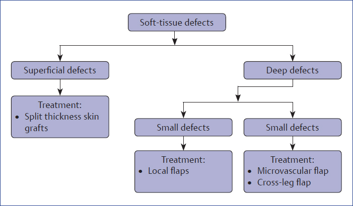
Flowchart 9.3 Overview of the soft-tissue defects.
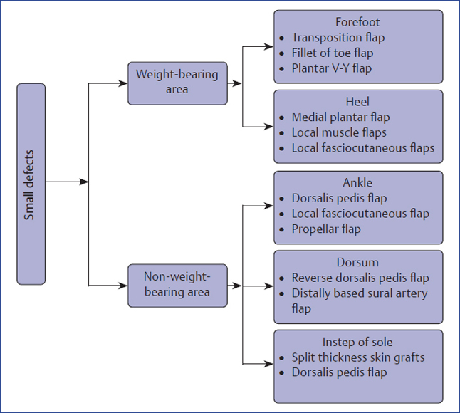
Flowchart 9.4 Overview of the soft-tissue coverage for small defects.
Region-Wise Soft-Tissue Coverage Options
Ankle
♦ Local flaps: used for smaller defects
♦ Distant flaps: used for larger defects
Commonly used locoregional flaps for ankle are as follows:
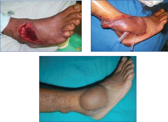
1. Distally based sural artery flap: It is a fasciocutaneous flap that is elevated from the posterior aspect of the calf along with sural nerve and small saphenous vein. It is based on a perforator of the peroneal artery that emerges 5 cm proximal to the lateral malleolus and then runs cephalad. It can be raised as an island flap along with neurovascular bundle or as a pedicle flap. It is a very useful flap for lower one-third of leg and ankle defects. Disadvantages are visible donor defect at the calf and loss of sensations at the lateral border of the foot (Fig. 9.2).
Related posts:

Stay updated, free articles. Join our Telegram channel

Full access? Get Clinical Tree


