Chapter
12
Lucid Approach and Simplistic Management of Diabetic Foot
India is the diabetic capital of the world!
Alarming Facts about Diabetic Foot
♦ More than 135 million diabetics worldwide
♦ Number would reach up to 300 million by 2025
♦ India has second largest (70 million) diabetic population after China
♦ India would overtake China by 2025
♦ India has 35 million prediabetes patients
♦ 15% of confirmed diabetic patients will get ulcers once in their lifetime
♦ 1% will undergo higher-level amputation
♦ 200,000 higher-level amputations per year are done in India for infected diabetic foot ulcers/wounds and gangrene
♦ 50% of people who have amputation of one limb would need amputation of the other limb within 5 years
♦ Mortality rate following amputation is
• 13% to 40% at 1 year
• 35% to 65% at 3 years
• 39% to 80% at 5 years
♦ 85% of these amputations can be completely prevented by simple cost-effective measures
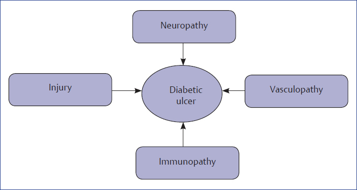
Strategy to prevent an ulcer is to preserve and protect epidermal barrier in the presence of advancing neuropathy, vasculopathy, and loss of pain sensations!
Fig. 12.1 shows the reasons for ulcers in diabetic foot.
Clinical Examination of Foot to Assess the Risk of Ulcer
Three types of clinical examination can be done as shown in Box 12.1.
Box 12.1 Types of clinical examination of diabetic foot.
♦ 2-minute examination
• Vascular examination
• Web space examination
• Status of foot skin
• Examination of ulcer
• Foot wear examination
♦ 5-minute examination
• All examinations as in 2 minutes
• Monofilament test (Fig. 12.2)
• Test to check status of intrinsic muscles
• Measurement of 1st toe extension with go niometer (Fig. 12.5)
♦ 10-minute examination
• All examinations as in 2 + 5 minutes
• Ankle–brachial index
• Test for heat andcold sensations
• Test for vibration perception
The following are the tests done in case of diabetic foot ulcer.
♦ Monofilament test: Monofilament pressure testing of the sole of the foot is the simplistic means of diagnosis of neuropathy. Lack of feeling of pressure is the neuropathy (Fig. 12.2).

♦ Test for vibratory sense: Biothesiometer measures vibratory sense. The probe is put at various areas of the sole of the foot to evaluate the perception of vibrations. Vibratory sensations are lost very early in the setting of neuropathy (Fig. 12.3).
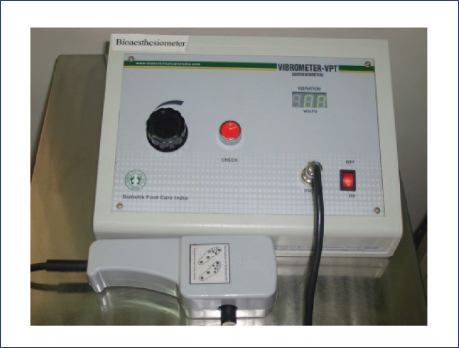
Fig. 12.3 Biothesiometer.
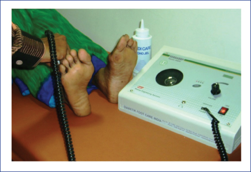
♦ Test for the evaluation of blood flow: Vascular Doppler will evaluate the blood flow patterns in dorsalis pedis and posterior tibial arteries of foot and ankle. The kind of flow waves and the pattern of flow give an idea about the vascular status of the limb (Fig. 12.4).
♦ Test to measure movements: Goniometer is used to measure the movements of forefoot joints. In diabetics, loss of these movements would be an early predictor of development of stiffness and deformities, which in turn may lead to ulcers. Fig.12.5 shows the method of measurement of first metatarsophalangeal (MTP) joint extension with a goniometer.
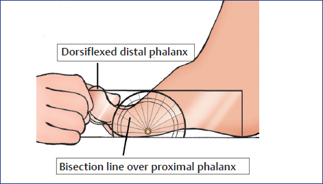
Fig. 12.5 Test to measure movements.
Radiology
♦ Plain X-rays:
• Active infection in soft tissues: Depicted in the form of swelling, fat plane obliteration, gas in cases with abscess or cellulitis due to gas-forming organisms (Fig. 12.6)
• Ulcer: A soft tissue defect
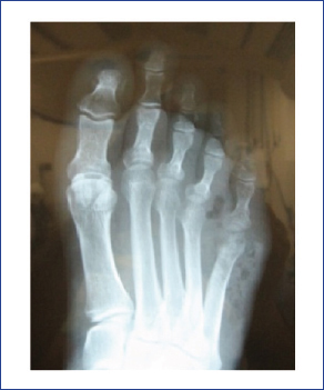
• Bony lesions: Bony erosions, osteomyelitis, Charcot neuroarthropathy, and arthritis of joint (Fig. 12.7–12.9).
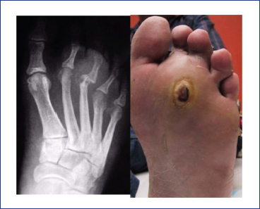
Fig. 12.7 Radiograph showing erosions in the head of third metatarsal in a patient with diabetic foot ulcer.
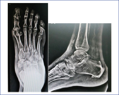
Fig. 12.8 X-ray showing Charcot midfoot.
Related posts:

Stay updated, free articles. Join our Telegram channel

Full access? Get Clinical Tree


