Fig. 36.1
The soft tissue mass appearances at the distal part of the forearm
Before the diagnosis of primary lesion, a portion of the patients may refer with signs of metastatic disease, due to silent or vague symptoms. In such cases, evaluation of the patients by algorithms for musculoskeletal tumors will be appropriate, while unnecessary tests and time loss can be avoided.
Physical Examination
Physical examination should be carried out following general systemic examination to assess the mass and its mechanical effects on the surrounding tissues. Skin alterations over the mass, peripheral edema, muscle atrophies, relation with contiguous anatomical structures, sensorial and motor deficiencies should be evaluated. In particular, the pulse abnormalities proximal or distal to the lesion, in comparison with the unaffected side, should be investigated by advanced tests.
The detection of distant metastases by physical examination may even be possible. While lungs are the most involved side of distant metastasis, meticulous evaluation of the respiratory system must be performed for the soft tissue sarcoma patients.
Laboratory Tests
Although there are no current specific biochemical markers for soft tissue sarcomas, indicators of muscle destruction like LDH and creatine kinase can be used. However, due to high levels of these markers after a trauma, alterations are not always that meaningful.
Metastases of STS may cause an increase in the biochemical parameters by dysfunction of the involved organ.
Imaging Methods
The role of imaging modalities in the evaluation of STS is unquestionable. Plain radiography, ultrasonography, computerized tomography (CT), magnetic resonance imaging(MRI), positron emission tomography(PET), angiography, and scintigraphy hold an important place in the radiological assessment of these lesions.
With the recent advances on imaging techniques, perfusion-diffusion imaging of lesions, three-dimensional anatomy and determination of relationship with the surrounding structures is possible.
As an inexpensive and noninvasive method, ultrasonography is used in the initial assessment of a soft tissue mass, by evaluation of liquid and solid structure and content of the vascularity.
Computerized tomography can be used for the determination of mediastinal and retroperitoneal placement in relation to lung and other organ systems and also useful in the evaluation of detecting metastases in these regions (Fig. 36.2).
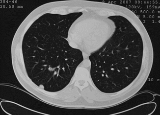
Fig. 36.2
Thoracic CT showing lung metastasis
Magnetic resonance imaging is used as a screening tool, for diagnostic and staging purposes and also for monitoring the treatment. MRI gives indirect information about the content and nature of the tissue by using various sequences, as well as the overall appearance of the lesion. Heterogeneity and the capsular structure of the tumoral tissue can be demonstrated with MRI. Intra-lesional heterogeneity can be observed in 95 % of T2-weighted images of the malignant tumors, while it was only 20 % in benign tumors. The presence of septations is another finding that gives a malignant impression [11]. While necrosis in the tumor can be demonstrated by contrast-enhanced images, the absence of necrosis does not rule out malignancy (Figs. 36.3, 36.4, and 36.5).
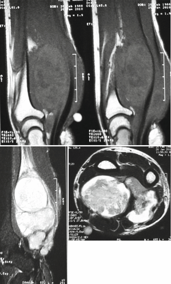
Figs. 36.3, 36.4, and 36.5
Malignant soft tissue tumor as hypointense in sagittal T1-, hyperintense in T2-weighted images, and heterogeneity in contrast-enhanced T1-weighted image
In recent years, information about tumor physiology can be obtained through the evaluation of perfusion and diffusion of contrast agents by dynamic MRI. Rapid contrast enhancement in dynamic MRI is meaningful in terms of malignancy. MRI and CT also play an important role in monitoring and follow-up of the treatment and detection of metastases. Scintigraphy is used both for determination of local invasion and distant metastases of the tumor. Gallium citrate can be used for the detection of non-pulmonary metastases and lymphatic involvement in pediatric rhabdomyosarcomas [12].
Biopsy
Biopsy is considered as the gold standard for the diagnosis of musculoskeletal lesions. However, using this precious diagnostic tool as a first-line method is not the right approach. Biopsy procedures must be applied after all diagnostic methods have been performed to confirm the presumptive diagnosis. An improper biopsy may affect the patient’s prognosis and treatment, as well as blurring the definitive diagnosis [13, 14]. Biopsy should be carried out in a tertiary center by the same team who will perform further treatment. Biopsy is an invasive approach that has to be applied through the shortest way to reach the tumor and that can be removed during the definitive surgery. To avoid bleeding that can cause dissemination of tumoral cells, cold application and short-term restriction of movements in the joints near by the biopsy site would be appropriate.
Biopsy Types
Fine needle aspiration biopsy (FNAB)
Core needle biopsy
Incisional biopsy
Excisional biopsy
Biopsy type is selected according to lesion size, location, and pathologist’s experience.
FNAB
Performed with 21–23 Gauge needles, it requires an experienced cytopathologist. Detection of sarcoma type and grade may fail with this method. Atraumatic and inexpensive application are the advantages [15]. In deep lesions, FNAB can be performed by guidance of CT. The risk of contamination is very low. It is a simple and useful method, particularly in retroperitoneal and intra-abdominal lesions. Very successful in the detection of distant metastases and recurrence of prediagnosed sarcomas.
Core Biopsy
Provides tissue 1–10 mm in thickness. It is usually performed by Tru-cut needles. The harvested tissue may be insufficient for diagnosis and grading. Enough material may not be achieved for special staining and electron microscopy [16]. It is still a commonly used method (Fig. 36.6).
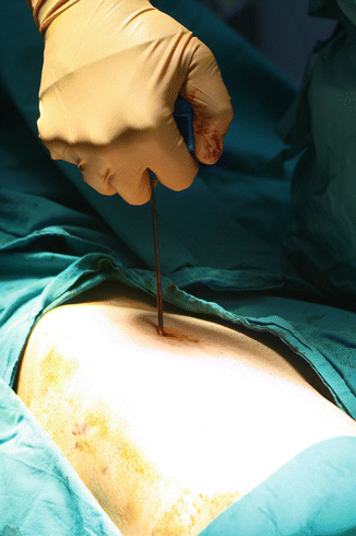
Fig. 36.6
Core biopsy
Incisional Biopsy
Applied in most soft tissue tumors. Longitudinal incisions should be used, especially in the extremities. Incisions must be carried out directly from the closest place to tumor. Appropriate hemostasis should be provided, drains should be avoided, if used it should be in or just near the incision side. If the lesion was diagnosed as malignant, drain pathway should also be excised during the definitive surgery.
Excisional Biopsy
Should be performed for lesions smaller than 3 cm, with lower risk of malignancy and for cases that would not risk the subsequent treatment even diagnosed as sarcoma. If it is done in deep lesions, the probability of contamination is very high and may adversely affect the subsequent treatment.
Staging of Soft Tissue Sarcomas
Staging of soft tissue sarcomas is very important in determining the treatment and prognosis. Therefore, various staging systems have been developed (Table 36.1). These are as follows:
Table 36.1
AJCC and Enneking staging systems for soft tissue sarcomas
American Joint Committee on Cancer (AJCC) staging system | ||||
|---|---|---|---|---|
Stage | Grade | Tumor size and localization | Lymph nodes | Metastases |
IA | Low | ≤5 cm | − | − |
IB | Low | >5 cm, superficial | − | − |
IIA | Low | >5 cm, deep | − | − |
IIB | High | ≤5 cm | − | − |
IIC | High | >5 cm, superficial | − | − |
III | High | >5 cm, deep < div class='tao-gold-member'>
Only gold members can continue reading. Log In or Register to continue
Related posts:Stay updated, free articles. Join our Telegram channel
Full access? Get Clinical Tree
 Get Clinical Tree app for offline access

| ||
