Fig. 9.1
Dorsoradial portal: radial to the APL tendon. Dorsoulnar portal: ulnar to the EPB tendon
Tower traction of about 10 lb is applied with the thumb secured by finger trap. Sterile adhesive tapes are applied to reinforce the trapping to avoid sudden giving way of the traction which may damage the delicate arthroscope. Further adhesive tapes are applied between the hand dorsum and the traction tower to stabilize the position of the hand so that the dorsum of the thumb (or the nail) is facing the surgeon.
PSLA (portal site local anaesthesia) using 2 % lignocaine with 1:200,000 adrenaline to infiltrate the skin and the joint capsule was injected with a 25 gauge needle. Around 2 ml of normal saline is injected through 1U portal to inflate the joint. A transverse 2 mm skin incision just passed through the dermis was made over 1U, and this is followed by puncture of the joint capsule using a haemostat. This will avoid injury to the cutaneous branches of radial nerve. A blunted-end trocar and cannula for the 1.9 mm scope was inserted and sweep transversely to dilate the joint space.
A tourniquet is not necessary. A 3L normal saline bag being hanged at 1.5 m high is connected to the cannula of the arthroscope. Continuous saline infusion through the cannula can allow clear visualization and adequate haemostasis.
We use a 1.9 mm arthroscope with 30° of visualization. Once the scope is inside the joint, the specific shape of the first CMCJ is appreciated. This joint had a double saddle shape, with the medial-lateral axis of trapezium in concave and the anteroposterior axis in convex. The articular surface on the first metacarpal base is just the opposite. The anterior oblique ligament (beak ligament) can be visualized from the 1U portal.
9.2.2 Indications
1.
Acute injury: fracture reduction and fixation
Arthroscopic-assisted reduction and fixation of intra-articular fracture involving first metacarpal base (e.g. Bennett’s fracture) is recommended. The quality of fracture reduction is difficult to be assessed under fluoroscopic guidance. Arthroscopy provides an advantage of more accurate reduction, and it allows the preservation of important ligamentous and capsular structures. The first K-wire should be driven obliquely from the base of the first metacarpal, aiming at the ulnar base fragment. The arthroscope should then be introduced under traction. Reduction was achieved by manipulation of the thumb (usually by axial traction and rotation), with the assistance by a 2 mm probe. When the reduction is satisfactory, the K-wire is further driven across to the smaller ulnar fragment, then fixing at the trapezium or trapezoid. An addition K-wire is inserted horizontally to transfix the first metacarpal shaft with second metacarpal shaft to enhance stability. The patient is then put in a thumb spica cast for 4 weeks or until fracture union.
2.
Post-traumatic arthritis
Post-traumatic arthritis is occasionally seen in active athletes. The causes of pain in such cases may be due to synovitis or loose bodies. Arthroscopy can be utilized in these cases for debridement of the synovitis and removal of loose bodies. Shaving of post-traumatic synovitis can be performed with the use of small diameter shaver. Caution about introducing iatrogenic trauma to the articular cartilage is very important when working in such a limited space, and shaving should start only when the shaver is visualized. Radiofrequency ablation probe can also be used for this purpose, but maintenance of continuous normal saline inflow is necessary to prevent thermal injury to the joint.
9.3 Metacarpophalangeal Joint (MCPJ) Arthroscopy
9.3.1 Surgical Technique
The procedure can be performed as an outpatient with portal site local anaesthesia. The patient is placed in a supine position with shoulder abducted and the arm supported on an arm board. No tourniquet is required. We use a 1.9 mm arthroscope with 30° angle of visualization. Other instruments include 2.0 mm shaver, mini VAPR, mini Vulcan and mini arthroscopic instruments. A finger trap is applied to the affected finger. Traction is applied at 5–10 lb of tension with a traction tower (Fig. 9.2). A two-portal technique is used, a dorsoradial portal and a dorsoulnar portal, each on either side of the central extensor tendon (Fig. 9.3). Once on traction, the vacuum sign becomes apparent and the joint space can easily be determined by thumb palpation. The intended port sites are marked with a marking pen. Local anaesthesia with 1–1.5 ml of 2 % lignocaine admixed with 1:200,000 adrenaline is injected to the port sites with fine 25G needle from skin down to joint capsule level. Intra-articular injection is optional and the needle can be used to confirm the joint space. Two millilitres of normal saline is injected via either port site for distention of the joint space. A longitudinal or oblique incision along skin crease of less than 0.5 cm is made with a No. 15 blade. The subcutaneous tissue is dissected and extensor expansion is perforated by blunt dissection with a haemostat forceps. Joint space is confirmed once gush of saline fluid egressed. Next, a cannula with blunted-end trocar is introduced into the left-sided portal (i.e. dorsoulnar for a left hand, dorsoradial for a right hand). Gentle force should be used to avoid scooping of articular cartilage of the metacarpal head, particularly in tight joint. The trocar is then removed. Continuous saline infusion using 3L normal saline bag under gravity is being connected to the cannula. Saline solution is allowed to fill up the cannula before the arthroscope was inserted into the joint to avoid trapping of air bubble inside the joint. The right-sided portal (i.e. dorsoradial for a left hand, dorsoulnar for a right hand) is used for instrumentation. The two portals are interchangeable. It is recommended to use the finger pivoting technique with the index or middle finger tip to fine control the lens motion as the joint is relatively small and shallow. It is particularly important in dealing with dorsal-sided pathology such as chondral lesion or synovitis to avoid the frequent dropping of the arthroscope out of the joint.
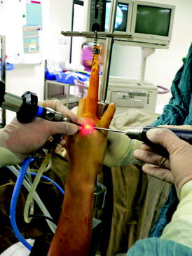
Fig. 9.2
Setting of MCPJ arthroscopy
Fig. 9.3
First MCPJ: dorsoradial portal – radial to EPB tendon, dorsoulnar portal: ulnar to EPL tendon. Second to fifth MCPJ: dorsoradial portal – radial to central slip, dorsoulnar portal – ulnar to central slip
The metacarpophalangeal joint is a relatively simple joint to examine arthroscopically as there is only one joint space. With the 30° arthroscope inside the joint, the dome-shaped cartilaginous surface of the metacarpal head should be apparent on the floor while part of the concave articular surface of the base of proximal phalanx can be seen in the upper part of the view. The volar plate can be seen between the two articular surfaces. However, the volar recess cannot be visualized normally due to its anterior and proximal location relative to the head. However, one can use a probe to palpate the recess. By rotating the lens in 360° manner, a much wider area of both articular surfaces can be reached. The dorsal part of the MC head and the accompanying dorsal synovial recess and capsular reflection can be seen with the lens facing downwards. This is an important part of the joint to be assessed as many painful chondral lesions tend to locate in this area, together with the associated synovitis. The synovitis needs to be debrided with a small shaver of 2 mm in order to reveal the true extent of any chondral lesion. Alternatively radiofrequency apparatus with mini-probe is equally effective. However, the use at the dorsal side of the joint should be with extreme caution due to the vicinity to the extensor mechanism. Over the volar plate area, the long flexor tendon is also at risk of thermal damage, and the use of thermal apparatus should be limited with caution. Towards the radial side, the arthroscope can follow the articular surface of the metacarpal head to sweep into the radial gutter to reach the radial synovial recess where the radial collateral ligament originates. The ligament can be assessed for any substance tear, avulsion and laxity and any loose body can be removed. It may be difficult to see the ulnar gutter and ulnar synovial recess from the radial side, and the arthroscope needs to be inserted from the dorsoulnar portal in order to have a more thorough assessment.
After the procedure, the skin wounds are apposed with sterile strip, and bulky dressing was applied for 2 days and then changed to a light dressing. The post-operative mobilization varies for different patients based on the underlying pathology. Patients with acute collateral ligaments tear required immobilization after the operation; otherwise, patients were encouraged to start free mobilization exercise as long as pain allowed.
9.3.2 Indications
Indications of MCPJ arthroscopy for athletes of ball sports injury can be separated into acute injury and chronic injury.
9.3.2.1 Acute Injury
1.
Acute collateral ligaments injury/gamekeeper’s thumb
The use of MCPJ arthroscopy for the treatment of gamekeeper’s thumb was first described by Ryu in 1995 [5]. Although the Stener lesion can be diagnosed with ultrasound or an MRI scan, these methods are less sensitive than direct visualization using the arthroscope. The role of MCPJ arthroscopy in cases of gamekeeper’s thumb includes detection of a Stener lesion and reduction of a Stener lesion if necessary. After the reduction of the collateral ligament to its original position, the thumb should be immobilized with a cast for 4 weeks. Badia [6] described the use of arthroscopy in the reduction of a bony gamekeeper’s avulsion fracture. With this technique, more precise reduction of the articular fragment is achieved with minimal capsular scarring and resulted in faster recovery.
2.


Reduction of intra-articular fracture
Metacarpal head fracture is uncommon but are occasionally seen in young, active athletes. Frequently patients may present with mild swelling and slight decrease in range of motion, and they may be missed if x-ray films are not well taken. Therefore, a true anteroposterior and lateral x-ray films must be included. As for all intra-articular fracture, the aim of treatment is to achieve anatomical reduction, stable fixation and early mobilization. In patients with metacarpal head intra-articular fracture, we can make use of MCPJ arthroscopy for direct visualization to achieve anatomical reduction. The fracture can be fixed in a retrograde manner with an absorbable pin.
Related posts:

Stay updated, free articles. Join our Telegram channel
Full access? Get Clinical Tree
