Radiographic measurement
Normal range
Dysplastic hip
Miscellaneous
Lateral center-edge angle
25–50°
<20°
AP radiograph
Anterior center-edge angle
24–45°
<20°
False profile
Acetabular angle
33–38°
>42°
AP radiograph
Tönnis angle
<10°
>10°
AP radiograph
% head coverage
80 %
<70 %
AP radiograph
Teardrop
Narrow teardrop
Widened “U” morphology
AP radiograph
Medial space
10–15 mm
>15 mm
AP radiograph
The most commonly utilized radiographs for evaluation of the hip include an anteroposterior (AP) view of the pelvis [8, 33], a cross-table lateral view [34], a false-profile view [35], a frog-leg lateral view [36], and a 45° or 90° Dunn view [37]. Each view provides important information about the pathomorphology. They are highly technique dependent, and standardized views must be provided in order to improve diagnostic accuracy and disease classification.
The anteroposterior pelvic radiograph can provide significant information when obtained using these standardized techniques. The subject’s lower extremities should be oriented in 15° of internal rotation in order to maximize the femoral neck length. The image should be centered between the superior border of the pubic symphysis and a line drawn connecting the anterior superior iliac spines to visualize the entire bony pelvis and to minimize scatter and parallax effect. A good AP pelvic radiograph should have neutral tilt, and the distance between the sacrococcygeal junction should be 2.5–4 cm above the superior end of the symphysis in males and between 4 and 5.5 cm in females [14]. Some authors prefer using the tip of the coccyx as opposed to the sacrococcygeal junction due to increased ease in identifying this anatomic landmark. In this scenario, the tip of the coccyx should be centered 1–3 cm above the pubic symphysis to ensure appropriate pelvic inclination [38]. This assures that pathology identified is true and not a rotational misinterpretation.
A dysplastic hip on AP radiograph can be represented by a shallow acetabulum with a widened teardrop and lateralized center of rotation, with anteversion or retroversion. The acetabulum is considered anteverted if there is no evidence of crossover sign. This is determined by the line of the anterior aspect of the rim not crossing the line of the posterior aspect of the rim before reaching the lateral aspect of the sourcil. If the acetabulum is retroverted, the line of the anterior aspect of the rim does cross the line of the posterior aspect of the rim before reaching the lateral edge of the sourcil (Fig. 1). Retroversion is also frequently associated with a prominent extension of the ischial spine into the pelvis [39]. There is often an upsloping sourcil with a dysplastic hip; the sourcil consists of the radiodense subchondral bone of the weight-bearing dome of the acetabulum. This finding can be quantified by the acetabular roof angle of Tönnis or the acetabular index of the weight-bearing zone; this is the angle formed between a horizontal line to the pelvis and a line extending from the medial to the lateral edge of the sourcil (Fig. 2). A Tönnis angle of 0–10° is considered normal, whereas an angle of >10° would be considered abnormally steep and consistent with dysplasia [40]. Similarly, the acetabular angle of Sharp, also referred to as the acetabular index or acetabular inclination (Fig. 3), identifies the inclination of the acetabulum by measuring the angle formed between a horizontal line spanning along the base of the two radiographic acetabular teardrops and a line from the ipsilateral inferior teardrop to the lateral sourcil. Dysplastic hips often have acetabular angles greater than 42° [41]. The lateral center-edge angle of Wiberg on the AP radiograph assesses the acetabular superolateral coverage (Fig. 3). This is the angle formed by the intersection of a vertical line through the center of the femoral head and a line extending through the center of the femoral head to the lateral sourcil. Hip dysplasia is defined as a lateral center-edge angle less than 20° [2].
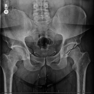
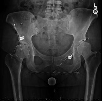
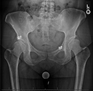
Fig. 1
Acetabular retroversion with a crossover and prominent ischial spines
Fig. 2
Demonstrates a lateral center-edge angle of the right hip of 15°and acetabular roof angle or Tönnis angle of 21°. These measurements are both considered abnormal and consistent with acetabular dysplasia
Fig. 3
Demonstrates a lateral center-edge angle of the right hip of 15° and Sharp’s acetabular angle of the left hip of 48°. These measurements are both considered abnormal and consistent with acetabular dysplasia
A false-profile view [35] is obtained by positioning the patient standing with the symptomatic hip against the cassette with the foot parallel to the cassette. The pelvis is rotated 65°, so the radiograph is centered at the femoral head. This view shows anterior acetabular coverage of the femoral head and can show anterior subluxation during weight bearing in the dysplastic hip. The examiner can measure the anterior center-edge angle of Lequesne by identifying the angle formed by the intersection of a vertical line through the center of the femoral head to the anterior sourcil. Dysplastic hips often have measurements less than 20° [35] (Fig. 4).
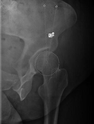
Fig. 4
Demonstrates an anterior center-edge angle of Lequesne on a false-profile radiograph
A cross-table lateral radiograph [42] is obtained with the patient supine on the x-ray table. The contralateral hip and knee are flexed up out of the field of the hip being imaged. The imaged extremity is internally rotated 15° to expose the full anterolateral and posterolateral femoral head-neck junction. The x-ray tube is orientated parallel to the table at a 45° angle to the imaged limb and centered on the femoral head.
The frog-leg lateral view [36] has the patient positioned supine on the x-ray table with the symptomatic hip abducted to 45° with the knee bent. The radiograph is centered midway between the pubic symphysis and the anterior superior iliac spine.
Finally, the Dunn lateral view [37] has been described with the radiograph taken either with the hip flexed to 45° or 90° and is referred to as the 45° Dunn lateral or the 90° Dunn lateral, respectively. The radiograph is obtained the same way despite the degree of hip flexion. The symptomatic hip is flexed to either 45° or 90° with 20° of abduction. The limb is placed in a neutral rotation. The beam is centered at a point midway between the pubic symphysis and the anterior superior iliac spine.
The cross-table lateral, the frog lateral, and the Dunn lateral views have slight projectional variations and highlight certain pathology on the proximal femur such as head sphericity, anterior and posterior outlines of the femoral neck, alpha angle, and head-neck offset. The radiograph of choice is often physician specific, however should be kept constant throughout the practice.
There are several described radiographic classification systems associated with acetabular dysplasia; Table 2 outlines the three more commonly used classifications. The first is the Crowe classification [43] in which Crow I describes minimal to no abnormal development, with <50 % subluxation of the hip joint and <10 % proximal femoral displacement. Crowe II describes dysplastic morphology with 50–75 % subluxation and 10–15 % proximal femoral displacement. Crowe III shows severe acetabular dysplasia often with the femur dislocated or near dislocated with 15–20 % proximal femoral displacement. Finally Crowe IV is considered a high dislocation with >20 % proximal migration. The second classification system noted in Table 2 is the Hartofilakidis classification, which was described to predict the bony deficiencies encountered during the operation from preoperative pelvis radiographs. Hartofilakidis type A is described as a dysplastic acetabulum, type B is a low lying dislocation, and type C describes a high riding dislocation.
Table 2
Hip dysplasia classification systems
Femoral head subluxation | Proximal displacement | |
|---|---|---|
Crowe classification [43] | ||
Crowe I | <50 % subluxation | <10 % |
Crowe II | 50–75 % subluxation | 10–15 % |
Crowe III | 75–100 % subluxation | 15–20 % |
Crowe IV | >100 % subluxation | >20 % |
Hartofilakidis classification [44] | ||
Type A (dysplasia) | Femoral head within acetabulum despite some subluxation. Segmental deficiency of the superior wall. Inadequate true acetabulum depth | |
Type B (low dislocation) | Femoral head creates a false acetabulum superior to the true acetabulum. There is complete absence of the superior wall. Inadequate depth of the true acetabulum | |
Type C (high dislocation) | Femoral head is completely uncovered by the true acetabulum and has migrated superiorly and posteriorly. There is a complete deficiency of the acetabulum and excessive anteversion of the true acetabulum | |
Tönnis classification of osteoarthritis by radiographic changes [40] | ||
Grade 0 | No signs of OA | |
Grade 1 | Increased sclerosis, slight joint space narrowing, no or mild loss of head sphericity
Related posts: Neuromuscular Hip Disorders: Focus on Cerebral Palsy Neuromuscular Hip Disorders: Focus on Cerebral Palsy
 Surgical Technique: Bone Graft for Avascular Necrosis of the Hip Surgical Technique: Bone Graft for Avascular Necrosis of the Hip
 Rehabilitation of Non-Operative Hip Conditions Rehabilitation of Non-Operative Hip Conditions
 Surgical Technique: Open Proximal Hamstring Repair Surgical Technique: Open Proximal Hamstring Repair
 Subspine Impingement and Surgical Technique Subspine Impingement and Surgical Technique
 Atraumatic Instability and Surgical Technique Atraumatic Instability and Surgical Technique
Stay updated, free articles. Join our Telegram channel
Full access? Get Clinical Tree
 Get Clinical Tree app for offline access

| |